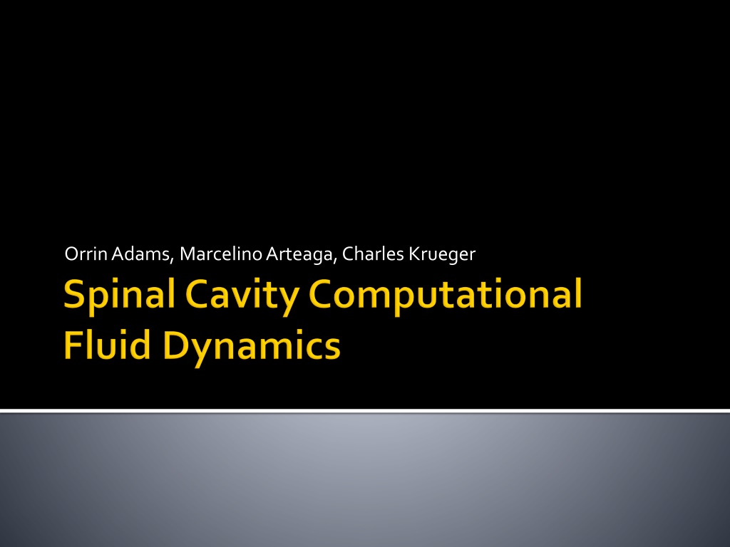
3D Printed Spinal Cord Cavity Model for Fluid Analysis
"Explore the process of building a model of the spinal cord cavity in CATIA for 3D printing and fluid analysis. From idealized nerve groups to creating a Dura cavity, learn how MRI data was used to place rootlets along the spine. Dive into the innovative project details and methodologies."
Download Presentation

Please find below an Image/Link to download the presentation.
The content on the website is provided AS IS for your information and personal use only. It may not be sold, licensed, or shared on other websites without obtaining consent from the author. If you encounter any issues during the download, it is possible that the publisher has removed the file from their server.
You are allowed to download the files provided on this website for personal or commercial use, subject to the condition that they are used lawfully. All files are the property of their respective owners.
The content on the website is provided AS IS for your information and personal use only. It may not be sold, licensed, or shared on other websites without obtaining consent from the author.
E N D
Presentation Transcript
The purpose of this project was to build a model of the spinal cord cavity in CATIA that could be 3D printed and used for fluid analysis. We started with STL files of the spinal cord and dura(the sheath around the cord that contains the fluid). Spinal Cord Dura
3D printed idealized nerve groups were used to support the spinal cord. Due to their size these nerves had to be idealized so they could be printed. Nerve rootlets form bundles and groups as they exit the dura and dimensions obtained from an MRI scan were used. Idealized Cervical Nerve Roots
Using Boolean subtraction a cavity in the shape of the Dura was created. Dura Cavity
Using MRI data, the idealized rootlets were placed along the spine. At the tail end simplified cylinders were used to provide support while still representing existing nerves. Spinal Cord + Rootlets


