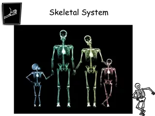
Overview of Microbiology in Bone and Joint Infections
Bone and joint infections can have devastating effects if not treated promptly. They can be caused by blood-borne spread or local trauma and are more common in infants and children. Acute osteomyelitis, a common type of bone infection, can last from a few days to several weeks or months. Understanding the risk factors associated with different routes of infection is crucial for early diagnosis and appropriate treatment. Recognizing the patient presentation, conducting a thorough differential diagnosis, and employing laboratory tests are vital steps in diagnosing bone and joint infections.

Download Presentation

Please find below an Image/Link to download the presentation.
The content on the website is provided AS IS for your information and personal use only. It may not be sold, licensed, or shared on other websites without obtaining consent from the author. Download presentation by click this link. If you encounter any issues during the download, it is possible that the publisher has removed the file from their server.
E N D
Presentation Transcript
Microbiology of Bone and Joint Infections MUSCULOSKELETAL BLOCK PROF.HANAN HABIB DEPARTMENT OF PATHOLOGY KSU
Introduction Bone & joint infections may exist separately or together. Both are more common in infants and children Usually caused by blood borne spread ,but can result from local trauma or spread from contiguous soft tissue infection. Often associated with foreign body at the primary wound site. If not treated lead to devastating effect
What is Acute Osteomyelitis Acute osteomyelitis is acute infectious process of the bone and bone marrow . Can have a short duration few days for hematogenously acquired infection last several weeks to months if secondary to contiguous focus of infection) In association with peripheral vascular disease diabetes mellitus ,severe atherosclerosis, vasculitis
How they reach Risk Group Primary Hematogenous route (Metaphysis of long bones ) Children common Adult less common (quiescent from childhood) Infant S.aureus, group B streptococci, E.coli. Children S.aureus, group A streptococci, H.influenzae. -S. Aureus septic arthritis diaphysis -Vertebral- GU infection -Candida Venous Catheter Contiguous soft tissue focus Post operative infection, contaminated open fracture, soft tissue infection , puncture wounds Gram positive cocci, Gram negative bacilli, anaerobes, and poly-microbial infection. Special clinical situations Prosthesis Coagulas -negative staphylococci, Propionebacterium, and S.aureus in foreign body infections Nosocomial infections and IV drug use Enterobacteriacea and Pseudomonas Fist injuries, and diabetic foot and dicubitus ulcers, Streptococci and anaerobes in sickle cell patients Salmonella or S. pneumoniae Human/ animal bites; Eikenella, Pasturella multocida AIDS. M.tuberculosis or M. avium
Patient Presentation Systemic manifestations occurs in less than 50% of patients. Acute onset of bone pain, fever with rigors and diaphoresis. Symptoms usually of less than 3 week s duration. Local signs : soft tissue swelling, erythema, warmth, point tenderness, percussion tenderness over the vertebral body & limited mobility of the involved extremity.
Differential diagnosis Primary and metastatic bone malignancies Trauma Acute rheumatic arthritis Hemarthrosis Ewing sarcoma Vertebral compression fracture.
Diagnosis Laboratory Radiological -CBC diff -Leukocytosis may or may not occur -Erythrocyte sedimentation rate ( ESR) elevated, but could be normal as well -Blood culture. -Aspiration of overlying abscess if blood cultures are negative -X-ray : normal early in disease, soft tissue swelling , subperiosteal elevation seen early. Bone destruction changes seen by 2-4 weeks. -MRI highly sensitive & specific. Preferred for vertebral osteomyelitis and cases associated with contiguous foci of infection or peripheral vascualr disease. -CT Scan used as alternative of MRI. -Technetium bone scan, Gallium and Indium -111- labelled WBC scan ( detection within 3 days of onset). Maximum effect to rule out osteomyelitis.
Organisms Antibiotics Duration/Surgery/complication and follow up MSSA: Cloxacillin, cefazolin or Clindamycin . Early treatment is critical Treat for 2-4 weeks parenteral followed by oral therapy for a total of at least 6 weeks. MRSA: Vancomycin followed by Clindamycin, Linezolid, or TMP- SMX Polymicrobial infection: Ampicillin-Sulbactam, Piperacillin- Tazobactam or Quinolone with Metronidazole. Surgery for neurological complications, para-vertebral abscess & hip joint involvement. Complications: septicemia, metastatic abscesses, septic arthritis, chronic osteomyelitis, loss of limb ,or paravertebral abscess. S.epidermidis: Vancomycin and Rifampicin Enterobacteriaca e: Ceftriaxone Other Gram negative bacilli: Quinolones Monthly ESR for 3 months and at 6 months useful to document treatment. P. aeruginosa: Cefepime, Meropenem, or Piperacillin +/- Aminoglycoside. Cases due to contiguous source more difficult to eradicate .Relapse common (50%) , surgery indicated. Anaerobes: Metronidazole or Clindamycin
Chronic Osteomyelitis A chronic infection of the bone and bone marrow usually secondary to inadequately treated or relapse of acute osteomyelitis. Management difficult , prognosis poor. Infection may not completely cured. May recur many years, decades, after initial episode. Most infections are secondary to a contiguous focus or peripheral vascular disease; chronic infection due hematological spread is rare. TB and fungal osteomyelitis clinically have indolent chronic course.
Etiology, Epidemiology & Risk factors Host risk factors: General risk factors: Peripheral vascular disease Peripheral neuropathy Sickle cell disease Diabetes mellitus Immunocompromised states. 1. 2. 3. 4. 5. Penetrating trauma 1. 2. Prosthetic devices 3. Animal bites 4. IV drug use Extent of disease and outcome depends on general nutritional status of involved tissues, degree of bone necrosis, virulence of pathogen.
Causative Agents The most common pathogen Other microorganisms Decubitus ulcers and diabetic foot infections. S.aureus S.epidermidis,enterococci ,streptococci,Enterobacte ricae,Pseudomonas, Acinetobacter spp., anaerobes (Bacteroides, anaerobic streptococci, Clostridium) Polymicrobial infection common
In immunosuppressed patients. Mycobacteria Tuberculosis (MTB) Fungi MTB osteomyelitis Primarily results from hemtogenous spread from lung foci or As an extension from a caseating lymph bone ( 50% in spine). Hematogenous osteomyelitis due to fungi eg. Candida spp., Histoplasma capsulatum, Aspergillus spp Other fungi may occur.
Clinical presentation and DD Differential Diagnosis Patient Presentation Acute symptoms and systemic manifestations are uncommon. Sinus tract Persistent wound drainage Chronic non-healing ulcer Local signs may be absent except during acute exacerbation. Overlying skin may be scarred and adherent to the involved bone. Osteoid osteoma Osteosarcoma Secondary bony metastases Paget s disease of the bone Gout
Diagnosis Radiological Laboratory WBC normal, ESR elevated but not specific. Blood culture not very helpful- because as bacteremia rare. Definite microbiological diagnosis by culture of bone biopsy or FNA & Histological examination) Surgery for diagnosis and therapeutic purposes Wound /sinus culture not reliable. Isolation of MRSA or vancomycin resistant enterococci should initiate infection control measures. Radiologic changes complicated by the presence of bony abnormalities MRI helpful for diagnosis and evaluation of extent of disease. Combined bone scan and Indium WBC scan.
Treatment and Management Medical Surgical Organisms Antibiotics Extensive surgical debridement with antibiotic therapy. Parenteral antibiotics for 3- 6 weeks followed by long term oral suppressive therapy. Some patients may require life long antibiotic ,others for acute exacerbations. Other bacteria treat as acute osteomyelitis. MSSA: parenteral cloxacillin followed by oral treatment MRSA & S.epidermid is: Vancomycin ( with added Rifampicin ) then oral Clindamycin or TMP- SMX. 4 drugs : INH,RIF ,Pyrazinamide & Ethambutol for 2 ms followed by RIF + INH for additional 4 ms. TB
Complications & Prognosis Complications: Prognosis Recurrence Loss of limb Pathological fractures Primary epidermoid carcinoma of sinus tract Malignant histocytoma Secondary amyloidosis Lymphoma & multiple myeloma( rare) Relapses are frequent
Blood culture & Bone images and cases http://t2.gstatic.com/images?q=tbn:o1PxYjGpU6gxMM:http://www.rch.org.au/specimen/images/specimen400/Blood-Culture-Bottles-all.jpg http://t2.gstatic.com/images?q=tbn:xwZqxgi7Iy6HVM:http://caribbean.scielo.org/img/revistas/wimj/v54n3/a10fig03.jpg http://t2.gstatic.com/images?q=tbn:THEEWTMnS8QxyM:http://farm1.static.flickr.com/200/495054326_dd31f8723e.jpg%3Fv%3D0 http://t2.gstatic.com/images?q=tbn:OOMEv8amyaMyWM:http://www.thachers.org/images/Chronic_osteomyelitis.JPG http://t2.gstatic.com/images?q=tbn:eWShvuUpUsluOM:http://www0.sun.ac.za/ortho/webct-ortho/osteitis/osteomyelitis-arthritis.jpg http://t2.gstatic.com/images?q=tbn:H0oRn4pfq0MuyM:http://www.medicalook.com/diseases_images/osteomyelitis.jpg
http://t0.gstatic.com/images?q=tbn:lBKUYLZE38Fo9M:http://patientsites.com/media/img/388/hand_finger_joint_intro01.jpghttp://t0.gstatic.com/images?q=tbn:lBKUYLZE38Fo9M:http://patientsites.com/media/img/388/hand_finger_joint_intro01.jpg Arthritis Infectious Arthritis is inflammation of the joint space secondary to infection. Generally affects a single joint and result in suppurative inflammation. Hematogenous seeding of joint is most common. Pain, swelling, limitation of movement common symptoms Diagnosis by arthocentesis to obtain synovial fluid for analysis Gram stain, culture & sensitivity Drainage & antimicrobial therapy important management.
Pathophysiology & Risk factors Risk factors Pathophysiology Age Diabetes Immunosuppresion IV drug use CV catheters Prior joint damage (rheumatoid arthritis) or procedure (arthroscopy) H/O sexually transmitted diseases. Results from introduction of organisms into joint space as a results of bacteremia or fungemia from infection at other body sites. Occasionally results from direct trauma, procedures (arthroscopy) or from contiguous soft tissue infection.
Etiology Other Organisms Common Organisms Streptococci and aerobic Gram negative bacilli. Lyme disease in endemic areas. Chronic arthritis may be due to MTB or fungi in Immunocopramized IV drug user Sternoclavecular or Sacroilliac due to P.aeruginosa S.aureus is most common cause. Gonococcal infection most common cause in young, sexually active adults Caused by Neisseria gonorrheae leads to disseminated infection secondary to urethritis/cervicitis. Nongonococcal arthritis occurs in older adults.
Patient Presentation Non-gonococcal arthritis Gonococcal arthritis Early disease: fever, rash, tenosynovitis ( especially of hands, wrists), polyarthralgia resulting from non- suppurative arthritis. 2. Late disease: monoarticular, suppurative arthritis. Monoarthicular suppurative Arthritis Knee and wrist are themost common, fever and pain Swollen and tender join with Joint effusion and limitation of joint movement 1.
Differential Diagnosis Crystal induced arthritis Gout, pseudogout Noninfectious inflammatory arthritis Acute rheumatoid arthritis Reactive arthritis Reiter syndrome, acute rheumatic fever Trauma Viral arthritis Parvovirus B19, Hepatitis B virus.
Samples Arthrocentesis should be done as soon as possible; Tests 1-Synovial fluid is cloudy and purulent 2- Leukocyte count generally > 50,000/mm3,with > 75 % PMN 3- Gram stain and culture are positive in >90% of cases 4-Exclude crystal deposition arthritis or noninfectious inflammatory arthritis. indicated Blood cultures - Culture of skin lesions can be performed Cervix, urethra, rectum & pharynx Swab or urine If gonococcal infection suspected for N.gonorrheae for culture and DNA testing for N.gonorrheae. Can be culture Skin Rash History/examination to exclude systemic illness. Note H/O tick exposure in endemic areas and sexual contact.
Treatment & Management Nongonococcal infectiuos arthritis: Gonococcal arthritis: MSSA: Cloxacillin or Cefazolin MRSA: Vancomycin Streptococci: Penicillin or Ceftriaxone or Cefazolin Enterobacetriacae: Ceftriaxone or Fluroquinolone Pesudomonas: Piperacillin and Aminoglycoside Animal bite : Ampicillin- Sulbactam Lyme disease arthritis: Doxycycline for 1 month. 1. 2. 3. IV Ceftriaxone ( or Ciprofloxacin or Ofloxacin) then switch to oral Quinolone or Cefixime for 7-10 days. 4. 5. 6. 7. Change the antibiotics according to sensitivity, Arthrocentesis can repeated and Surgery rarely required
Prognosis & Complications Complications Prognosis Gonococcal arthritis has an excellent outcome Risk factors for long term adverse sequellae include: Age Prior rheumatoid arthritis Poly-articular joint involvement Hip or shoulder involvement Virulent pathogens Delayed initiation or response to therapy Nongonococcal arthritis: can result in scarring with limitation of movement, ambulation is affected in 50% of cases.
Arthritis http://t0.gstatic.com/images?q=tbn:eW4cuvhqCCKUnM:http://ajs.sagepub.com/content/35/7/1059/F1.large.jpg http://t1.gstatic.com/images?q=tbn:3SE14OFBbJRkFM:http://www.agrabilityproject.org/images/clip_image005_0003.jpg
See full size image http://t0.gstatic.com/images?q=tbn:v4Yc_nqj7_k3IM:http://i.ytimg.com/vi/I_byiWb21Bw/0.jpg http://t1.gstatic.com/images?q=tbn:BTY9B4jqSIr5UM:http://www.vetmed.wsu.edu/resources/Techniques/images/arthro_carpus.jpg http://t1.gstatic.com/images?q=tbn:ZrHR2ZkSLqIB2M:http://www.mendmeshop.com/_img/arthrocentesis.jpg http://t2.gstatic.com/images?q=tbn:vEOaO9pLIY7cvM:http://www.aurorahealthcare.org/healthgate/images/si55550575.jpg http://t2.gstatic.com/images?q=tbn:-NzeokeRKjJK3M:http://www.netterimages.com/images/vtn/000/000/010/10437-150x150.jpg
TABLE FINDINGS IN SYNOVIAL FLUID IN VARIOUS FORMS OF ARTHRITIS TABLE FINDINGS IN SYNOVIAL FLUID IN VARIOUS FORMS OF ARTHRITIS Septic Bacterial Arthritis Laboratory Test Laboratory Test Normal Clarity and color Septic Bacterial Arthritis Degenerative Joint Disease Trauma, Trauma, Degenerative Joint Disease Rheumatic Arthritis Rheumatic Arthritis Gout Gout Normal Clear Opaque, yellow Clear, yellow Translucent to green or opalescent Viscosity High Variable High Low White blood cells/mm <200 25,000-100,000 200-2000 2000-20,000 Polymorphonuclear cells (%) <25 >75 25-50 50 Glucose level (relative to Nearly <25% Nearly equal 50+80% simultaneous blood glucose level) equal
Infections of Joint Prosthesis 1-5% of total joint replacement. Most infections occurs within 5 years of joint replacement. Often caused by skin flora Diagnostic aspiration of joint fluid necessary Result in significant morbidity and health care costs. Successful outcomes results from multidisciplinary approach.
Etiology, Epidemiology& Risk factors Results from contamination during surgery or post op. wound infection adjacent to the prosthesis. Factors delay healing ( hematoma, ischemia) Occasionally result from bacteremia Prosthesis & bone cement predispose to infection Occurs at the prosthesis-bone interface Bacteria adhere to biomaterials and develop a biofilm that protect them from host defenses and antimicrobial agents. Mostly caused by coagulase negative staph., or S.aureus.
Occasional pathogens: streptococci, enterococci ,and anaerobes Usually single pathogen ,occasionally polymicrobial Risk factors: H/O superficial wound infection, post surgical complications, underlying illness, any source of bacteremia. Differential diagnosis: Aseptic loosening or dislocation of prosthetic joint Prosthetic debris induced cynovitis & hemarthrosis
Patient Presentation Subacute onset S.aureus, streptococci,Gram negative rods can cause acute ,rapidly progressive infection Joint pain ,swelling most common Fever with acute ,early postsurgical infections Cellulitis, cutaneous wound, or discharging sinus overlying the joint.
Diagnosis of Prosthetic Arthritis ESR and C-reactive protein( CRP ) may be high. Aspiration & surgical exploration to obtain specimen for culture & sensitivity testing & histopathology. Skin flora regarded as pathogens if isolated from multiple deep tissue cultures. Plain X-ray may not be helpful Arthrography may help define sinus tracts Bone scan-not specific for infection
Treatment & Management Surgical debridement and prolonged antimicrobial therapy Surgery: removal of prosthesis Antibiotic impregnated cement during re- implantation Antimicrobial for 6 weeks: Begin empiric IV antibiotic to cover MRSA and Gram negative rods ( Vancomycin+ Cefepime, Ciprofloxacin,or Aminoglycoside) Chronic therapy with oral drug if removal of prosthesis not possible.