Understanding the Formation and Structure of Brachial Plexus

Learn about the formation, divisions, trunks, cords, and branches of the brachial plexus in this detailed lecture with clear objectives and diagrams, guiding students to describe and list the main components of the brachial and lumbosacral plexuses. Discover the stages of the plexus, from roots to branches, aiding in understanding the applied anatomy related to this crucial nerve network.

Download Presentation

Please find below an Image/Link to download the presentation.
The content on the website is provided AS IS for your information and personal use only. It may not be sold, licensed, or shared on other websites without obtaining consent from the author. Download presentation by click this link. If you encounter any issues during the download, it is possible that the publisher has removed the file from their server.
E N D
Presentation Transcript
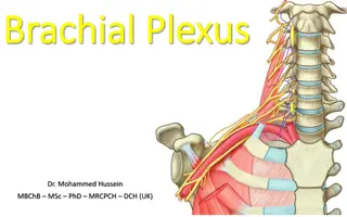
Dr.Sanaa Dr.Sanaa Alshaarawy Alshaarawy
Objectives At the end of this lecture, the students should be able to : Describe the formation of brachial plexus (site, roots) List the main branches of brachial plexus Describe the formation of lumbosacral plexus (site, roots) List the main branches of lumbosacral plexus Describe the important Applied Anatomy related to the brachial & lumbosacral plexuses.
FORMATION OF BRACHIAL PLEXUSES It is formed in the posterior triangle of the neck. It is the union of the anterior rami of the 5th ,6th,7th,8thcervical and the 1stthoracic spinal nerves
DIVISIONS (STAGES) The plexus is divided into : Roots Trunks Divisions Cords Terminal branches
TRUNKS Upper trunk Union of the roots of C5 & 6 Middle trunk Continuation of the root of C7 Lower trunk Union of the roots of C8 & T1
DIVISIONS & CORDS Each trunk divides into anterior and posterior division Posterior cord: From the 3 posterior divisions of the 3 trunks. Lateral cord: From the anterior divisions of the upper and middle trunks. Medial cord : It is the continuation of the anterior division of the lower trunk.
CORDS & BRANCHES Branches All three cords will give branches in the axilla, those will supply their respective regions
The Plexus can be divided into 5 stages: Roots: in the posterior Trunks: in the posterior Divisions: behind the clavicle Cords: in the axilla Branches: in the axilla The first 2 stages lie in the posterior triangle, while the last 2 sages lie in the axilla.
The Brachial Plexus Long Thoracic (C5,6,7) Nerve to Subclavius(C5,6) Dorsal Scapular(C5) Suprascapular(C5,6) upper trunk roots lateral Cord (2LM) C5 C6 middle trunk C7 lower trunk C8 posterior Cord (ULTRA) T1 medial Cord (4MU) Anterior divisions Posterior divisions
BRANCHES (A) From Roots: 1. C5: Nerve to rhomboids (dorsal scapular nerve). 2. C5,6 &7: Long thoracic nerve (supplies serratus anterior). (B) From Trunk (upper trunk): 1. Nerve to subclavius 2. Suprascapular nerve (supplies supraspinatus & infraspinatus)
(C)BRANCHES From Cords Lateral Cord (2LM) .Lateral pectoral n .Lateral root of median n .Musculocutaneous n C5 C6 C7 C8 T1 Medial cord (4MU) .Medial pectoral n. .Medial root of median n. .Medial cutaneous n of arm. .Medial cutaneous n of forearm. .Ulnar n. Posterior Cord (ULTRA) .Upper subscapular n .Lower subscapular n .Thoracodorsal n .Radial n .Axillary n
http://t3.gstatic.com/images?q=tbn:ANd9GcR4j2HPMhlPQBMYRVqLW6hxNDEZwfxI2QJSEuhTtmVR3g0jsJ3LeQhttp://t3.gstatic.com/images?q=tbn:ANd9GcR4j2HPMhlPQBMYRVqLW6hxNDEZwfxI2QJSEuhTtmVR3g0jsJ3LeQ Erb-Duchenne s paralysis due to injury of Upper Trunk of Brachial Plexus.
http://t2.gstatic.com/images?q=tbn:ANd9GcRAfIMNzAqb-nV3pG1kh4RdDhjzqXahidyzBEmrxiK3Wu043dDLhttp://t2.gstatic.com/images?q=tbn:ANd9GcRAfIMNzAqb-nV3pG1kh4RdDhjzqXahidyzBEmrxiK3Wu043dDL Claw Hand http://classconnection.s3.amazonaws.com/574/flashcards/598574/png/picture41311464890819.png Hand of Benediction or Pop s Blessings (APE HAND) will result from median nerve injury.
LUMBAR PLEXUS Formation: By ventral rami of L1,2,3 and most of L4 Site: In the substance of psoas major muscle Main branches: Iliohypogastric & ilioinguinal: to anterior abdominal wall Obturator: to medial compartment of thigh Femoral: to anterior compartment of thigh
SACRAL PLEXUS Formation: By ventral rami of a part of L4 & whole L5 (lumbosacral trunk) + S1, 2, 3 and most of the S4 Site: In front of piriformis msucle
SACRAL PLEXUS Main branches: Pelvic splanchnic nerve (from sacral): preganglionic parasympathetic to pelvic viscera & hindgut Pudendal nerve (from sacral plexus): to perineum Sciatic nerve (from Lumbosacral plexus: L4&5+S1,2,3): to lower limb
FEMORAL NERVE Origin: A branch from lumbar plexus (L2,3,4) Course: Descends lateral to psoas major & enters the thigh behind the inguinal ligament Passes lateral to femoral artery & divides into terminal branches.
FEMORAL NERVE INJURY Motor effect: Wasting of quadriceps femoris Loss of extension of knee Weak flexion of hip (psoas major is intact ; because it takes supply from other fibers of the lumbar plexus) Sensory effect: loss of sensation over areas supplied antero- medial aspect of thigh & medial side of leg & foot (injury of Saphenous br.of femoral)
SCIATIC NERVE The largest nerve of the body Origin: from sacral plexus (L4, 5, S1, 2, & 3) It is one of the terminal branch of sacral plexus. Course: Leaves the pelvis through greater sciatic foramen, below piriformis & passes in the gluteal region (between ischial tuberosity & greater trochanter) then to posterior compartment of thigh Ischial tuberosity Divides into tibial & common peroneal (fibular) nerves
TIBIAL NERVE Course: Descends through popliteal fossa to posterior compartment of leg, accompanied with posterior tibial vessels Passes deep to flexor retinaculum to reach the sole of foot where it divides into 2 terminal branches
COMMON PERONEAL (FIBULAR) NERVE Course: Leaves popliteal fossa & turns around the lateral aspect of neck of fibula. Then divides into: Superficial peroneal: descends into lateral compartment of leg 1. Deep peroneal: descends into anterior compartment of leg 2.
SUMMARY The lumbar plexus is formed by ventral rami of L1,2,3 and most of L4, in substance of psoas major muscle The sacral plexus is formed by ventral rami of a part of L4 & whole L5 (lumbosacral trunk) plus the S1,2,3 and most of S4, in front of piriformis msucle. The femoral nerve, a branch of lumbar plexus (L2,3,4). Its injury will affect the flexion of hip & extension of knee as well as loss of sensation of skin of anteromedial aspects of the thigh, medial side of knee, leg and foot (Saphenous br.of femoral). The sciatic nerve is a branch of sacral plexus (L4,5, S1,2,3) Its injury will affect the flexion of knee, extension of hip, all movements of leg & foot, as well as loss of sensation of skin of leg & foot (except areas supplied by saphenous branch of femoral nerve)
1. Lesion of the upper trunk of the brachial plexus leads to : Klumpke palsy. Erb-Duchenne palsy Drop wrist & hand. Ape hand. 2. Which one of the following nerves is a branch of posterior cord of brachial plexus? Ulnar Radial Median Musclocutanous
QUESTION 1 The femoral nerve supplies: Extensors of hip. Skin of dorsum of foot. Hamstrings. Extensors of knee a. b. c. d.
QUESTION 2 Injury of common peroneal nerve leads to: Loss of dorsiflexion of ankle Loss of inversion of foot Loss of extension of knee Loss of flexion of toes a. b. c. d.


")


BRANCHES From Cords")


 NERVE")
 NERVE")