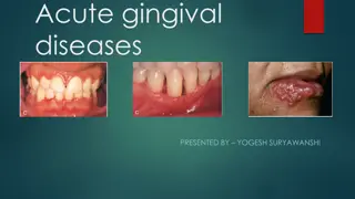
Understanding Clinical Features and Types of Gingivitis

The clinical features of gingivitis include redness, sponginess, bleeding, contour changes, and the presence of calculus or plaque. Gingivitis can manifest as chronic or acute, with localized or generalized involvement. Learn about the different types and presentations of gingivitis to better recognize and manage this common gum disease.
Download Presentation
Please find below an Image/Link to download the presentation.
The content on the website is provided AS IS for your information and personal use only. It may not be sold, licensed, or shared on other websites without obtaining consent from the author. Download presentation by click this link. If you encounter any issues during the download, it is possible that the publisher has removed the file from their server.

Presentation Transcript
Clinical features of gingivitis
Learning objectives Types of gingivitis Clinical presentation of different types of gingivitis Bleeding on probing Colour changes in gingiva Changes in consistency of gingiva Surface texture changes in gingivitis Position of gingiva
Clinical features of gingivitis can be characterized by any of the following clinical signs: redness and sponginess of the gingival tissue, bleeding on provocation, changes in contour, the presence of calculus or plaque with no radiographic evidence of crestal bone loss.
Gingivitis can develop with sudden onset and have a short duration, and it can be painful. A less severe phase of this condition can also occur. Recurrent gingivitis reappears after having been eliminated by treatment or after disappearing spontaneously.
Chronic gingivitis develops slowly and has a long duration. It is painless, unless it is complicated by acute or subacute exacerbations, and it is the type that is most often encountered . Chronic gingivitis is a fluctuating disease in which inflammation persists or resolves and normal areas become inflamed.
Localized gingivitis is confined to the gingiva of a single tooth or group of teeth. Generalized gingivitis involves the entire mouth. Marginal gingivitis involves the gingival margin, and it can include a portion of the contiguous attached gingiva.
Papillary gingivitis involves the interdental papillae, and it often extends into the adjacent portion of the gingival margin. Papillae are involved more frequently than the gingival margin the earliest signs of gingivitis often occur in the papillae. Diffuse gingivitis affects the gingival margin, the attached gingiva, and the interdental papillae.
Localized marginal gingivitis is confined to one or more areas of the marginal gingiva
Localized papillary gingivitis is confined to one or more interdental spaces in a limited area
Generalized marginal gingivitis involves the gingival margins in relation to all the teeth. The interdental papillae are usually affected
Gingival Bleeding on Probing Two earliest signs of gingivitis are crevicular fluid production and bleeding from the gingival sulcus on gentle probing
Bleeding on probing (BOP) is easily detected clinically. BOP appears earlier than a change in color or other visual signs of inflammation. BOP is widely used by clinicians to measure disease prevalence and progression, measure outcomes of treatment, and motivate patients to perform necessary home care.
Gingival Bleeding Caused by Local Factors Factors that contribute to plaque retention and may lead to gingivitis include anatomic and developmental tooth variations, caries frenum pull iatrogenic factors, malpositioned teeth mouth breathing, overhangs, partial dentures, lack of attached gingiva, and recession. Orthodontic treatment and fixed retainers are associated with increased plaque retention and increased BOP.
Chronic and Recurrent Bleeding The most common cause of abnormal gingival BOP is chronic inlammation. The bleeding is chronic or recurrent, and it is provoked by mechanical trauma (e.g., toothbrushing, toothpicks, food impaction) by biting into solid foods (e.g., apples).
Color Changes With Gingivitis Change in color is an important clinical sign of gingival disease. The normal gingival color is coral pink, and it is produced by the tissue s vascularity and modified by the overlying epithelial layers. Color changes in acute gingival inflammation differ in both nature and distribution from those in patients with chronic gingivitis
Changes in Gingival Consistency Chronic and acute inflammations produce changes in the normal firm and resilient consistency of the gingiva.
Changes in Gingival Surface Texture The surface of normal gingiva usually exhibits numerous small depressions and elevations that give the tissue an orange-peel appearance referred to as stippling. Stippling is restricted to the attached gingiva and is predominantly localized to the subpapillary area, but it extends to various degrees into the interdental papilla.
With chronic inflammation, the gingival surface is smooth and shiny or firm and nodular, and stippling is lost
Changes in Gingival Position 1] Traumatic Lesions- chemical, physical, or thermal 2] Gingival recession- Gingival recession is a common finding. The prevalence, extent, and severity of gingival recession increase with age, and this condition is more prevalent among males.
Changes in Gingival Contour Changes in gingival contour are primarily associated with gingival enlargement but changes may also occur with other conditions. Stillman cleft-specific type of gingival recession that consists of a narrow, triangular shaped gingival recession. McCall festoon-rolled, thickened band of gingiva that is usually seen adjacent to the cuspids when recession approaches the mucogingival junction.


 is easily detected clinically.")