Understanding the Ultrastructure of Spermatozoa and Their Components

Explore the intricate details of spermatozoa and their structures, including the head, nucleus, acrosome, and more. Learn about the dimensions of mammalian spermatozoa and the principal parts that make up these essential reproductive cells.

Download Presentation

Please find below an Image/Link to download the presentation.
The content on the website is provided AS IS for your information and personal use only. It may not be sold, licensed, or shared on other websites without obtaining consent from the author. Download presentation by click this link. If you encounter any issues during the download, it is possible that the publisher has removed the file from their server.
E N D
Presentation Transcript
ULTRASTRUCTURE OF SPERMATOZOA Dr Alok Kumar
Structure of spermatozoa Structure of spermatozoa http://www.fastbleep.com/assets/notes/image/9876_1.jpg
Structure of spermatozoa of farm Structure of spermatozoa of farm animal and other vertebrates animal and other vertebrates
Dimensions ( Dimensions ( m m ) of Mammalian Spermatozoa Spermatozoa of Mammalian species Head Length 9 Mid piece Length 14 Principle piee Total length width 4.5 Width Bull 42 50-70 Boar 8.5 4.2 10 36.1 54.6 Ram 8.2 14 42 60-70 Horse 6.41 3 50 Rat 7.9 3.2 18.4 1.3 96 123 Dog 5.9 3.9 10.6 45.8 62 Cat 5.76 2.5 8.34 0.77 46.25 59
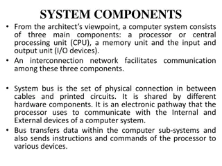
Principal part of spermatozoa HEAD Nucleus Acrosome Connecting piece Middle piece Principal piece End piece TAIL
Head Head It is the anterior most part of the spermatozoa. The head of the spermatozoa is elongated and ovoid in shape. The head length and width of spermatozoa is varied species to species. The head is formed of acrosome and nucleus.
Nucleus Nucleus The head nucleus is entirely filled with nearly homogenous nuclear material. Haploid chromosome are present in the nucleus. DNA surrounded by the nuclear membranes.
Acrosome Acrosome The anterior 60 percent of the nucleus covered by acrosome cap or head cap. The acrosome is formed by golgi body and it is bounded by unit membrane. Outer membrane of acrosome cap is identical with galea capitis. Acrosome cap thickness is 0.1 microns. Following enzymes are present in the acrosome Acid hydrolases, Acid phosphatase, Hyaluronidase, Acrosin etc.
The anterior part of the nuclear membrane or the inner membrane of acrosome, is modified to form the perforatorium. Cell membrane encloses the head, body and tail of the spermatozoon. The membrane is closely applied to the cell at the anterior portion of the acrosome cap, the postnuclear cap and at Jensen s ring.
Detachment occurs by rupture of cell membrane and outer membrane of the acrosome cap in the equatorial zone. The detached outer membranes of the acrosome may break into two halves, or remain joined and appear as bathing cap shaped structure.
Structure of sperm head Structure of sperm head
Tail Tail It is the longest part of sperm. It contains axial filaments. Axial filament is covered by a sheath, which is made up of 9 singlet fibers. The morphological point of view tail consist Neck, middle piece, principal piece and end piece.
Neck Neck It is the smallest part of spermatozooa. In neck of sperm 2 centrioles are present. Proximal centriole Distal centriole Proximal centriole lies in a depression in the posterior surface of the nucleus. Proximal centriole is perpendicular to main axis of the sperm.
The proximal Centriole has no active function in the spermatozoon but is a potential activist within an egg during first cleavage division of the fertilized egg Distal centriole is along longitudinal axis of the sperm. Distal centriole acts as basal body and gives rise to axoneme of the sperm-tail. Axial filament of sperm structure as 9 + 2 manner.
Middle piece Middle piece Middle piece is located between the neck and the annulus. It contains the mitochondrial sheath, a helix 80 to 100 mitochondria wrapped around the axoneme. The axoneme of the mammalian sperm is surrounded by nine outer dense fibers which are also called the coarse or accessary fibres. The mitochondrial sheath is believed to be the source of energy (ATP) for sperm motility.
Middle piece of sperm have 9+9+2 fibrous pattern. At the junction of mid piece and principal piece is present the annulus which is also known as the ring centriole or Jensen s ring. This ring centriole prevent the mitochondria toward the tail reason.
Principal piece Principal piece The Principal piece continues posteriorly from the annulus and extend to the near the end of tail . The principal piece of mammalian spermatoza is surrounded by a fibrous sheath. Fibrous sheath is composed of a series of circumferentially oriented ribs that extend half way around the tail end in two longitudinal columns.
End piece End piece The end piece or terminal portion of the tail is about 3 to 4 microns in length and consist of the terminal portion of the fibrils covered by the cell membrane but fibrillar coil sheath of the tail is absent.
Axenome Axenome filament filament Runs throughout the tail. The core of the axenome consists of two central microtubules surrounded by a row of nine doublet microtubules. one microtubule of each doublet is complete, having 13 protofilaments, the other is C-shaped and has only 11 protofilaments . which are made exclusively of the dimeric protein tubulin.
Dynein arm motor complexes allow microtubule to slide against each other. This causes the synchronised shortening and extension of the microtubule on opposite site of the axoneme allowing the flagellar to bend. Alteration of this bending action creates the beating motion of a swimming sperm. Radial spoke regulate the motion of axoneme.
AXENOME AXENOME Principle piece Middle piece
Points to remember Points to remember The head and tail are principle part of spermatozoa. Acrosomal cap and nucleus are part of head. Tail are subdivided in to the four part Neck, middle piece, principle piece and end piece. Outer coarse fiber are present on the surface of mid piece and principle piece only. Mid piece and principle piece have 9+9+2 fibrous arrangement. End piece have 9+2 fibrous arrangement. Tubulin and dynein are the axonemal protein.