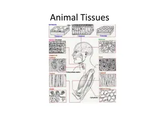
Understanding Muscle Tissue: Structure and Function
Muscle tissue is a vital component in most animals, facilitating movement and maintaining posture. Derived from embryonic cells, muscles come in three types: skeletal, cardiac, and smooth, each serving specific functions in the body. While skeletal muscles are under voluntary control, cardiac and smooth muscles operate involuntarily. The intricate interplay of actin and myosin filaments within muscle cells enables contractions that power locomotion, digestion, and other bodily processes.

Download Presentation

Please find below an Image/Link to download the presentation.
The content on the website is provided AS IS for your information and personal use only. It may not be sold, licensed, or shared on other websites without obtaining consent from the author. Download presentation by click this link. If you encounter any issues during the download, it is possible that the publisher has removed the file from their server.
E N D
Presentation Transcript
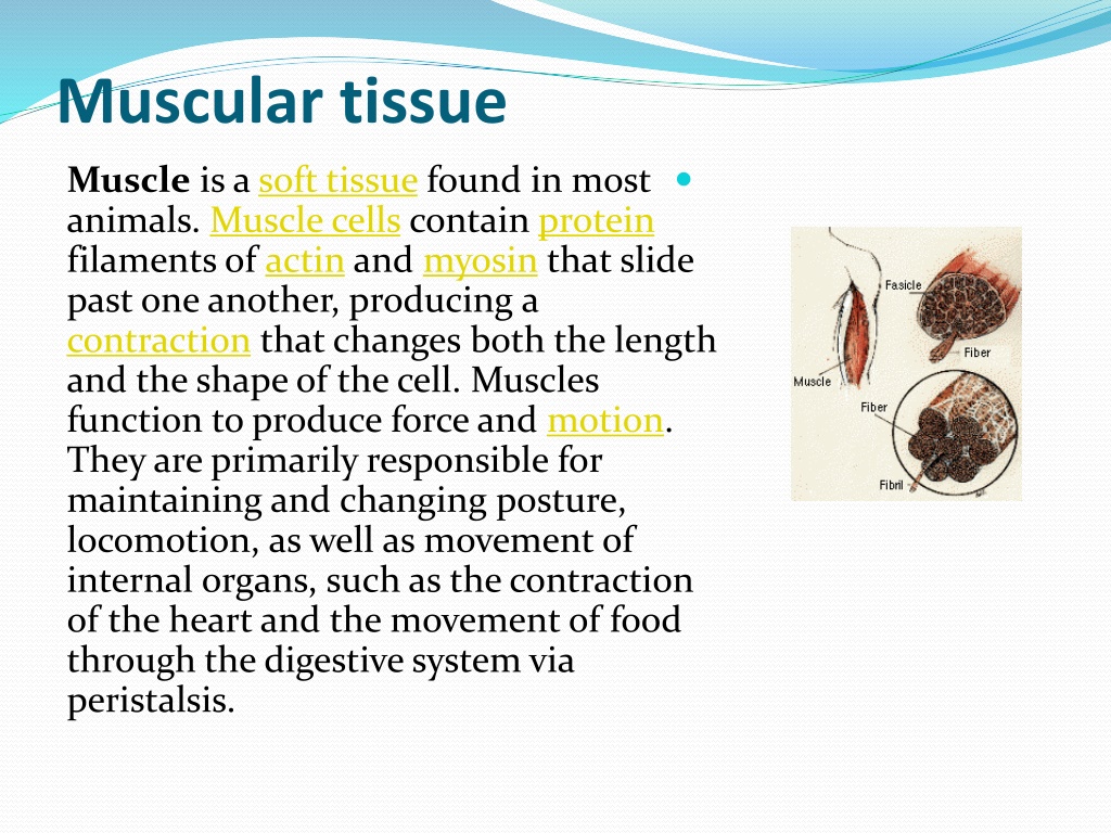
Muscular tissue Muscle is a soft tissue found in most animals. Muscle cells contain protein filaments of actin and myosin that slide past one another, producing a contraction that changes both the length and the shape of the cell. Muscles function to produce force and motion. They are primarily responsible for maintaining and changing posture, locomotion, as well as movement of internal organs, such as the contraction of the heart and the movement of food through the digestive system via peristalsis.
Skeletal muscle.jpg https://upload.wikimedia.org/wikipedia/commons/thumb/9/9c/1022_Muscle_Fibers_%28small%29.jpg/220px-1022_Muscle_Fibers_%28small%29.jpg
Muscle tissuesare derived from the mesodermal layer of embryonic germ cells in a process known as myogenesis. There are three types of muscle, skeletal or striated, cardiac, and smooth. Muscle action can be classified as being either voluntary or involuntary. Cardiac and smooth muscles contract without conscious thought and are termed involuntary, whereas the skeletal muscles contract upon command Skeletal muscles in turn can be divided into fast and slow twitch fibers.
The term muscle is derived from the Latin musculus meaning "little mouse" perhaps because of the shape of certain muscles or because contracting muscles look like mice moving under the skin... A body tissue composed of elongated cells (called muscle fibers) that contract to produce movement. In vertebrate animals, voluntary movement is produced by the action of muscles on bone. Movement of the muscles of the heart and other organs is involuntary and controlled by the autonomic nervous system
The 3 types of muscle tissue are cardiac, smooth, and skeletal.. Cardiac muscle cells are located in the walls of the heart, appear striated, and are under involuntary control. Smooth muscle fibers are located in walls of hollow visceral organs, except the heart, appear spindle-shaped, and are also under involuntary control. Skeletal muscle fibers occur in muscles which are attached to the skeleton. They are striated in appearance and are under voluntary control..
Structure We know that living organisms can move on their own or can perform other types of movement. Muscle tissue has a ability to relax and contrast and so bring about movement and mechanical work in various parts of the body. There are other movements in the body too which are necessary for the survival of the organism such as the heart beat and the movements of the alimentary canal. Muscles can be divided into three main groups according to their structure, e.g.: Smooth muscle tissue. Skeletal muscle tissue. Cardiac (heart) muscle tissue.
Muscles are predominantly powered by the oxidation of fatsand carbohydrates, but anaerobic chemical reactions are also used, particularly by fast twitch fibers. These chemical reactions produce adenosine triphosphate (ATP) molecules that are used to power the movement of the myosin heads.
https://upload.wikimedia.org/wikipedia/commons/thumb/e/e5/414_Skeletal_Smooth_Cardiac.jpg/220px-414_Skeletal_Smooth_Cardiac.jpghttps://upload.wikimedia.org/wikipedia/commons/thumb/e/e5/414_Skeletal_Smooth_Cardiac.jpg/220px-414_Skeletal_Smooth_Cardiac.jpg https://upload.wikimedia.org/wikipedia/commons/thumb/d/dd/1007_Muscle_Fibes_%28large%29.jpg/300px-1007_Muscle_Fibes_%28large%29.jpg
Smooth Muscle Tissue. Smooth muscle tissue is made up of thin-elongated muscle cells, fibres. These fibres are pointed at their ends and each has a single, large, oval nucleus. Each cell is filled with a specialised cytoplasm, the sarcoplasm and is surrounded by a thin cell membrane, the sarcolemma. Each cell has many myofibrils which lie parallel to one another in the direction of the long axis of the cell. They are not arranged in a definite striped (striated) pattern, as in skeletal muscles - hence the name smooth muscle . Smooth muscle fibres interlace to form sheets or layers of muscle tissue rather than bundles. Smooth muscle is involuntary tissue, i.e. it is not controlled by the brain. Smooth muscle forms the muscle layers in the walls of hollow organs such as the digestive tract (lower part of the oesophagus, stomach and intestines), the walls of the bladder, the uterus, various ducts of glands and the walls of blood vessels .
Functions of Smooth Muscle Tissue Smooth muscle controls slow, involuntary movements such as the contraction of the smooth muscle tissue in the walls of the stomach and intestines. The muscle of the arteries contracts and relaxes to regulate the blood pressure and the flow of blood.
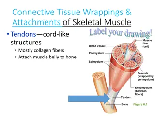
Skeletal muscle Skeletal muscle is the most abundant tissue in the vertebrate body. These muscles are attached to and bring about the movement of the various bones of the skeleton, hence the name skeletal muscles. The whole muscle, such as the biceps, is enclosed in a sheath of connective tissue, the epimysium. This sheath folds inwards into the substance of the muscle to surround a large number of smaller bundles, the fasciculi. These fasciculi consist of still smaller bundles of elongated, cylindrical muscle cells, the fibres. Each fibre is a syncytium, i.e. a cell that have many nuclei. The nuclei are oval in shaped and are found at the periphery of the cell, just beneath the thin, elastic membrane (sarcolemma). The sarcoplasm also has many alternating light and dark bands, giving the fibrea striped or striated appearance (hence the name striated muscle).
With the aid of an electron microscope it can be seen that each muscle fibre is made up of many smaller units, the myofibrils. Each myofibril consists of small protein filaments, known as actinand myosin filaments. The myosin filaments are slightly thicker and make up the dark band (or A-band). The actin filaments make up the light bands (I-bands) which are situated on either side of the dark band. The actin filaments are attached to the Z-line. This arrangement of actin and myosin filaments is known as a sacromere. During the contraction of skeletal muscle tissue, the actin filaments slide inwards between the myosin filaments. Mitochondria provide the energy for this to take place. This action causes a shortening of the sacromeres (Z-lines move closer together), which in turn causes the whole muscle fibre to contract. This can bring about a shortening of the entire muscle such as the biceps, depending on the number of muscles fibres that were stimulated. The contraction of. skeletal muscle tissue is very quick and forceful
Functio ns of Skeletal Muscle Tissue Skeletal muscles function in pairs to bring about the co-ordinated movements of the limbs, trunk, jaws, eyeballs, etc. Skeletal muscles are directly involved in the breathing process. https://upload.wikimedia.org/wikipedia/commons/thumb/c/c6/1006_Sliding_Filament_Model_of_Muscle_Contraction.jpg/220px-1006_Sliding_Filament_Model_of_Muscle_Contraction.jpg
Cardiac (Heart) Muscle Tissue. This is a unique tissue found only in the walls of the heart. Cardiac (Heart) Muscle Tissue shows some of the characteristics of smooth muscle and some of skeletal muscle tissue. Its fibres , like those of skeletal muscle, have cross-striations and contain numerous nuclei. However, like smooth muscle tissue, it is involuntary. Cardiac muscle differ from striated muscle in the following aspects: they are shorter, the striations are not so obvious, the sarcolemma is thinner and not clearly discernible, there is only one nucleus present in the centre of each cardiac fibre and adjacent fibres branch but are linked to each other by so-called muscle bridges. The spaces between different fibres are filled with areolar connective tissue which contains blood capillaries to supply the tissue with the oxygen and nutrients.
Functions of Cardiac (Heart) Muscle Tissue Cardiac muscle tissue plays the most important role in the contraction of the atria and ventricles of the heart. It causes the rhythmical beating of the heart, circulating the blood and its contents throughout the body as a consequence.


 Muscle Tissue.")