Overview of Muscle Disorders and Muscular Dystrophies
The human body consists of over 600 muscles, divided into skeletal, smooth, and cardiac muscles. Diseases of skeletal muscle encompass myopathy, myositis, and muscular dystrophy. Muscle disorders are categorized into genetic muscular dystrophies, channelopathies, inflammatory myopathies, and endocrine myopathies. Muscular dystrophies are genetically inherited disorders characterized by muscle degeneration, with Duchenne Muscular Dystrophy being the most common and severe. The pathophysiology involves the role of dystrophin, a crucial protein in muscle stability and flexibility.
Download Presentation

Please find below an Image/Link to download the presentation.
The content on the website is provided AS IS for your information and personal use only. It may not be sold, licensed, or shared on other websites without obtaining consent from the author.If you encounter any issues during the download, it is possible that the publisher has removed the file from their server.
You are allowed to download the files provided on this website for personal or commercial use, subject to the condition that they are used lawfully. All files are the property of their respective owners.
The content on the website is provided AS IS for your information and personal use only. It may not be sold, licensed, or shared on other websites without obtaining consent from the author.
E N D
Presentation Transcript
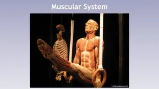
The human body has over 600 muscles; their bulk comprises about 40% of the total body weight. Muscles are divided into skeletal muscles, smooth muscles and cardiac muscles Each muscle type has distinct morphologic and biochemical characteristics that separate them and enable diseases to involve one or more muscle types
Diseases of skeletal muscle are called by several general names: myopathy, implying all types of muscle disease; myositis, implying inflammation in the muscle; muscular dystrophy, implying degeneration of muscle, often hereditary.
Table -1 Common Features of Primary Skeletal Muscle Diseases
Muscle diseases are divided into 4 broad categories: 1. muscular dystrophy due to genetic abnormalities 1. channelopathies with abnormal sodium, calcium, or potassium membrane ion channels; 1. inflammatory myopathies 1. secondary endocrine myopathies.
Muscular dystrophies Muscular dystrophies are genetically determined disorders that have a wide variation in age of onset, sex distribution, and location of maximal muscle atrophy.
Duchenne Muscular Dystrophy Is the most common and most serious muscular dystrophy , associated with a marked deficiency or absence of dystrophin, a large protein which is encoded by a gene on the X chromosome. As DMD is transmitted by X-linked recessive inheritance, nearly all patients are male. About 10% of female carriers have mild muscle weakness. The incidence of DMD is 30/100,000 male births.
Pathophysiology Muscle dystrophin is a large molecular weight protein that is found primarily within skeletal, smooth, and cardiac muscle. In the muscle, dystrophin links myofibrillar elements with the sarcolemma, affording stability and flexibility to the muscle fiber.
Dystrophin gene mutations that cause DMD result in either the absence of dystrophin protein production or markedly truncated proteins. The absence of dystrophin leads to membrane instability, myofiber leakiness of creatine kinase (CK), and susceptibility to injury from normal muscle contractions.
Over time, fibrosis and scarring develop in the muscle, and fat cells invade, replacing the degenerating muscle cells. The net process may transiently give rise to enlarged doughy muscles that have a pseudohypertrophic appearance.
Clinical Features Although children with DMD have disease activity in the neonatal period (elevated serum CK and necrosis on muscle biopsy) they rarely have clinical symptoms until age 3 to 4 years. Parents usually report difficulty in running or climbing, frequent falls, and enlargement of the calf muscles (pseudohypertrophy), which feel firm and rubbery.
By 4 to 5 years of age, the gait becomes wide- based and waddling and the child often walks on his toes because of heel cords contractures. The weakness is greatest in proximal muscles, producing a Gowers maneuver (placing hands on the knees and climbing up the thighs to stand). In the early school years, the limb weakness progresses and is accompanied by excessive lordosis.
By age 10 to 12 years, the child is unable to walk and is confined to a wheelchair. Deep tendon reflexes are lost. By late teens the weakness is profound, scoliosis is marked, and joint contractures are frequent. Intellectual impairment in DMD is common. Cardiomyopathy, slowly develops. Kyphoscoliosis and weakness of respiratory muscles produce a decreasing lung vital capacity The terminal stages of DMD are characterized by recurrent pulmonary infections and often congestive heart failure. The age of death ranges from 10 to 30 years, with a mean of 18 years.
Laboratory Findings Serum CK levels are elevated to between 20 and 100 times normal. The levels are abnormal at birth but decline late in the disease because of inactivity and loss of muscle mass. The electrocardiogram is abnormal in 2/3 of patients. EMG demonstrates features typical of myopathy. The muscle biopsy shows muscle fibers of varying size as well as small groups of necrotic and regenerating fibers. Connective tissue and fat replace lost muscle fibers. A definitive diagnosis of Duchenne dystrophy can be established on the basis of dystrophin deficiency in a biopsy of muscle tissue or mutation analysis on peripheral blood leukocytes.
Management Glucocorticoids, administered as prednisone in a dose of 0.75 mg/kg per day, significantly slow progression of Duchenne dystrophy for up to 3 years. Management involves use of joint bracing and prevention or release of contractures to maintain walking for as long as practical. Considerable research is underway to replace the mutated dystrophin gene, but to date all results have been disappointing.
Becker Muscular Dystrophy This less severe form of X-linked recessive muscular dystrophy results from defects of the same gene responsible for Duchenne dystrophy. Becker muscular dystrophy is ~10 times less frequent than Duchenne, with an incidence of about 3 per 100,000 live-born males.
Clinical Features The pattern of muscle wasting in Becker muscular dystrophy closely resembles that seen in Duchenne. Proximal muscles, especially of the lower extremities, are prominently involved. As the disease progresses, weakness becomes more generalized. Hypertrophy of muscles, particularly in the calves, is an early and prominent finding. Most patients with Becker dystrophy first experience difficulties between ages 5 and 15 years, although onset in the third or fourth decade or even later can occur.
By definition, patients with Becker dystrophy walk beyond age 15, while patients with Duchenne dystrophy are typically in a wheelchair by the age of 12. Patients with Becker dystrophy have a reduced life expectancy, but most survive into the fourth or fifth decade. Mental retardation may occur in Becker dystrophy, but it is not as common as in Duchenne. Cardiac involvement occurs in Becker dystrophy and may result in heart failure.
Laboratory Features Serum CK levels, results of EMG, and muscle biopsy findings closely resemble those in Duchenne dystrophy. Treatment The use of glucocorticoids has not been adequately studied in Becker dystrophy.
Facioscapulohumeral (FSH) Muscular Dystrophy This form of muscular dystrophy has An autosomal dominant inheritance pattern with a prevalence of ~1 in 20,000.
Clinical Features The condition typically has an onset in childhood or young adulthood. In most cases, facial weakness is the initial manifestation, appearing as an inability to smile, whistle, or fully close the eyes. Loss of scapular stabilizer muscles makes arm elevation difficult. Scapular winging becomes apparent with attempts at abduction and forward movement of the arms. Biceps and triceps muscles may be severely affected, with relative sparing of the deltoid muscles. Weakness of the anterior compartment muscles of the legs may lead to footdrop.
In most patients, the weakness remains restricted to facial, upper extremity, and distal lower extremity muscles. In 20% of patients, weakness progresses to involve the pelvic girdle muscles, and severe functional impairment and possible wheelchair dependency result.
Laboratory Features The serum CK level may be normal or mildly elevated. EMG usually indicates a myopathic pattern. The muscle biopsy shows nonspecific features of a myopathy. Treatment No specific treatment is available; ankle-foot orthoses are helpful for footdrop. Scapular stabilization procedures improve scapular winging but may not improve function.
Myotonic Dystrophy Myotonic dystrophy is also known as dystrophia myotonica (DM) is an autosomal dominant disorder.
Clinical Features The clinical expression of myotonic dystrophy varies widely and involves many systems other than muscle. Affected patients have a typical "hatchet- faced" appearance due to temporalis, masseter, and facial muscle atrophy and weakness. Frontal baldness is also characteristic of the disease. Neck muscles, including flexors and sternocleidomastoids, and distal limb muscles are involved early.
Weakness of wrist extensors, finger extensors, and intrinsic hand muscles impairs function. Ankle dorsiflexor weakness may cause footdrop. Proximal muscles remain stronger throughout the course. Palatal, pharyngeal, and tongue involvement produce a dysarthric speech, nasal voice, and swallowing problems.
Myotonia, which usually appears by age 5 years, is demonstrable by percussion of the thenar eminence, the tongue, and wrist extensor muscles. Myotonia causes a slow relaxation of hand grip after a forced voluntary closure. Advanced muscle wasting makes myotonia more difficult to detect.
Cardiac conduction defects occur commonly. Other associated features include: intellectual impairment hypersomnia, cataracts, gonadal atrophy, insulin resistance, decreased esophageal and colonic motility.
Laboratory Features The diagnosis of myotonic dystrophy can usually be made on the basis of clinical findings. Serum CK levels may be normal or mildly elevated. EMG evidence of myotonia is present in most cases of cases. Muscle biopsy shows muscle atrophy, which selectively involves type 1 fibers in 50% of cases. Treatment Phenytoin and mexiletine are the preferred agents for the occasional patient who requires an antimyotonia drug; other agents, particularly quinine and procainamide, may worsen cardiac conduction.
