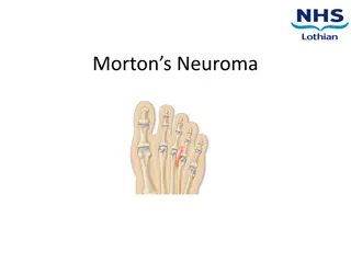
Understanding Acoustic Neuroma: Diagnosis and Management
Vestibular schwannoma, also known as acoustic neuroma, is a common tumor in the cerebellopontine angle. It can be sporadic or associated with neurofibromatosis type 2. Patients typically present with hearing loss and tinnitus, which may be gradual or sudden. Grading systems like Koos, Ojemann, and Jackler help classify tumor sizes and stages. Management involves observation, surgery, or radiation therapy depending on the tumor's size and symptoms.

Download Presentation

Please find below an Image/Link to download the presentation.
The content on the website is provided AS IS for your information and personal use only. It may not be sold, licensed, or shared on other websites without obtaining consent from the author. Download presentation by click this link. If you encounter any issues during the download, it is possible that the publisher has removed the file from their server.
E N D
Presentation Transcript
ACOUSTIC NEUROMA ACOUSTIC NEUROMA DIAGNOSIS AND MANAGEMENT DIAGNOSIS AND MANAGEMENT
Introduction Vestibular schwannoma is the most common tumor occurring in the CP angle (about 85-90%) 6 % of all intracranial tumors Incidence in US: 10 per million / year Peak incidence in 4thto 6thdecade M:F= 2:3 95% Sporadic (unilateral) 5% Neurofibromatosis type 2 (bilateral) Slow growing tumors = Average 1.8 mm/year (0.2 to 4.0 mm)
Introduction Neither Neuroma nor Acoustic (auditory) Schwannoma : arising from vestibular nerve Schwann cells at transition zone of the peripheral and central myelin Obersteiner Redlich zone (at the lateral CPA/medial IAC) Majority originate within the IAC Equal frequency on Superior and Inferior vestibular nerves ???? Rarely occur on the cochlear division of the 8thCN. VS occurs as a result of mutations in a tumor suppressor gene Merlin Located on chromosome 22q12 VS requires both copies to be mutated People with NF2 inherit one mutated gene
Cerebello-pontine angle Superior limb of cerebellopontine fissure Inferior limb of cerebellopontine fissure Apex-located laterally where superior & inferior limbs meet Floor- Middle cerebellar peduncle Anterior: posterior surface of temporal bone Posterior: anterior surface of the cerebellum Medial: lateral surface of brainstem Lateral: petrous bone
Cerebello-pontine angle Cranial nerves: V VII & VIII IX, X, XI Important structures: Flocculus Lateral aperture of 4thventrical AICA
Grading Koos: (Grade 1-4) upto 1, 2, 3, >3 cm (intracanalicular+cisternal) Ojemann: (small, med, large)<2, 2-3, >3cm (intracisternal) Samii: >3 x 2 cm large, rest small (both intra + extrameatal also T1, T2, T3 AB, T4 AB Sekhar: (small, med, large) <2, 2-3.9, >3.9 cm (only intracisternal) Stage Tumor Size Intracanalicular I (small) II (medium) III (Large) IV (Giant) Tumor confined to IAC < 10 mm 11-25 mm 25-40 mm > 40 mm Jackler Staging System
What do patients complain of ? Hearing loss 95% of patients Most have slowly progressive loss 20% have sudden HL Level of hearing loss is NOT a predictor of size Tinnitus 65% of patients Usually constant with a high buzzing pitch Disequilibrium 60% of patients Usually well-compensated
Hearing Loss Most frequent initial symptom Most common symptom ~ 95% AN patients Asymmetric SNHL High Frequency Decreased Speech Discrimination Lack of conclusive correlation between tumor size and hearing * * Stipkovits EM et al., Am. J. Otology 1998: 19; 834-9 Pathophysiology of Hearing Loss Exact etiology is unknown Compressive effect on cochlear nerve Vascular occlusion of internal auditory artery
Gardener Robertson Scale Grade I (good-excellent) PTA (dB) : 0-30 SD (%) : 70-100 Grade II (serviceable) PTA (dB) : 31-50 SD (%) : 50-69 Grade III (non-serviceable) PTA (dB) : 51-90 SD (%) : 5-49 Grade IV (poor) PTA (dB) : 91-max SD (%) : 1-4 Grade V (none) PTA (dB) : not testable SD (%) : 0 TABLE 1. Hearing on affected side in 190 patients with vestibular schwannoma Gardner-Robertson class Distribution of Hearing in AN %age of patients A B C D Total 21.1 27.9 15.3 35.8 100. 0 *Forty-nine percent of patients had serviceable hearing. Myrseth: Neurosurgery, Volume 59(1).July 2006.67-76
More complaints. Facial and trigeminal nerve dysfunction Usually V2 numbness Sensory component of CN VII is usually involved first Hitselberger sign numbness of the posterior EAC Facial weakness or spasm occurs in 17% of patient Cerebellar: Wide gait, Falling to side of lesion Brainstem: Headache, altered MS, nausea, Visual Loss, Other Cranial nerves: IX dysphagia (large tumors, J F S) X hoarseness, aspiration (large tumors, J F S) XI shoulder weakness (large tumors, J F S)
Diagnostic Tests Audiometric Testing. Electrophysiologic Testing. Vestibular Testing.- ENG Computerized dynamic posturography. Rotary chair testing. CT & MRI.
Audiometric Testing Pure-tone testing: SNHL- most commonly high frequency (65%). Normal hearing (5%). Speech discrimination: Scores out of proportion with pure-tone thresholds. Some may score well. Rollover phenomenon improve the sensitivity. Acoustic reflex thresholds: typically elevated or absent. If present then reflex decay measured. The sensitivity is 85% for detecting retrocochlear problem.
BAER: Retrocochlear Pathology Most sensitive & specific audiologic test. Increased interpeak intervals I-to-III interval of 2.5 ms, III-to-V interval of 2.3 ms, and I-to-V interval of 4.4 ms Interaural wave V latency difference (IT5) Greater than 0.2 ms (40-60%). Poor waveform morphology i.e. only some of the waves are discernible Absent waveform in 20-30%. Wave 1 present but all remaining waves are absent in 10-20%. Normal in 10-15%. Fraysse B et al. First International Conf. on Acoustic Neuroma. 1992
BAER: Diagnostic Efficiency Generally, Efficiency increases with Size Sensitivity: > 90 % for tumor > 3 cm False negative Rate: 15 % (Wilson 1992 6/40) 33 % (5/15) for Intracanalicular Tumor False positive Rate: > 80 % (Jackler 2005) Positive predictive value: 15 % (Weiss 1990 4/26) 12 % (Walsted 1992 23/185)
Cost Cost- -Effective Initial Screening for Vestibular Schwannoma: Effective Initial Screening for Vestibular Schwannoma: Auditory Brainstem Response or Magnetic Resonance Imaging? Auditory Brainstem Response or Magnetic Resonance Imaging? V Rupa, A Job, M George, V Rajshekhar. Dept of ENT & NSx , CMC, Vellore Otolaryngol Head Neck Surg , June 1, 2003 vol. 128 no. 6 823-828 90 patients with asymmetric audiovestibular symptoms, investigated prospectively with both ABR and gadolinum-enhanced magnetic resonance imaging (GdMRI). 6 were diagnosed with VS on GdMRI. On ABR testing, 4 patients with VS had retrocochlear pathology and 2 with profound sensorineural hearing loss had no responses. ABR was found to have a sensitivity of 100% and specificity of 61.9%. A protocol involving screening of all patients with asymmetric audiovestibular symptoms using ABR and only subjecting those patients with no responses or retrocochlear pathology to GdMRI would effect a savings of $1200 for every patient detected to have a VS. CONCLUSIONS: Including ABR as the preliminary screen for patients with asymmetric audiovestibular symptoms is a cost-effective strategy.
MRI is the Gold Standard T1: T2: T1 -Gad: Isointense to brain, hyperintense to CSF Hyperintense to brain, hypointense to CSF Enhancing
MRI characteristics of other lesions Lesion T1WI T2WI Enhancem ent No Suggestive feature Epidermoid Hypo Hyper Hyper on DWI Dermoid Hyper Hypo No Fat and calcium Arachnoid cyst Anuerysm Hypo Hyper No Iso to CSF, hypo on DWI Hypo Hypo Possible Well circumscribed hypo- on T2 Hypo- rim on T1 & T2 Cholestrol gran Chondroma Hyper Hyper No Hypo Hyper Variable Origin from synchondrosis Chordoma Hypo Hyper Yes Intra tm septa
CT Brain with contrast Heterogeneous enhancement on contrast Indicated in: Contraindication to MRI (metallic implants), claustrophobic patients May not be able to detect small tumor < 1.5cm Radiation risks Pre-op Thin cut CT of post fossa (Samii: Essen In Nsx) Identify bone destruction Expansion of IAC Position of labyrinth-relation to fundus Position of sigmoid sinus and emmisary vein
Management options - No strict guidelines Surgery Radiosurgery or Fractionated RT Observation (with careful audiologic and radiologic monitoring) Management of Preoperative Hydrocephalus Asymptomatic Steroids/Intraop EVD No special treatment Obviously sick; gross hydrocephalus with symptoms of raised ICT like headache, vomiting, papilloedema Ventriculoperitoneal shunt
Historical perspective Sandifort-1777, earliest description of AN Sir Charles Balance, 1894 finger enucleation Annadale-1895, first true AN removal (Cushing) Cushing 1905- subtotal intracapsular removal Hemostasis: silver clips, bone wax, electrocautery Mortality: 20 % (1917) 4% (1931) Dandy, 1925- first total removal unilat SOC (advocated- ventricular tapping, open cisterna magna, resect lateral third cerebellum, unroof IAC for complete resection) . Mortality 10% William House (1960) Translabyrinthine approach using surgical drill and operating microscope Givre & Olivecrona pioneered facial nv. Preservation Rand and Kurze 1968-cochlear + facial n. preservation Delgado- intraop VII nv monitoring, 1979
Surgical approaches Surgical approaches Retromastoid suboccipital transmeatal approach Middle fossa approach Translabyrinthine approach
Retromastoid suboccipital transmeatal approach Position Surgeon s preference Lateral oblique o Comfort of the surgeon o Excellent visualization of CPA, direct visualization of vessels o Ease of tumor removal o Prevention of hypotension o No concern about air embolism Semisitting/sitting-air embolism, hypotension, surgeon discomfort, but clean field Prone Lateral : BPI Supine oblique: Cervical spondylosis
Retromastoid suboccipital transmeatal approach Incision Vertical linear ( 1 cm medial to the mastoid process ) S / Lazy S Inverted J -shaped/ Hockey-stick Anatomical variants- Dolichoectatic VA/Occipital artery Hypoplastic VA (20 %)- Avoid extreme flexion
Retromastoid suboccipital transmeatal approach Landmarks of Cerebellopontine angle and extent of craniotomy Extradural landmarks Transverse sinus superiorly Intradural landmarks Tentorium superiorly Sigmoid sinus & tranverse sigmoid junction laterally Petrous bone laterally Mastoid tip/ digastric groove inferiorly Flat floor of posterior fossa inferiorly
Retromastoid suboccipital transmeatal approach Maintain the arachnoidal plane & dissect it from the tumor superiorly, inferiorly & medially Dissect sup pole first in large tm- Vth nerve is easily identifiable V nerve- typical flat appearance with prominent fascicles, located at the junction between the tentorium & temporal bone Arachnoidal bands b/w tumor capsule & the nerve cut by meticulous technique Superior petrosal vein. coagulated, if needed
Retromastoid suboccipital transmeatal approach Internal decompression (CUSA/laser/biopsy forceps) Push arachnoid with vessels back Debulk Tumor Tumour capsule separates from the arachnoid Sharp dissection Cut arachnoidal bands between tumor and cranial nerves, dissect into the cleft between the tumor & brainstem Identify VII & VIII nerve All the pressure to be placed on the tumor capsule while separating it from the cr. nerves & brainstem
Retromastoid suboccipital transmeatal approach Facial N displacement: Ant 70% Sup 10% Inf 13% Post 7% Shape: Thin bundle 2/3 Splayed over capsule 1/3 nerves join the pontomedullary sulcus Silvery white, very shiny vs. dull yellow ( Vestibular nv) - Facial nerve landmarks- Lateral end of pontomedullary sulcus, 1-2mm ant to VIII n VII n arises 2-3 mm above the most rostral rootlet of IX n Choroid plexus protruding from the foramen of Luschka- VII n lies just anterosuperior Flocculus- lies just posterior to the site where VII& VIII
RMSOC Intrameatal part Drill the posterior meatal wall Continuous irrigation to prevent thermal injury Prevent bone dust dissemination in subarachnoid space High projection of the jugular bulb, mastoid air cells. Identify neural structures in IAC after opening the dura Care taken not to enter the labyrinth The dissection along the eighth nerve is done in a medial to lateral direction Meticulous dissection to prevent VII n injury as it turns the corner over the medial and ant lip of IAC to enter the subarachnoid space
Retromastoid suboccipital transmeatal approach SUBTOTAL REMOVAL Hearing preservation in large tumors. Very thin 7th nerve with thick adhesions to tumor. Elderly debilitated pt with brainstem compression.
Retromastoid suboccipital transmeatal approach Advantages: Good exposure of the CPA cistern Good for medial tumors Even large Tumor Facial preservation Hearing preservation (50% in tumors <2cm) Direct visualization of vessels Disadvantages: Poor exposure of lateral end of the internal auditory canal Cerebellar retraction CSF leak (7-21%) Persistent postop headache
Complications of RMSOC approach Post-operative cranial nerve dysfunction V, VII, VIII, LCN VII nerve paresis- Artificial tears, Lubricant eye gel, Lateral tarsorrhaphy, reanimation LCN paresis- RT feeds/ Feeding gastrostomy CSF leak from the wound Rule out Hydrocephalus Stitch + Temporary LP drain CSF oto-rhinorrhoea Meningitis Wound infection Post-operative hematoma
Complication avoidance VIII nerve dysfunction- Intra-op BAER Identify cochlear division at the transverse crest Arachnoid over the cochlear nerve to be preserved Minimal manipulation of the nerve Preservation of auditory artery VII nerve dysfunction- Intraoperative monitoring Identify nerve at the nerve root exit zone and at the transverse crest Sharp dissection Minimal manipulation of the nerve V nerve dysfunction- Debulk large mass-remove tumor from the nerve, not nerve from the tumor LCN dysfunction- Minimal manipulation of the nerve Leave arachnoid over the nerves Intact Protect with gelfoam Sharp dissection
Complication avoidance Injury to AICA- Never coagulate any vessel until proximal/distal directions and supply is determined CSF otorhinorrhoea (Paradoxical CSF rhinorrhoea)- Knowledge of pneumatized temporal bone and porus Waxing of mastoid air cells Cerebellar swelling/infarction- Adequate size of craniotomy CSF to be released from cisterna magna Periodic release of pressure from retractor Craniectomy Lasix/mannitol CSF leak from the wound- Clean sharp incision Careful handling of wound edges Meticulous closure of the fascial layers, esp. along the inferior aspect of the wound overlying the mastoid tip, where clear fascial layers are not always present Strict antisepsis, minimizing chances of wound infection
Middle fossa approach House, 1961 Indication: Small intracanalicular tumor, especially in lat part with the aim of facial nerve & hearing preservation. Extradural subtemporal approach with microneurosurgical unroofing of IAC
Middle fossa approach IAC exposed by following GSPN to the geniculate ganglion The bone is then drilled off the arcuate eminence until only a thin layer of bone remains over superior semicircular canal Posterior boundary of the dissection of IAC VII nv. followed from geniculate ganglion to the lateral end of the IAC
Middle fossa approach Advantages- Extradural dissection Complete exposure of the IAC Avoid blind dissection in lateral IAC Total removal of Tumor even the lateral part- good for small tumors. Hearing preservation (50-70%) No risk of CSF leak Disadvantages: Facial nerve comes first- more manipulation Limited access to post fossa, esp. if there is bleeding Only small intracanalicular tumor Elderly patients with thin dura are less tolerant to temporal lobe retraction Postop Complications: Bleeding Stroke SIADH CSF leak Meningitis
Translabyrinthine approach Panse, 1904 House, 1964 Exposes posterior fossa in the retromeatal trigone ( Trautmann s triangle) Sigmoid sinus Jugular bulb Superior petrosal sinus Moderate sized tumor 1-2.5cm Auditory function lost
Translabyrinthine approach Adv: Early identification of facial nverve 97 % preservation Direct approach to the CPA, absence of significant cerebellar retraction Short distance between surface and tumor Excellent exposure of the lateral end of IAC Less postop headaches Disadv: Deafness Reduces exposure More CSF leak 27 % Middle ear infection is a contraindication. Limited in patients with anterior sigmoid sinuses and high-riding jugular bulbs
Choice of approach ACOUSTIC TUMOR > 2.5 cms <= 2.5cm Hearing preservation Suboccipital approach No Yes Translabyrinthine 1-2.5 cms Suboccipital <= 1cm Middle fossa
Facial N preservation 1st- Cairns 1931 Olivecrona- 1st to attempt in a large series of patients VII nerve monitoring EMG monitoring of muscles innervated by VII nerve Displayed on an oscilloscope connected to an audio amplifier. Statistically significant difference in anatomical & functional VII nerve preservation Facial Motion sensor
Ebersold (1992): amplitude of CMAP on stimulation of facial N in lateral IAC predicts outcome. Others- Intraop change in threshold of stimulation reflects amount of damage. Threshold of stimulation at REZ at brainstem reflects outcome. Delayed Facial weakness: edema/inflammation- usually complete recovery by 6 month Outcome assessed at one year
House and Brackmann Grading 1- normal 2- close eyelids with min efforts 3- no functional impairment, close eyes with max efforts, obvious synkinesis/ contracture/ hemifacial spasm 4- Normal symmetry and tone at rest, can t close eyes, severe synkinesis etc 5-Asymmetry at rest, decreased/absent nasolabial folds, minimal movment of eyelids 6- No motion, loss of tone Outcome Ojemann 1993: Tm size 1cm-100% 1-2 m- 95% 2-3cm- 80% 3-4cm- 60% >4 cm- 50-55% Samii: better results for same size
Microsurgical management of giant acoustic neuromas: An institutional series of 400 cases Sumit Sinha, B S Sharma, Asian Journal of Neurosurgery 2008; 11: 47-56 Facial nerve anatomically preserved in 78%, last follow up- 82% patients showed acceptable facial function. GTR in 24.2%, NTR 47.2% and STR 28.6%. The preoperative tumor size was not statistically related to the extent of resection. Meningitis and Chest infection-major causes of morbidity. Mortality 2.2%. Intraoperative facial nerve monitoring: definite adv in anatomical preservation. Learning curve of surgeons, large tumor size, preoperative lower cranial nerve involvement, altered sensorium at the time of admission, cystic nature of the tumors and low general condition at the time of surgery were the main factors contributing to morbidity and mortality.
Hearing preservation: Monitoring BAER- monitors pathways central to tm ECoG- CAP of auditory nerve monitors pathways distal to tm, cochlear microphonics indicates status of cochlea Cochlear N direct recording of CNAP Elliot and McKissock first reported hearing preservation in 1954
Wave V is most prominent monitored Disadv: BEAR unrecordable pre op in 1/3 patients Delay in response of upto 20-60 sec due to signal averaging BAER Monitors CAP of auditory nerve near the cochlea and cochlear microphonics, generated from hair cells. Adv: Rapid feed back of N1. Not affected by anesthetic agents. Almost always detectable. Direct recording of CNAP from nerve- good predictor, but impairs surgical field, 8th nerve must be visible before it can be used Problems: dislodgement of electrode, fluid in middle ear may block sound transmission. ECoG
Recent Advances Direct recording of potentials from cochlear nucleus in lateral recess (Jannetta et al) Fast BAER response (10 sec) by using electrodes attached to cerebellar retractor (Samii et al)
Mech of hearing loss: Direct damage to cochlear nerve Involvement of cochlear nerve by tm Interruption of blood supply to cochlea/nerve Injury to Labyrinth Delayed hearing loss: Nerve edema Impairment in vasa- nervosum circulation Increased permeability of endoneural vessels after mech. compression trauma Progressive scarring of IAC with compression of cochlear N or microvasculature
Technical points avoiding cochlear nerve injury Jannetta et al: Elevate cerebellum, avoid medial retraction ! Sharp dissection with scissors (No CUSA/laser/forceps) Alternate dissection in all directions Preserve even small vessels going into the IAC
Hearing preservation: Prognostic factors GOOD Small tm (<2cm) Good pre op hearing Lack of lateral tm extension to fundus of IAC Absent caloric response (tm from sup vestibular N.) POOR Sudden intra-op loss of potentials is a poor prognostic factor Intraoperative presence of severe adhesions between nerve and tm- m imp. factor : Moriyama et al JNS 2002
Results Gormley, Shekhar et al, NS 1997 (179 patients, 5yr FU, 99% complete removal) Size Facial(Grade 1/2) <2 96% 2-3.9 74% >4 38% Hearing (Grade 1/2) 48% 25% 0


: amplitude of CMAP on stimulation of")