Common Complications of Exodontia in Dental Practice

Undesirable situations can arise during dental exodontia, including perioperative and postoperative complications. Perioperative complications may include crown fractures, soft tissue injuries, and failure to obtain anesthesia, while postoperative complications can involve trismus, hematoma, and wound infections. Recognition and proper management of these issues are essential for successful outcomes in dental procedures.

Download Presentation

Please find below an Image/Link to download the presentation.
The content on the website is provided AS IS for your information and personal use only. It may not be sold, licensed, or shared on other websites without obtaining consent from the author. Download presentation by click this link. If you encounter any issues during the download, it is possible that the publisher has removed the file from their server.
E N D
Presentation Transcript
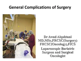
Complication of exodontia Dr.Mohammed Alaraji B.D.S,F.I.B.M.S
Undesirable situations are often encountered in dental practice, may caused by a dentist s mistake, culpability of the patient, or other unstable factors; I. Perioperative complications are the complications that occur during the surgical procedure. II. postoperative complications occur during the postoperative period.
Perioperative Complications. 1. Fracture of the crown of the adjacent tooth or luxation of the adjacent tooth 2. Soft tissue injuries 3. Failure to obtain anesthesia. 4. Fracture of the alveolar process 5. Fracture of the maxillary tuberosity 6. Fracture of the mandible 7. Broken instrument in tissues 8. Dislocation of the temporomandibular joint 9. Bleeding 10. Displacement of the root or root tip into soft tissues 11. Displacement of an impacted tooth, root or into the maxillary sinus 12. Oroantral communication 13. Nerve injury
Postoperative Complications 1. Trismus 2. Hematoma 3. Ecchymosis 4. Edema 5. Postextraction granuloma 6. Painful postextraction socket 7. Fibrinolytic alveolitis (dry socket) 8. Infection of wound 9. Disturbances in postoperative wound healing
Fracture of Crown or Luxation of Adjacent Tooth The fracture of the crown of an adjacent tooth that presents extensive caries or a large restoration. Luxation or dislocation of an adjacent tooth occurs when a great amount of force is exerted during the luxation attempt. Treatment when an adjacent tooth is inadvertently luxated or partially avulsed, the tooth is stabilized for approximately 40 60 days. If there is still pain during percussion even after this period, then the tooth must be endodontically treated. If the tooth is dislocated, it must be repositioned and stabilized for 3 4 weeks.
Failure to obtain anesthesia A. Faulty technique. B. Insufficient dosage of anesthesia. C. Expired anesthesia. D. The presence of acute infection.
Soft Tissue Injuries Soft tissue injuries are a common complication and most times are due to the: A. Inadvertent manipulation of instruments (e.g., slippage of elevator) during the removal of teeth. The areas most often injured are the cheeks, the floor of the mouth, the palate, and the retromolar area. B. burn may occur on the lower lip if an overheated surgical handpiece comes into contactwith the lip . C. Abrasions also happen when the shank of a rotating bur comes into contact with the area. D. tearing of the flap during reflection, as well as tearing of the gingiva during extraction
Treatment 1. When injuries are small and localized at the region of the cheek, tongue, or lips, then no particular treatment is considered necessary. 2. In certain cases healing is facilitated if the lesion is covered with petrolatum (Vaseline) (e.g., lip injury). 3. When the injury is extensive, though, and there is also hemorrhaging, the surgical procedure must be postponed and the dentist must control the bleeding and proceed with suturing of the wound.
Fracture of Alveolar Process This complication may occur if extraction movements are abrupt and awkward, or if there is ankylosis of the tooth in the alveolar process, where upon part of the labial, buccal, palatal or lingual cortical plate may be removed together with the tooth.
Treatment A. When the broken part of the alveolar process is small and has been reflected from the periosteum, then it is removed with forceps and the sharp edges, if any, of the remaining bone are smoothed. Afterwards, the area is irrigated with saline solution and the wound is sutured. B. If the broken part of the alveolar process is still attached to the overlying soft tissues, then it may remain after stabilization and suturing of the mucoperiosteum.
Fracture of Maxillary Tuberosity Fracture of the maxillary tuberosity is a grave complication, which, depending on its extent, may create problems for the retention of a full denture in the future. This complication may occur during the extraction of a posterior maxillary tooth and is usually due to the following reasons: 1. Weakening of the bone of the maxillary tuberosity. due to the maxillary sinus pneumatizing into the alveolar process. 2. Ankylosis of a maxillary molar that presents great resistance to movements during the extraction attempt.
Treatment A. When the fractured segment has not been reflected from the periosteum, it is repositioned and the mucoperiosteum is sutured. B. If the bone segment has been completely reflected from the tissues and oroantral communication occurs, the tooth is first removed and the bone is then smoothed and the wound is tightly sutured. Broad-spectrum antibiotics and nasal decongestants are then prescribed.
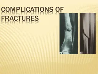
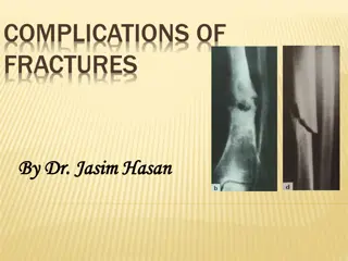
Fracture of Mandible Fracture of the mandible is a very unpleasant, but fortunately rare, complication that is associated almost exclusively with the extraction of impacted mandibular third molars. This may occur during the use of excessive force with the elevator, when an adequate pathway for removal of the impacted tooth has not been created. Treatment Stabilization of fracture by way of intermaxillary fixation or rigid internal fixation of the jaw segments is applied for 4 6 weeks and broad-spectrum antibiotics are administered.
Broken Instrument in Tissues Breakage the anesthesia needle or bur may break during the removal of the bone surrounding the impacted tooth or root. In these cases, after precise radiographic localization, the broken pieces are removed surgically at the same time as extraction of the tooth or root.
Dislocation of Temporomandibular Joint This complication may occur during a lengthy surgical procedure on patients who present (previeous history of TMJ dislocation)a shallow mandibular fossa of the temporal bone, low anterior articular tubercle, and round head of condylar process. In unilateral dislocation the mandible deviates towards the healthy, while in bilateral dislocation, the mandible slides forward in a gaping prognathic position. The patient is unable to close their mouth (open bite) and movement is restricted. In order to avoid such a complication, the mandible must be firmly supported during an extraction and patients must avoid opening their mouth excessively, especially those with a history of habitual temporomandibular joint luxation.
Treatment Immediately after the dislocation, the thumbs are placed on the occlusal surfaces of the teeth, while the rest of the fingers surround the body of the mandible right and left. Pressure is then exerted downward with the thumbs and simultaneously upwards and posteriorly with the rest of the fingers, until the condyle is replaced in its original position After repositioning, the patient must limit any movement of the mandible that may lead to excessive opening of the mouth for a few days. When luxation is habitual, themandible is often repositioned in its original position spontaneously.
Bleeding Hemorrhage is a common complication in oral surgery, and may occur during a simple tooth extraction or during any other surgical procedure. In all cases, hemorrhage may be due to: Either Local or systemic cause 1. trauma of the vessels in the region as well as to problems related to blood coagulation. Profuse hemorrhage may occur as a result of injury or severance of the inferior alveolar vessels or the palatal artery.local 2. Severe hemorrhagic diatheses (e.g., hemophilia, etc.) should be ascertained by taking a thorough medical history, and management must be planned before the surgical procedure.systemic Postoperative bleeding in healthy patients may be the result of poor hemostasis of the wound due to insufficient compression, or to inadequate removal of inflammatory and hyperplastic tissue from the surgical field
Treatment 1. The main means of arresting bleeding are: 2. compression, 3. ligation 4. suturing, 5. electrocoagulation and the use of various hemostatic agents.
Displacement of Root or Root Tip into Soft Tissues This complication may occur when the buccal or lingual cortical plate, as well as the root tips region of maxillary posterior teeth is eroded. In this case, the root or root tip may easily be displaced during luxation towards the buccal soft tissues or the floor of the mouth, or between the bone and mucosa of the maxillary sinus, respectively. Treatment Removal of the root tip, especially from buccal soft tissues, is not particularly difficult if its exact position has been localized. This localization is achieved with careful palpation of the area suspected of containing the displaced root tip.
Displacement of Impacted Tooth, Root, or Root Tip into Maxillary Sinus This complication may occur particularly during an attempt to luxate an impacted maxillary molars, when the tooth is close to the maxillary sinus
Treatment Caldwell Luc approach
Oroantral Communication Oroantral communicationmay be the result of: 1. Displacement of an impacted tooth or root tip into the maxillary sinus during a removal attempt. 2. Closeness of the root tips to the floor of the maxillary sinus. In this case the bony portion above the root tips is very thin or may even be absent. 3. The presence of a periapical lesion that has eroded the bone wall of the maxillary sinus floor 4. Extensive fracture of the maxillary tuberosity (during the extraction of a posterior tooth), whereupon part of the maxillary sinus may be removed together with the maxillary tuberosity 5. Extensive bone removal for extraction of an impacted tooth or root.
It is identified easily by I. The periapical curette enters to a greater depth than normal during debridement of the alveolus, which is explained by its entering the maxillary sinus. II. Oroantral communication may also be confirmed by observing the passage of air or bubbling of blood from the postextraction alveolus when the patient tries to exhale gently through their nose while their nostrils are pinched (Valsalva test).
Preventive Measures In order to avoid oroantral communication as well as displacement of an impacted tooth or root into the maxillary sinus, the following preventive measures are recommended: 1. Radiographic examination of the region surrounding the tooth to be extracted 2. Careful manipulations with instruments, especially during the luxation of a root tip of a maxillary posterior tooth 3. Careful debridement of periapical lesions that are close to the maxillary sinus
Treatment 1. For a small-sized oroantral communication, which is perceived immediately after the extraction, treatment consists of suturing the gingiva with a figure eight suture after filling the alveolus with collagen. 2. there are enough soft tissues, in which case placement of tight sutures over the wound is preferred. 3. When the soft tissues do not suffice, a small portion of the alveolar bone is removed with a bone rongeur so that the buccal and palatal mucosa can be reapproximated more easily, facilitating closure of the oroantral communication. 4. The patient is informed of the situation, and given appropriate instructions (e.g., avoiding sneezing, blowing nose), and is rescheduled for examination in 15 days. 5. A large oroantral communication or one that has remained open for 15 days or longer must be treated using other techniques (such as the closure with flap procedure using pedicle mucoperiosteal flaps (buccal, palatal)
Nerve Injury Nerve injury, especially the severance of large nerve branches, is one of the most serious complications that may occur during oral surgical procedures. The most common nerve injuries are of the inferior alveolar, mental, and lingual nerves. Nerve trauma may cause sensory disturbances (anesthesia or hypesthesia1), paresthesia2), dysesthesia3)) in the innervated area, resulting in various undesirable situations, such as a burning sensation, tingling, needles and pins, biting of the tongue and lips, abnormal chewing, burns through consumption of hot foods
classification of nerve injuries Neurapraxia: This type of damage has the most favorable prognosis and may occur even after simple contact with the nerve. Nerve conduction failure is usually temporary and there is complete recovery, without permanent pathologic and anatomic defects. Recovery is quite rapid and occurs gradually within a few days to week
Neurotmesis: This is the gravest type of nerve injury, resulting in discontinuation of conduction due to severance of the nerve or due to the formation of scar tissue at the area of trauma. This type of injury may cause permanent damage to nerve function, including paresthesia or even anesthesia. The formation of scar tissue may also prevent axon regeneration
Etiology 1. During administration of a nerve block 2. While creating an incision that extends to the region of the mental foramen and the lingual vestibular fold 3. removal of impacted teeth, roots and root tips that are deep in the bone and are close to the mental or inferior alveolar nerves 4. compression of the lingual nerve, due to excessive retraction of the tongue with a retractor during the surgical procedure. 5. compression and strangulation of a nerve, after inadvertent suturing of the nerve during the suturing of a flap.
Treatment 1. No particular therapy is indicated for neurapraxia unless there is a root tip or other foreign body compressing the nerve, 2. Treatment is usually palliative, including the administration of analgesics in painful situations, and multi-vitamin supplements of the vitamin B complex to restore sensation more rapidly. 3. Damage to the nerve as a result of neurotmesis must be treated as soon as possible; often, a graft must replace the injured nerve segments or the severed segments must be sutured.