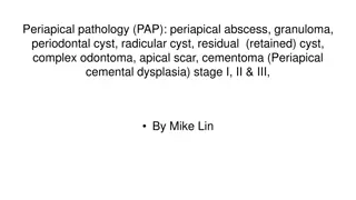
Understanding Periapical Pathology in Dental Hygiene

 undefined


Periapical
Pathology
(PAP)
New York City College of Technology
Dental Hygiene Department
DEN 1218
–
E 612
April 1, 2019
Presented by :
Jashlie Sanchez & Diana Usuga
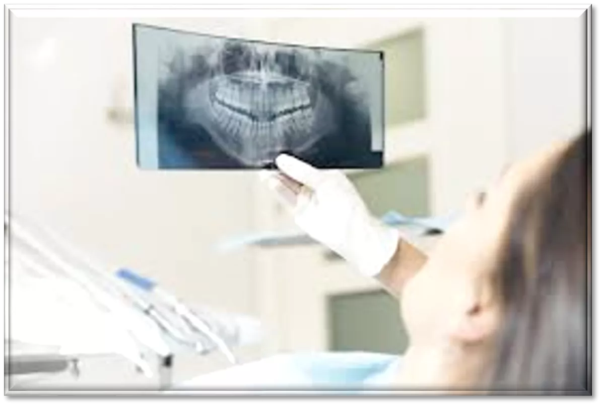


What is Periapical Pathology?
Periapical pathology
–
disease occurring around the apex of a tooth.
In this slide show we will be identifying several different types of
Periapical pathology (PAP) such as:
Periapical abscess
Granuloma
Periodontal cyst
Radicular cyst
Residual (retained) cyst
Complex odontoma
Apical scar
Cementoma
Stages of Periapical Cemental Dysplasia

Periapical Abscess
(
Dento-alveolar abcess)
Results from an infection of the pulpal tissue causing the pulp
to become necrotic. It is formed when pus escapes from walls of
the pulp chamber and the root canal(s) through the
apical
foramen
.
An area of pus and fluid accumulation
forms in the bone
surrounding the apex of the tooth. There is acute
inflammation
of
apical periodontium. Affected teeth are painful.
Radiolucent area at apex of root.
Widened periodontal ligament space.
Shama, S. A. (2013). Periapical abscess of the maxillary teeth and its fistulizations: Multi-detector CT
study.
Alexandria Journal of medicine
,
49
(1), 273-
279.
https://www.sciencedirect.com/science/article/pii/S2090506812001066
Periapical Abscess cont..
Without a microscopic
diagnosis, a clinician
is frequently unable to
differentiate between
a periapical
granuloma, a
radicular cyst, and an
apical abscess.
Periapical abscess
Granuloma
Localized mass of
chronic granulation
tissue
permeated by diverse inflammatory
cells (lymphocytes, plasma cells,
macrophages, mast cells)
formed as an
attempt of the periapical tissues to neutralize
and confine the irritating toxic products
escaping from the root canal.
Radiographically appear as a well-defined
radiolucent area of variable size seemingly
attached to root apex. Demarcated from
surrounding bone. Induce resorption of
supporting bone adjacent to this area.
A granuloma
may evolve into a radicular
cyst or an apical abscess
. The lesion is
usually asymptomatic but may sometimes
exhibit mild pain and sensitivity to
percussion.
Bajaj, A. (2018). Acme, Pathosis, Furuncle: the Periapical
Granuloma.
Journal of Gastrointestinal Disorders and Liver
function
,
4
(1), 11-13.
https://www.ommegaonline.org/article-
details/Acme-Pathosis-Furuncle-the-Periapical-Granuloma/1885
(Lateral) Periodontal Cyst (LPC)
Non-keratinized and
non-inflammatory
developmental cysts located adjacent or lateral to
the root of a vital tooth.
Arise along the lateral
periodontium or within the bone between the
roots of erupted vital teeth. Are generally
asymptomatic.
The most frequent sites are the premolar region of
the mandible and the anterior segment of the
maxillary alveolar process.
Radiologically, a LPC is a round or slightly oval-
shaped, well-defined radiolucent area, frequently
with a sclerotic margin. Usually does not exceed a
maximum in diameter of 10 mm.
Friedrich, R. E., Scheuer, H. A., & Zustin, J. (2014). Lateral periodontal
cyst.
in vivo
,
28
(4), 595-598.
iv.iiarjournals.org/content/28/4/595.short
Radicular Cyst (Periapical Cyst)
Arise from epithelial residues (cell rests of Malassez) in the
periodontal ligament as a consequence of inflammation, usually
following the death of the dental pulp. Approximately 20% of
periapical granulomas progress to periapical cysts.
Radiographically, the lesion is usually seen as a round or oval, well-
circumscribed radiolucent image involving the apex of the roots.
Often, the associated tooth has a deep restoration or large carious
lesion.
Over the years, the cyst may regress, remain static or grow in size.
Cyst may be greater than granuloma (> 1.5 cmm). Occasionally
exhibits thin, radiopaque line around the periphery of radiolucent
area which indicates reaction of bone to slowly expanding mass.
Narula, H., Ahuja, B., Yeluri, R., Baliga, S., & Munshi, A. K. (2011). Conservative non-
surgical management of an infected radicular cyst.
Contemporary clinical
dentistry
,
2
(4), 368. https://www.ncbi.nlm.nih.gov/pmc/articles/PMC3276870/
Residual Cyst
A cyst that
may persist after
the extraction of the
causative tooth or
incomplete removal of
periapical
granuloma/periapical cyst
.
Usually asymptomatic.
Round to ovoid
radiolucency in alveolar
ridge.
Continued growth can
cause significant bone
resorption.
Complex Odontoma
Odontomas are the most common
benign odontogenic tumor.
Abnormal proliferation of cells of
the enamel organ, they give rise
from the odontogenic epithelium
and mesenchyme that produce
enamel and dentin. There are two
types of odontomas: compound
and complex.
A compound tumor represents
multiple toothlike structures, where
a
complex odontoma has
irregularly shaped masses of
enamel (amorphous) showing no
anatomic resemblance to a tooth.
Retrieved from:
https://www.dentalcare.com/en-us/professional-
education/ce-courses/ce513/case-study-4-odontoma
Apical scar
An
apical scar
is caused by the
body’s exuberant attempt at
healing itself
after a root canal or
similar endodontic procedure.
Sometimes the body overreacts
and sends more tissue than is
needed to heal a certain sized
space, so a scar is left behind that
is thicker than normal. People are
unaware an apical scar has
formed unless it is accompanied
by an infection.
Retrieved from: https://omegadentists.com/blog/apical-scarring-and-
root-canal/
Cementoma
Cementoma
is an
odontogenic tumor of
cementum. It is usually
observed as a benign
spherical mass of hard tissue
fused to the root of a tooth. It is
found most commonly in the
mandible in the region of the
lower molar teeth.
Reference:
Ben Z. Pilch (2001). Head and Neck Surgical Pathology.
Lippincott Williams & Wilkins. pp. 222–. ISBN 978-0-397-51727-5
Stages of Periapical Cemental Dysplasia
Periapical cemental dysplasia is a benign condition
mostly seen in patients over 20 years of age and is
more common in women. The lesion occurs in and
near the periodontal ligament around the apex of a
tooth, usually a mandibular incisior. Most cases
usually present with multiple lesions involving the
apices of several mandibular anterior teeth or
bicuspids. Since the lesion is asymptomatic, it is
often accidentally discovered during routine
intraoral roentgenographic examination.
1.
In the early stage of development, the lesion appears
as a periapical radiolucency resembling periapical
granuloma or cyst.
2.
The second stage in the development of the lesion is
the beginning of calcification in the radiolucent area.
3.
The third stage appears on the roentgenogram as a
well-defined radiopacity that is usually bordered by
a thin radiolucent line.
No treatment is required, as it is harmless, and only
periodic observation is required. It is difficult to radio
graphically distinguish this lesion from a periapical
granuloma. Hence to make this distinction, vitality
testing of the pulp should be done. No treatment is
required for this condition.
Retrieved by: https://screening.iarc.fr/atlasoral_list.php?cat=E21&lang=1
Photo retrieved by: https://www.semanticscholar.org/paper/A-clinical-and-radiographic-study-of-104-cases-with-Hwang-Lee dcb19585c38b667e5e5f5eda0db7014a1f9c5f4f
Periapical pathology involves diseases around the tooth apex, such as abscesses, granulomas, cysts, and more. This presentation delves into identifying and differentiating these conditions, highlighting symptoms and radiographic features of each. Understanding these pathologies is crucial for dental professionals to provide accurate diagnoses and appropriate treatment interventions.

Download Presentation

Please find below an Image/Link to download the presentation.
The content on the website is provided AS IS for your information and personal use only. It may not be sold, licensed, or shared on other websites without obtaining consent from the author. Download presentation by click this link. If you encounter any issues during the download, it is possible that the publisher has removed the file from their server.
E N D
Presentation Transcript
+ Periapical Pathology (PAP) New York City College of Technology Dental Hygiene Department DEN 1218 E 612 April 1, 2019 Presented by : Jashlie Sanchez & Diana Usuga
+What is Periapical Pathology? Periapical pathology disease occurring around the apex of a tooth. In this slide show we will be identifying several different types of Periapical pathology (PAP) such as: Periapical abscess Granuloma Periodontal cyst Radicular cyst Residual (retained) cyst Complex odontoma Apical scar Cementoma Stages of Periapical Cemental Dysplasia
+Periapical Abscess (Dento-alveolar abcess) Results from an infection of the pulpal tissue causing the pulp to become necrotic. It is formed when pus escapes from walls of the pulp chamber and the root canal(s) through the apical foramen. An area of pus and fluid accumulation forms in the bone surrounding the apex of the tooth. There is acute inflammation of apical periodontium.Affected teeth are painful. Radiolucent area at apex of root. Widened periodontal ligament space. Shama, S. A. (2013). Periapical abscess of the maxillary teeth and its fistulizations: Multi-detector CT study.Alexandria Journal of medicine,49(1), 273- 279.https://www.sciencedirect.com/science/article/pii/S2090506812001066
+ Periapical Abscess cont.. Without a microscopic diagnosis, a clinician is frequently unable to differentiate between a periapical granuloma, a radicular cyst, and an apical abscess. Periapical abscess
+ Granuloma Localized mass of chronic granulation tissue permeated by diverse inflammatory cells (lymphocytes, macrophages, mast cells) formed as an attempt of the periapical tissues to neutralize and confine the irritating toxic products escaping from the root canal. plasma cells, Radiographically appear as a well-defined radiolucent area of variable size seemingly attached to root apex. Demarcated from surrounding bone. Induce resorption of supporting bone adjacent to this area. A granuloma may evolve into a radicular cyst or an apical abscess. The lesion is usually asymptomatic but may sometimes exhibit mild pain percussion. and sensitivity to Bajaj, A. (2018). Acme, Pathosis, Furuncle: the Periapical Granuloma.Journal of Gastrointestinal Disorders and Liver function,4(1), 11-13. https://www.ommegaonline.org/article- details/Acme-Pathosis-Furuncle-the-Periapical-Granuloma/1885
+(Lateral) Periodontal Cyst (LPC) Non-keratinized and non-inflammatory developmental cysts located adjacent or lateral to the root of a vital tooth. Arise along the lateral periodontium or within the bone between the roots of erupted vital teeth. Are generally asymptomatic. The most frequent sites are the premolar region of the mandible and the anterior segment of the maxillary alveolar process. Radiologically, a LPC is a round or slightly oval- shaped, well-defined radiolucent area, frequently with a sclerotic margin. Usually does not exceed a maximum in diameter of 10 mm. Friedrich, R. E., Scheuer, H. A., & Zustin, J. (2014). Lateral periodontal cyst.in vivo,28(4), 595-598.iv.iiarjournals.org/content/28/4/595.short
+Radicular Cyst (Periapical Cyst) Arise from epithelial residues (cell rests of Malassez) in the periodontal ligament as a consequence of inflammation, usually following the death of the dental pulp. periapical granulomas progress to periapical cysts. Approximately 20% of Radiographically, the lesion is usually seen as a round or oval, well- circumscribed radiolucent image involving the apex of the roots. Often, the associated tooth has a deep restoration or large carious lesion. Over the years,the cyst may regress,remain static or grow in size. Cyst may be greater than granuloma (> 1.5 cmm). Occasionally exhibits thin, radiopaque line around the periphery of radiolucent area which indicates reaction of bone to slowly expanding mass. Narula, H., Ahuja, B., Yeluri, R., Baliga, S., & Munshi, A. K. (2011). Conservative non- surgical management of an infected radicular cyst.Contemporary clinical dentistry,2(4), 368. https://www.ncbi.nlm.nih.gov/pmc/articles/PMC3276870/
+ Residual Cyst A cyst that may persist after the extraction of the causative tooth or incomplete removal of periapical granuloma/periapical cyst. Usually asymptomatic. Round to ovoid radiolucency in alveolar ridge. Continued growth can cause significant bone resorption.
+ Complex Odontoma Odontomas are the most common benign odontogenic Abnormal proliferation of cells of the enamel organ, they give rise from the odontogenic epithelium and mesenchyme enamel and dentin. There are two types of odontomas: compound and complex. tumor. that produce A compound multiple toothlike structures,where a complex irregularly shaped enamel (amorphous) showing no anatomic resemblance to a tooth. tumor represents odontoma has of masses Retrieved education/ce-courses/ce513/case-study-4-odontoma from: https://www.dentalcare.com/en-us/professional-
+ Apical scar An apical scar is caused by the body s exuberant healing itself after a root canal or similar endodontic procedure. attempt at Sometimes the body overreacts and sends more tissue than is needed to heal a certain sized space, so a scar is left behind that is thicker than normal. People are unaware an apical formed unless it is accompanied by an infection. scar has Retrieved root-canal/ from: https://omegadentists.com/blog/apical-scarring-and-
+ Cementoma Cementoma is an odontogenic tumor of cementum. It is usually observed as a benign spherical mass of hard tissue fused to the root of a tooth. It is found most commonly in the mandible in the region of the lower molar teeth. Reference: Ben Z. Pilch (2001). Head and Neck Surgical Pathology. Lippincott Williams & Wilkins. pp. 222 . ISBN 978-0-397-51727-5
+ Stages of Periapical Cemental Dysplasia Periapical cemental dysplasia is a benign condition mostly seen in patients over 20 years of age and is more common in women. The lesion occurs in and near the periodontal ligament around the apex of a tooth, usually a mandibular incisior. Most cases usually present with multiple lesions involving the apices of several mandibular anterior teeth or bicuspids. Since the lesion is asymptomatic, it is often accidentally discovered intraoral roentgenographic examination. during routine 1. In the early stage of development,the lesion appears as a periapical radiolucency resembling periapical granuloma or cyst. 2. The second stage in the development of the lesion is the beginning of calcification in the radiolucent area. 3. The third stage appears on the roentgenogram as a well-defined radiopacity that is usually bordered by a thin radiolucent line. No treatment is required, as it is harmless, and only periodic observation is required.It is difficult to radio graphically distinguish this lesion from a periapical granuloma. Hence to make this distinction, vitality testing of the pulp should be done. No treatment is required for this condition. Retrieved by: https://screening.iarc.fr/atlasoral_list.php?cat=E21&lang=1 Photo retrieved by: https://www.semanticscholar.org/paper/A-clinical-and-radiographic-study-of-104-cases-with-Hwang-Lee dcb19585c38b667e5e5f5eda0db7014a1f9c5f4f


")


 Periodontal Cyst (LPC)")
")