Structure and Function of a Serous Compound Tubulo-acinar Gland
A detailed description of a compound tubulo-acinar gland with serous-secreting nature is provided, highlighting its cellular composition, arrangement of ducts, and connective tissue organization. The glandular structure, including adenomeres, centroacinar cells, and myoepithelial cells, is discussed, along with the role of various components within the gland. The presence of Glisson's capsule and the organization of lobules within the parenchyma are outlined, with a focus on interlobular septations. Additionally, the hepatic architecture, hepatocytes' arrangement towards sinusoids, and their cellular characteristics are elucidated.
Download Presentation

Please find below an Image/Link to download the presentation.
The content on the website is provided AS IS for your information and personal use only. It may not be sold, licensed, or shared on other websites without obtaining consent from the author.If you encounter any issues during the download, it is possible that the publisher has removed the file from their server.
You are allowed to download the files provided on this website for personal or commercial use, subject to the condition that they are used lawfully. All files are the property of their respective owners.
The content on the website is provided AS IS for your information and personal use only. It may not be sold, licensed, or shared on other websites without obtaining consent from the author.
E N D
Presentation Transcript
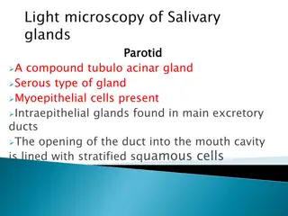
It is a compound tubulo acinar gland of serous secreting nature The capsule is made up of connective tissue which divides the gland through its trabeculi into several lobes and lobules The glandular adenomeres are made up of Pyramidal cells with parabasally placed spherical nucleus The granules containing enzymes cytoplasm presents eosinophilic cytoplasmic
The main excretory duct is lined with simple columnar epithelium The centroacinar cells which are extended epithelial cells of intercalated ducts Myoepithelial cells are absent central lumen of the acini presents
Irregular connective tissue known as Glissons capsule is covered to its maximum extent by peritoneum From this capsule the CT descends towards the center of the gland which are predominated with reticular fiber These septations divide the parenchyma into roughly hexagonal lobules The interlobular septations are quite prominent in pig and made up of collagen fibers and are least developed in other animals
The arranged towards The sinusoids vein The is is mitochondria,polyribosomes,ribosomes,lysosomes,SER,lipid and The cells arranged in towards the The hepatic sinusoids which vein The hepatocytes spherical mitochondria,polyribosomes,ribosomes,lysosomes,SER,lipid and glycogen cells of in radiating the periphery hepatic cell which start of the radiating columns periphery of cell column start from the liver liver are columns from of the column are from periphery are known known as from outer the lobule are separated periphery and as hepatocytes outer margin hepatocytes which margin of which are of central are vein central vein lobule separated from and opens from each opens in each other in the other by the central by central hepatocytes are spherical are usually usually mononucleated and mononucleated cells cytoplasm cells and and nucleus rich nucleus and cytoplasm rich in in glycogen granules granules
The stellate known The hepatocyte The sinusoidal stellate shaped known as The hepatocyte is is called sinusoidal wall shaped phagocytic as Kuffer s space called space wall is is associated phagocytic cells Kuffer s cells space space of associated with cells popularly with large popularly large cells between between the of Disse the sinusoids Disse sinusoids and and
