Understanding Mediastinal Masses: A Comprehensive Visual Guide
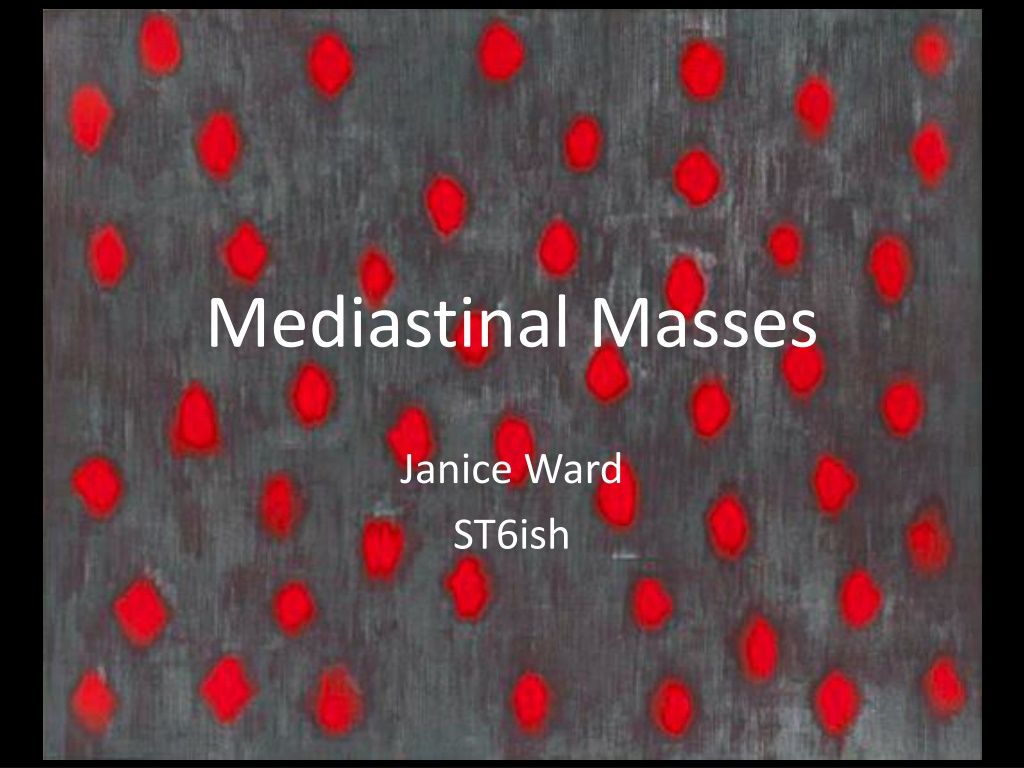


Mediastinal Masses
Janice Ward
ST6ish

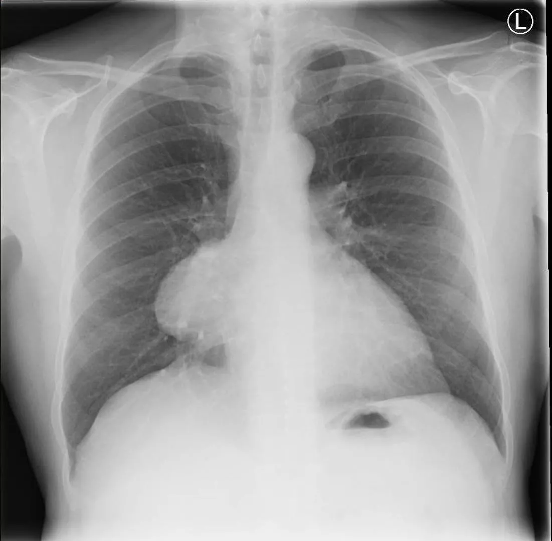
RV
LV
Fat pad
Left diaphragm
Right diaphragm
Mediastinal compartments
4 densities of xray
Sihouette Sign
An interface is not
visible when two areas
of similar radiodensity
touch.
Hilum overaly sign
There is aerated lung
infront or behind the
mass making it less
dense than the hilar
–
therefore it is not on
the same plane as
the hilar vessels.
Note angle
–
not
mediastinal
Anterior
4Ts
1.
Thymoma
2.
Thyroid
3.
Teratoma
4.
Terrible lymphoma
Others
•
Hernia of Morgagni
•
Pericardial cyst
•
Fat pad
•
Ascending aorta aneurysm
Medial
•
Hilar lymph nodes
•
Bronchogenic cyst
•
Foregut cyst
•
Oesophageal tumour
•
Pericardial/cardiac tumour
•
Aortic arch or pulmonary artery aneurysm
•
(Thyroid)
Posterior
•
Neural tumours and
cysts
•
Lipoma
•
Descending aorta
aneurysm
•
Gastric pull through
•
Extramedullary
haematopoesis
•
Bochdalek posterior
diaphragmatic hernia
Management of patient
History
•
?mass effect
–
E.g. SVCO
•
?functional effect
–
E.g myasthenia gravis
•
Systemic enquiry
–
metastasis
•
Examination
Maybe some reassurance
Imaging
•
Cxr and lateral
•
CT with contrast
…
CT
•
Anatomy
–
online
–
Thymus reduces in size
and disappears by age
40
Density
•
Fluid
–
Water hounsefield unit 0
–
Blood 30
•
Fat
–
Hounsefield 80
•
Air
–
Hounsfield -1000
•
Bone
–
Housefield 2000
•
Enhancement
Necrotic lymph nodes
Teratoma
Next Investigations
Case
•
Middle age man
•
Cough
•
Obs normal
•
Covid swab
•
A+E wanted to admit, GIM consultant
discharged with urgent o/p CT and resp
referral
Summary
•
Use of logical approach to plain film to localise
mass
•
List of differentials
•
Assessment of patient
–
mass effect or functional effect
•
CT to characterise
•
Some further specific tests
Bibliography
•
https://radiologyassistant.nl/chest/mediastinum-
masses
•
https://www.radiologycafe.com/medical-
students/radiology-basics/chest-anatomy
•
BTS short course radiology
•
Felsons Principles of Chest Roentgenology. Lawrence
Goodman. Second edition. 1999
•
Thoracic Imaging: Illustrated clinical cases. Copley et al.
second edition. 2014.
•
Oxford Handbook of respiratory medicine.
Sophie
West.
Second edition 2009.
Explore detailed visuals and descriptions of mediastinal masses, compartments, x-ray densities, and key signs in diagnosis. Learn about different types of masses and their management, including common differential diagnoses and imaging techniques.

Download Presentation

Please find below an Image/Link to download the presentation.
The content on the website is provided AS IS for your information and personal use only. It may not be sold, licensed, or shared on other websites without obtaining consent from the author. Download presentation by click this link. If you encounter any issues during the download, it is possible that the publisher has removed the file from their server.
E N D
Presentation Transcript
Mediastinal Masses Janice Ward ST6ish
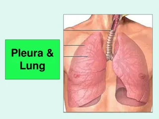
RV LV Left diaphragm Fat pad Right diaphragm
Mediastinal compartments Thoracic duct Heart Trachea oesophagus Nerve roots Lymph nodes Sympathetic and parasympathetic chains Aortic arch Thyroid Thymus Vena cava Ascending aorta Lymph nodes Lymph nodes Descending aorta Pulmonary artery Phrenic Nerve vertebrae
Sihouette Sign An interface is not visible when two areas of similar radiodensity touch.
Hilum overaly sign There is aerated lung infront or behind the mass making it less dense than the hilar therefore it is not on the same plane as the hilar vessels. Note angle not mediastinal
Anterior 4Ts 1. Thymoma 2. Thyroid 3. Teratoma 4. Terrible lymphoma Others Hernia of Morgagni Pericardial cyst Fat pad Ascending aorta aneurysm
Medial Hilar lymph nodes Bronchogenic cyst Foregut cyst Oesophageal tumour Pericardial/cardiac tumour Aortic arch or pulmonary artery aneurysm (Thyroid)
Posterior Neural tumours and cysts Lipoma Descending aorta aneurysm Gastric pull through Extramedullary haematopoesis Bochdalek posterior diaphragmatic hernia
Management of patient History ?mass effect E.g. SVCO ?functional effect E.g myasthenia gravis Systemic enquiry metastasis Maybe some reassurance Imaging Cxr and lateral CT with contrast Examination
CT Anatomy online Thymus reduces in size and disappears by age 40
Density Fluid Water hounsefield unit 0 Blood 30 Fat Hounsefield 80 Air Hounsfield -1000 Bone Housefield 2000 Enhancement Teratoma Necrotic lymph nodes
Next Investigations Possible diagnosis Blood test Specific Imaging Functioning thymoma AChR ab Seminoma US testes Non-seminomatous germ cell tumours bHCG and AFP CT for distant metastasis Thyroid TSH, T4, T3 Radioisotope scan Flow-volume loop Lymphoma FBC, ESR, LDH, HIV PET Surgical biopsy Neural tumours and cysts MRI
Case Middle age man Cough Obs normal Covid swab A+E wanted to admit, GIM consultant discharged with urgent o/p CT and resp referral
Summary Use of logical approach to plain film to localise mass List of differentials Assessment of patient mass effect or functional effect CT to characterise Some further specific tests
Bibliography https://radiologyassistant.nl/chest/mediastinum- masses https://www.radiologycafe.com/medical- students/radiology-basics/chest-anatomy BTS short course radiology Felsons Principles of Chest Roentgenology. Lawrence Goodman. Second edition. 1999 Thoracic Imaging: Illustrated clinical cases. Copley et al. second edition. 2014. Oxford Handbook of respiratory medicine. Sophie West. Second edition 2009.


![textbook$ What Your Heart Needs for the Hard Days 52 Encouraging Truths to Hold On To [R.A.R]](/thumb/9838/textbook-what-your-heart-needs-for-the-hard-days-52-encouraging-truths-to-hold-on-to-r-a-r.jpg)