Insights into DTI and Advanced MRI Measures for Cognitive Impairment
Comparison of DTI protocols and measures in 317 participants revealed strong associations with age and cognitive impairment. MD and TDF-FA emerged as best measures, with robust connections to age. NODDI in advanced MRI allows for differentiation of A-beta positive and negative groups, offering greater insight into underlying microstructure compared to standard DTI measures. Associations across NODDI implementations indicate potential for increased understanding of neurodegenerative processes through diffusion MRI measures.

Download Presentation

Please find below an Image/Link to download the presentation.
The content on the website is provided AS IS for your information and personal use only. It may not be sold, licensed, or shared on other websites without obtaining consent from the author. Download presentation by click this link. If you encounter any issues during the download, it is possible that the publisher has removed the file from their server.
E N D
Presentation Transcript
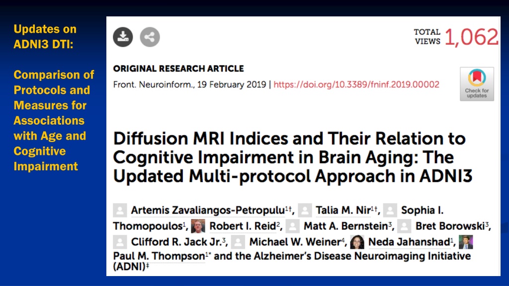
Updates on ADNI3 DTI: Comparison of Protocols and Measures for Associations with Age and Cognitive Impairment
ADNI3 DTI Data from 317 participants 6 DTI protocols compared In all protocols, all 4 standard DTI measures showed strong associations with: - age - sobCDR, MMSE, ADAS-Cog Best measures: MD (mean diffusivity, cingulum hippocampal bundle), and TDF- FA (fractional anisotropy derived from the tensor distribution function)
All standard DTI measures show robust associations with Age: ADNI3 DTI Data from 317 participants; diffusivity shows stronger associations than DTI-FA; TDF-FA adjustment also boosts signal for FA Data from Artemis Zavaliangos-Petropulu, Talia Nir et al., Frontiers in Neuroinformatics, Feb. 2019
High-end Multi-shell Diffusion MRI parameters (NODDI, in blue) compared to best measures from standard DTI protocol (Mean Diffusivity, or MD, in green) NODDI distinguishes intracellular volume fraction (index of axonal loss) and isotropic volume fraction (generally increased diffusivity, inflammation) Data from Talia Nir et al., OHBM 2019
Both Standard (DTI) and Advanced (NODDI) Diffusion MRI Measures separate A-beta positive and A-beta negative groups
High-end Multi-shell Diffusion MRI measures (NODDI, in blue) compared to best measures from standard DTI protocol (Mean Diffusivity, or MD, in green) Similar associations were detected across NODDI implementations; AMICO slightly better for A-beta associations. For MCI v. CTL contrast, AMICO ICVF detected significant effects in the greatest number of ROIs, while MD showed the largest effect sizes overall. NODDI may offer greater insight into underlying microstructure. Like MD, the largest ISOVF effect sizes were detected in the genu of the corpus callosum (GCC; MD Cohen s d=1.66; ISOVF AMICO d=1.35), suggesting increased free-water from CSF or inflammation; for ICVF the largest effects were in the cingulum (CGC DMIPY d=-1.55), a region showing no group difference with MD or ISOVF, suggesting neuronal loss. dMRI measures distinguished groups separated by A + / A - status strongest effects in the superior frontal-occipital fasciculus (SFO) across measures (MD d=2.37; DMIPY ODI d=-1.99; AMICO ISOVF d=2.37).


 and Advanced (NODDI) Diffusion MRI")
")