Comprehensive Lung Examination and Assessment Techniques
This detailed guide provides insights into conducting a thorough lung examination, including objectives, equipment needed, preparation steps, subjective data assessment, chest landmarks identification, inspection of the thorax's anterior, posterior, and lateral aspects, and evaluation of breathing patterns. It emphasizes safe and accurate assessment techniques to evaluate respiratory health effectively.

Download Presentation

Please find below an Image/Link to download the presentation.
The content on the website is provided AS IS for your information and personal use only. It may not be sold, licensed, or shared on other websites without obtaining consent from the author. Download presentation by click this link. If you encounter any issues during the download, it is possible that the publisher has removed the file from their server.
E N D
Presentation Transcript
Prof Dr. Salma Khadim Jehad Dr. Ali Faris M.Sc Hassanain Mohammed Kadhim Lecture -6-
Lung Examination Objectives: At the end of this lab, the students will be able to: 1. Demonstrate the ability to safely & accurately complete thorax & lung assessment. 2. Demonstrate the ability to accurately document thorax & lung assessment data in organized manner. Equipment Needed 1. Stethoscope 2. Small ruler, marked in centimeters 3. Marking pen 4. Alcohol swab
Preparation 1. Ask the client to sit upright & the male to disrobe to the waist. 2. For female, leave the gown on & open at the back. 3. When examining the anterior chest, lift up the gown & drape it on her shoulders rather than removing it completely. 4. For farther comfort: a warm room, a warm diaphragm end piece. 5. Private examination time with no interruption.
Subjective data: Cough Past history of respiratory infections Self-care behaviors Shortness of breath Smoking history Chest pain with breathing Environmental exposure
Chest Landmarks Anterior : Right anterior axillary line , Right midclavicular line , Mid sternum line left midclavicular line , left anterior axillary line Mid axillary line Posterior: L . posterior axillary line , L .mid scapular line ,mid spinal line , R. mid scapular line and R. posterior axillary line ,
Inspect anterior, posterior, & lateral thorax for the following: Color : Pink Intercostals spaces : Even Chest symmetry: Equal Rib slope : Less than 90 degree downward Respiration (rate, depth, rhythm) ,Even, 12-20/min, unlabored Anterior-posterior to lateral diameter 1 : 2 ratio Shape & position of sternum : level with ribs Position of trachea Midline
Breathing Pattern 3-Tachypnea: Rapid shallow breathing is a rate above 20 breaths per minute, associated with increased activity or a disease process 1-Eupnea: Normal breathing is relaxed, effortless, and regular at 14-16 breath\minute
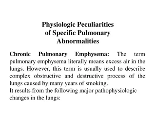
4-Bradypnea: slow breathing is a rate blow 12 breath per minute with normal depth and rhythm , associated with Sedation , anesthesia 5-Hypoventilation : Shallow irregular breathing hypercapnia and hypoxemia such as in COPD 6-Hyperventilation increased depth and rate of breathing (kussmaul s respiration caused by diabetic ketoacidosis
7-Cheyne-Stokes is the term for cycles of breathing characterized by deep, rapid breaths for about 30 seconds, followed by absence of respirations for 10 to 30 seconds. Cheyne-Stokes respirations constitute a serious symptom and precedes death in cerebral hemorrhage, uremia, or heart disease 8- Biot's respiration rapid, short breathing with pauses of several seconds, indicating increased intracranial pressure.
Inspection Normal chest Slight retraction of intercostal spaces 2x as wide as deep Anterior/posterior diameter 1:2
Inspection Barrel chest anterior-posterior diameter 2:2
Inspection Pigeon chest Sternum protrudes outward anterior-posterior diameter
Inspection Scoliosis Lateral curvature of thoracic spine Assessment Shoulders elevated? Complications Lung & heart damage Back problems Body image
Inspection Kyphosis Abnormal curvature of the thoracic spine
Inspection Lordosis Sway-back Abnormal curvature of the lumbar spine
Inspection Uniform expansion of the chest Pneumonia Pleural effusion Pneumothorax Bulging intercostal spaces Obstruction Emphysema
Palpate thorax at three levels for the following: Sensation : no pain or tenderness Vocal fremitus ( tactius) as client says 99Use either the palm base (the ball) of fingers, or the ulnar edge of one hand. - Touch the client's chest-Ask the client to repeat a resonant phrases that generate strong vibration Like 99. Start over the lung apices & palpate from side to another.- Avoid palpating over the scapulae. Vibration decreased over periphery of lungs & increased over major airways . - -
- Palpate chest expansion : Posterior : placing your warmed hand on the poster lateral chest wall - The thumbs should be at level of T9 or T10. - Slide your hands medially to pinch up a small fold between your thumbs. -Ask the client to take deep breath. - Your thumb should move with respiration. Anterior: placing your warmed hand on the anterolateral wall. - Thumbs should be along the costal margins & pointing toward the xiphoid process. -Ask the client to take deep breath. - Watch your thumbs move with respiration.
2 to 3-inch symmetrical thoracic expansion. Symmetrical expansion (thumbs move apart equal distance in both directions).
Percussion (Diaphragmatic Excursion) Posteriorely : ask the client to exhale & hold it. - Percuss down the scapular line until the sound changes from resonant to dull each side. - Mark the level where the sound changed to dull. -Ask the client to take deep breath & hold it. - Continue percussion from the mark down ward. Mark the level the sound changed to dull on deep inspiration. - Normal Finding : It should be equal bilaterally, & measure about 3-5cm in adult, although it may be up to 7-8cm. . -
Auscultation Purpose Asses normal and abnormal air flow through bronchial tree by using Diaphragm of stethoscope Compare R to L
Auscultation: normal lung sound 1-Bronchial : Trachea , high , inspiration shorter than expiration 2-Bronchovesicular : Moderate , Between scapulae Side of sternum intercostals space , inspiration equal with expiration 3-Vesicular : Lung field , inspiration longer than expiration is it soft and low
Abnormal Lung sound 1-Crackles (fine): high, short, popping sound heard during inspiration not clear with cough Caused by: inhaled air sudden open of the small deflated air passage with sticky with exudates , can be associated with pneumonia , congestive heart failure or bronchitis and asthma
2-Coarse crackles low pitch bubbling , moist sound that may persist from early inspiration to early expiration air comes into contact with secretions in the large bronchi and trachea may indicated pneumonia , pulmonary edema client with COPD
3-Wheeze(sibilant): high in pitch ,musical sound heard in expiration or may be inspiration ,air pass through constricted passage as secretion or tumor heard asthma or emphysema 4-Wheeze (sonorous): low pitch snoring or moaning sound heard during expiration clear with cough , heard in bronchitis , sleeping apnea . 5-Stridor: harsh honking wheeze heard with broncholaryngospasm as in croup
6-Pleural friction rub: low pitch grating sound superficial occur during I&E result of rubbing of two inflamed pleural surface as pleuritis Best heard anterior, Lower, lateral area


")


: high in pitch ,musical sound heard in")