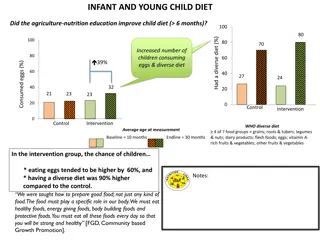
Case Study: Infant with Persistent Fever and Cry
Aditi Malakar, a 2-month-old female infant, presented with a remittent fever persisting for 9 days unresponsive to antipyretics, along with persistent crying and decreased feeding for the last 2 days. She was referred to the hospital after being diagnosed with sepsis elsewhere. Aditi had a history of low birth weight, Neonatal sepsis, and normal development. On examination, she appeared toxic with enlarged lymph nodes and bulged fontanelle. Her vital signs showed tachycardia, increased respiratory rate, low BP, and elevated temperature. Further evaluation revealed normal chest and abdominal findings and neurological assessment.

Download Presentation

Please find below an Image/Link to download the presentation.
The content on the website is provided AS IS for your information and personal use only. It may not be sold, licensed, or shared on other websites without obtaining consent from the author. Download presentation by click this link. If you encounter any issues during the download, it is possible that the publisher has removed the file from their server.
E N D
Presentation Transcript
By Dr. Sayani Pan 2ndyear PGT, Dept. of Paediatrics, BMCH
Name : Aditi Malakar Age : 2 months 15 days Sex : Female Address : Kotulpur, Bankura Date of admission : 22/06/2020 Duration of stay in the hospital : 18 days Informant : Mother
Fever for last 9 days Persistent cry for last 2 days
Fever since last 9 days, remittent in nature, unresponsive to antipyretics. Persistent cry since last 2 days associated with decreased feeding. No history of chills or rigor, jaundice, convulsion, any recent drug intake or blood transfusion. Baby was rushed to Arambagh SDH on the 5thday of fever and was provisionally diagnosed as Sepsis. Child was admitted there for 4 days. But fever did not subside despite antipyretics and several intravenous antibiotics, rather the child became more irritable, was unable to take feed properly and there was incessant cry. Referred to BMCH on 22/6/2020.
Normal vaginal delivery at hospital/ Term/ low birth weight - 2.25 kg History of SNCU admission at 6 days of age for Neonatal sepsis and respiratory distress. Received past immunizations as per date. No history of adverse reaction to 1stdose of pentavalent vaccine. No history suggestive of any delayed development. No history of hypertension tuberculosis, asthma or diabetes in the family.
General survey General look of the baby was toxic. Baby was alert but irritable. No pallor, cyanosis, icterus, clubbing, edema B/l cervical lymph nodes : enlarged, size->1.5 cm, non-tender, non-matted. No other lymph nodes palpable in the body. Ant. fontanelle : Bulged, pulsatile
Vitals : Pulse : 150/min, regular, normovolemic Respiration : 48/min, regular BP : 82/50 mm of Hg Temperature : 102.1* F CRT : <3 seconds SpO2 : 96%
Chest : B/l air entry good, VBS + CVS : Apex in left 4thICS, just lateral to the mid- clavicular line S1, S2 + No murmur P/A : Soft, non-tender, no organomegaly CNS : Head circumferance : 40 cm Pupils : B/l reactive Plantar reflex : B/l flexor Tone : Normal
On 23/6/2020 : Complete blood count : Hb : 10.3g% TLC : 20,200 DLC : N15L76M06E03B0 Platelets : 6,30,000 ESR : 36mm/1sthour CRP : 82.1 Blood culture : sent
Urine R/E, M/E : Appearance : clear Albumin : trace Sugar : nil Pus cells : 5-8/hpf Epithelial cells : 7-10/hpf RBCs : nil Urine Culture: No growth CSF study : App. : slightly hazy Cell count : 30/cmm Cell type : PMN- 40%, Lymphocytes- 60% Sugar : 57.8mg/dl, Protein : 61.2mg/dl
LFT : Total bilirubin- 1.2, ALT- 34, AST- 36, ALP- 102, Serum Albumin- 3.6g/dl RFT : Blood urea 17, Creatinine 0.6 Serum sodium : 134 mEq/L Serum potassium : 3.6 mEq/L Dengue NS1 antigen ELISA: Negative Widal test : Negative Malaria parasite : Not found Scrub typhus IgM : Negative Chest X-ray : No abnormality detected COVID-19 RT-PCR : Negative. Rapid antibody test could not be done due to lack of diagnostic kit.
Sepsis Sepsis
Baby was initially started on : Inj Ceftriaxone (100mg/kg/day) Inj Amikacin (15mg/kg/day) Syr Paracetamol On the next day(23/6/2020): Maculopapular rashes appeared, first on the trunk, then progressing to buttocks and lower limbs. On 24/6/2020, cracking of lips and buccal erythema was noted. Fever was still persistent. Blood culture report came to be negative.
24/06/2020 : Received Intravenous immunoglobulin (IVIg) at the dose of 2g/kg Oral Aspirin started at the dose of 80mg/kg/day Echocardiography done on 25/6/2020. Findings were normal. Child still febrile even 36 hours after the IVIg infusion So, on 27/06/2020, another 2g/kg IVIg was infused Child finally became afebrile on 28/06/2020 Low dose Aspirin (5mg/kg/day) was started on 30/6/2020
2D Echocardiography (25/6/2020) : Good biventricular systolic function Diameters of coronary arteries are within normal limit Normal study
Echocardiography (03/07/2020) : Medium aneurysm of left main coronary artery(LMCA) Diameter 4.7 mm, Z score +8.97 and Proximal right coronary artery(RCA) Diameter 3mm, Z score +6.34 Normal sized LAD, LCA and distal RCA Trivial MR Mild TR Intact IAS/IVS Good biventricular function
2D Echocadiography (3/7/2020)
Low-dose aspirin(5mg/kg/day) was continued. There was no recurrence of fever. Periungal desquamation appeared 2 weeks after onset of fever. Child was stable and kept under observation for another 7 days. Discharged on 10/7/2020 with the advice to continue low dose aspirin(5mg/kg/day) and repeat Echocardiography after 3 weeks.
Child on discharge (10/07/2020)
A case of IVIg-resistant Kawasaki disease with initial presentation of Sepsis, complicated with medium aneurysm of LMCA and proximal RCA in a 2.5 month-old child
There is no further episodes of fever Child is doing well 2D echocardiography was repeated on 03/08/2020 and these were the findings : Medium aneurysm of LMCA (diameter 3.6mm, Z score +5.53) Dilated LAD (Z score +2.47) No significant dilatation of RCA and LCA Mild TR No MR Tiny echogenic shadow in LMCA lumen, ?thrombus Good biventricular function
Child was advised to continue low dose aspirin (5mg/kg/day) and started on oral warfarin (o.1mg/kg/day) by Paediatric cardiologist. 2D Echocardiography(03/08/2020)
Continue low-dose aspirin Maintain target INR between 2-2.5 during warfarin therapy Influenza vaccination Repeat Echocardiography at regular intervals Assessment for inducible ischaemia Cardiac CT or MRI if needed Physical activity counselling Cardiovascukar risk factor assessment and management
Kawasaki disease, an acute self-limiting systemic vasculitis involving medium and small-sized arteries, has been a challenge for paediatricians for several decades. Occasionally Kawasaki disease initially presents with fever and lymphadenopathy (Node-first Kawasaki disease). Children with Node-first KD tend to be older and have more days of fever and higher CRP levels. In our case, we did find the long-standing fever and high CRP, but the age of the child was only 2.5 months. If left untreated, KD can lead to various complications like coronary artery aneurysm, thrombosis, stenosis and even sudden death. In fact, Kawasaki disease is considered as the leading cause of acquired heart diseases in developed countries [1]. Cardiovascular complications are a major cause of morbidity, the primary concern being Coronary artery abnormalities, which can develop in 15 25% of untreated children, usually in the subacute phase of the disease [2]. Fortunately, timely treatment with IVIg can reduce coronary complications to <5% with only 1% risk of aneurysm[3,4] .
Contd. Duration of fever is a powerful risk factor. Younger patient age, particularly less than 1 year of age, male sex, delayed diagnosis and treatment, and IVIg resistance have also been associated with development of coronary artery aneurysms. Laboratory-detected conditions, including leukocytosis, thrombocytopaenia, lower haemoglobin or haematocrit and lower serum albumin, are also prominent risk factor. [2] Patients with coronary artery aneurysms require frequent and long-term monitoring, with individualised care that may require anticoagulation, stress testing, coronary artery imaging by angiography, CT or MRI, beta blockade, and occasionally coronary artery revascularizaton. Future research is needed to optimise approaches to monitoring, management and intervention strategies for these high risk patients.
Coronavirus disease 2019 (COVID-19) emerged as a small outbreak in Wuhan rapidly progressing into the deadliest pandemic since the Spanish flu of 1918. The disease was deemed trivial in children. However, since late April 2020, multiple American and European as well as Indian institutions have brought attention to an enigmatic phenomenon known as multisystem inflammatory syndrome in children (MIS-C) with seeming connections and/or overlapping phenotype to KD. [5,6] The clinical presentation of MIS-C includes fever, severe illness, and the involvement of two or more organ systems, in combination with laboratory evidence of inflammation and laboratory or epidemiologic evidence of SARS-CoV-2 infection. In our institution also we have noticed a surge in number of cases of Kawasaki disease since March, 2020 and also a higher incidence of Coronary involvement in those patients, although none of them tested positive for COVID-19.
Clinicians should keep a high index of suspicion for Kawasaki disease in children who are unresponsive to antibiotics and have prolonged fever. Early institution of IVIg therapy is crucial in reducing the incidence of coronary artery abnormalities. Patients with diagnosed coronary artery aneurysm require antithrombotic therapy with close monitoring via echocardiography and other means.
1. Chung CJ, Stein L. Kawasaki disease: a review. 1998 Jul; 208(1):25-33 2. Robert M K, Nathan J B, Samir S S, Joseph St G, Robert C T, Karen M W, et al. Nelson Textbook of Paediatrics, 21st edition, volume 1:1310-6 3. Alexoudi I, Kanakis M, Kapsimali V, Vaiopoulos G. Autoimmun Rev. 9. Vol. 10. Elsevier B.V.; 2011. Kawasaki disease: Current aspects on aetiopathogenesis and therapeutic management; pp. 544 7 4. Galeotti C, Bayry J, Kone-Paut I, Kaveri SV. Autoimmun Rev. 6. Vol. 9. Elsevier B.V; 2010. Kawasaki disease: Aetiopathogenesis and therapeutic utility of intravenous immunoglobulin; pp. 441 8. 5. Riphagen S, Gomez X, Gonzalez-Martinez C, Wilkinson N, Theocharis P. Hyperinflammatory shock in children during COVID-19 pandemic. Lancet. May 2020;395(10237):1607 1608 6. McCrindle BW, Manlhiot C.Kawasaki disease and SARS-CoV-2 related multisystem inflammatory syndrome in children. 2020;3.


")
 :")
 :")

 was continued.")


 emerged as a small")