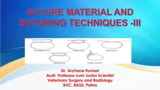
Treatment of Tongue Affections in Veterinary Surgery by Dr. Archana Kumari
In this educational material, Dr. Archana Kumari discusses various tongue affections in animals such as strangulation, smooth tongue, tumors, and ranula. Treatment methods for each condition are described in detail, providing valuable insights for veterinary surgeons. From managing venous engorgement in strangulation cases to addressing mucoceles in ranula, the document offers practical guidance for effective treatment strategies.

Download Presentation

Please find below an Image/Link to download the presentation.
The content on the website is provided AS IS for your information and personal use only. It may not be sold, licensed, or shared on other websites without obtaining consent from the author.If you encounter any issues during the download, it is possible that the publisher has removed the file from their server.
You are allowed to download the files provided on this website for personal or commercial use, subject to the condition that they are used lawfully. All files are the property of their respective owners.
The content on the website is provided AS IS for your information and personal use only. It may not be sold, licensed, or shared on other websites without obtaining consent from the author.
E N D
Presentation Transcript
Affection of Tongue and their Treatment Regional Veterinary Surgery VSR-421(2+1) Dr. Archana kumari Asstt. Prof Deptt. of Veterinary Surgery And Radiology
Strangulation Strangulation of the free portion of the tongue has been made to control vicious horse by tying a string round it or by fixing it in cord round the lower jaw. The free portion of tongue in front of ligature becomes swollen and cyanotic from venous engorgement. If the ligature is not removed within 24 hours, the portion will undergo necrosis due to arrest of blood supply to the area. Treatment : To remove ligature and scarify the swelling on the dorsal part to prevent pressure necrosis. To control venous bleeding due to incision, application of a thick layer of flour on the dorsal linguae is necessary and subsequently keeping the jaws strapped together by a tight nose-band for a few hours.
Smooth tongue Smooth tongue is a congenital anomaly and occasional cases are reported in Holstein-Friesian and Brown Swiss cattle. This condition is associated with either presence of small or absence of horne papillae on the dorsal surface of tongue, leading smooth appearance. The calves feel difficulty in prehension particularly when they are put to grazing field. As a result, the animal gradually looses body condition and growth is retarded. Treatment of this condition is not promising and hence not indicated.
Tumours of tongue Tumours of tongue are rarely observed in ruminants and pet animals. In rare instances, fibroma, angioma and lipoma may affect the tongue. Epithelioma usually also affects the tongue and in dog, contagious papilloma of the mouth appear in the tongue. Clinical signs : difficulty in mastication, Salivation Dysphagia quidding (partially chewed food materials mixed with saliva, drop out from mouth). Treatment : malignant tumours should not be interfered as there is a chance of recurrence. If the tumour involves the anterior portion of the tongue, amputation of free portion of tongue is indicated.
Ranula / Honey cyst Ranula is a mucocele occurring on the floor of the mouth alongside the tongue close to frenum linguae. frequently seen in dogs, less frequently in other animals It originates from the ducts of the sublingual salivary gland. The presence of cyst interferes with mastication and swallowing. The cyst is non-inflammatory and there is salivation.
Treatment incise the cyst and place a drain in the area of mucocele to allow fluid to escape from the area until it has a chance to heal. Tincture iodine may be touched in the cyst wall to destroy its lining and to prevent further accumulation of cystic fluid.
Trauma Trauma to the tongue usually occurs in cattle, buffaloes and dogs caused by thorns, barbed wire, sharp teeth and accidental ingestion of certain foreign bodies. The affected animals feel pain and reluctant to eat. There is excessive salivation which is often mixed with blood. Treatment of this condition includes through washing of the mouth with light potassium permanganate solution and consequent application of local antimicrobial solutions. Soft liquid diet at least for a week is recommended.
Suturing the traumatic lesion Trauma in tongue of horse After suturing photograph of tongue Suturing the traumatic lesion
Gangrene Gangrene of the tongue is sometimes seen in animals especially in dogs accompanying with gangrenous stomatitis. it is restricted to the anterior portion of the tongue. Clinical signs: offensive odour from mouth, greenish and shrivelled appearance to tongue. Treatment: includes frequent irrigation of the mouth with hydrogen peroxide or potassium permanganate solution until the dead part is separated.
Sublingual abscess Sublingual abscess is occasionally seen beneath the tongue. There is a swelling on the floor of the mouth in the intermaxillary space. Clinical signs salivation difficulty in eating. Treatment: consists of opening the abscess in the intermaxillary space and draining the pus material by washing the mouth with light anti- septic solutions.
Glossoplegia Glossoplegiaor paralysis of the tongue may occur due to involvement of hypoglossal nerve, but is a rare incidence. The condition may develop during the course of an infectious disease, such as distemper, chronic pleuropneumonia or in association with acute pharyngitis. Besides, it may occur due to trauma resulting from a wound or excessive traction of tongue. The prognosis in deplegia cases is grave. Signs The tongue becomes flaccid and hangs out of the mouth. In unilateral paralysis, the tongue is deviated towards the non-affected side. Treatment In true glossoplegia, treatment should be made as for paralysis in general. The response of treatment in this case will take longer time. If it is due to acute pharyngitis, systemic, administration of antibiotics and corticosteroid produces promising recovery within a few days.
Actinobacillosis Actinobacillosisis the most common clinical condition in cattle and buffaloes and occasional in sheep, goat and other domestic animals. This condition is characterized by one or more swelling in the submandibular region along with inflammation of the affected tissue. There may be involvement of tongue in cattle, rarely in other ruminants. Signs In advanced cases there is difficulty in mastication and swallowing. In more advanced cases, there is swelling entire throat region along with inflammation of pharyngeal lymph node leading to dysphgia and dyspnoea. Discharge of yellowish pus material from the ruptured swelling.
Treatment Maturation of hard abscess with counter-irritant and hot fomentations is indicated followed by draining of matured abscess. Systemic administration of potassium iodine is still considered effective therapeutic protocol of this condition. In bovine @ 6-10 grams orally for 7-10 days or 10% sodium iodine (1gm/12kg) as single intravenous injection is routinely used. Cont .
Surgical excision of large timorous growth is indicated. A course of antibiotic like Streptopenicillin for 7-10 days should be administered. Iodized glycerine or boroglycerin may be smeared in involvement of gum, lips and tongue.
Milk suckling (surgical treatment) Milk suckling is the interface with the ability to suckle milk and may enter the nasal cavity due to involvement of lip and premaxilla (primary cleft palate).
Surgical treatment Position of animal: The animal should be placed in ventral recumbency and the head elevated on a cushion under the mandible which provides good surgical access to the lips. Oral speculum may be used in involvement of premaxilla. Operation technique: The hair on the muzzle is clipped and the skin is prepared for surgery.
The edges of the cleft defect are incised to a depth of 2 to 3 mm along the entire margin of the defect to create an inner mucosal layer and outer cutaneous layer. The mucosal edges are apposed starting from the most dorsal point with interrupted absorbable sutures. Attempt should be made to appose the tissue without tension. Skin should be closed starting from the lip margin to avoid step deformity using non-absorbable suture material in interrupted fashion. In case of involvement of premaxilla, closure is troublesome. Mucosal flaps based on nasal or oral mucosa are elevated from each side of the defect and are sutured together with fine absorbable suture material. Generally two layer closures are preferred but due to insufficient tissue, one layer closure is performed. In one layer closure, the nasal epithelial side should be reconstructed and the oral mucosal side is allowed to heal by second intension.
Cleft palate(Palatoschisis) congenital disorder occurs in all species commonly observed in brachycephalic breeds of canine. chronic nasal discharge and pneumonia lead its diagnosis as a neonate or young foal
Etiology Nutritional, hormonal and mechanical factors Genetic and stress factors are also responsible for this condition. Intrauterine infection and exposure to toxins at specific periods of gestation may also lead to this situation.
Signs Food material passes to the nostrils on expiration and to the pharynx on inspiration. In early stages of life, rhinitis or pneumonia occurs. Milk or food in the nasal cavity frequently causes sneezing or gaggling. In some cases, the animal is unable to create a negative pressure in the mouth and may lead to death by starvation. Treatment- Surgical reconstruction of cleft palate condition is indicated by pedicle flaps of palatine mucosa.


")


")