Overview of Paramphistomidae in Veterinary Helminthology at Bihar Animal Sciences University

D
r
.
A
J
I
T
K
U
M
A
R
D
e
p
a
r
t
m
e
n
t
o
f
V
e
t
e
r
i
n
a
r
y
V
e
t
e
r
i
n
a
r
y
P
a
r
a
s
i
t
o
l
o
g
y
B
i
h
a
r
V
e
t
e
r
i
n
a
r
y
C
o
l
l
e
g
e
B
i
h
a
r
A
n
i
m
a
l
S
c
i
e
n
c
e
s
U
n
i
v
e
r
s
i
t
y
P
a
t
n
a
-
8
0
0
0
1
4
F
a
m
i
l
y
:
P
a
r
a
m
p
h
i
s
t
o
m
i
d
a
e
V
e
t
e
r
i
n
a
r
y
H
e
l
m
i
n
t
h
o
l
o
g
y
–
I
(
V
P
A
-
6
0
1
)
D
a
t
e
:
1
2
.
0
2
.
2
0
2
1


Image source: Google image
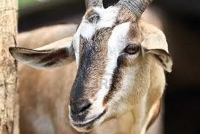
Thick fleshy flukes commonly
called amphistomes
Oral sucker is visible at the
tip of
cone
and the ventral sucker
situated at the
subterminally
posterior end called acetabulum.
Due location of posterior sucker
(acetabulum) at posterior end
called as amphistomes.
Pharynx absent and tegument is
spineless.
Intestinal caeca are simple.
Testes are frequently lobed and
usually anterior to the small
ovary
Paramphistomum
sp.
is the
commonest species
The live adult specimens are
light
red /pink in colour.
Slightly concave ventrally and
converse dorsally
Slightly lobed testes are tandem
in position and
lie anterior to the
ovary
.
Oral sucker is visible at the
tip of
cone
and the ventral sucker
situated at the
sub-terminally
posterior end called acetabulum.
Cross-sections
of the flukes are
almost
circular
.
It is
closely resembles
to
P. cervi
but there is a
Genital sucker
surrounding the
genital pore.
Genital sucker
O
t
h
e
r
N
a
m
e
:
P
a
r
a
m
p
h
i
s
t
o
m
u
m
e
x
p
l
a
n
a
t
u
m
Body is conical with dorsal side strongly
convex.
Oral sucker small but Posterior sucker is
very large, deep and located ventrally
.
Testes are lobed and diagonally placed.
Rests of morphological characters are
similar to
Paramphistomum
sp.
Eggs are yellowish in colour
.
A series of superficial haemorrhage may
be found in bile ducts and gall-bladder.
I
m
m
a
t
u
r
e
a
n
d
m
a
t
u
r
e
f
l
u
k
e
c
a
u
s
e
d
b
i
l
i
a
r
y
a
m
p
h
i
s
t
o
m
o
s
i
s
i
n
b
u
f
f
a
l
o
e
s
.
Large deep
ventral sucker
T
e
s
t
e
s
o
o
v
a
r
y
Body of
Gastrodiscus
aegyptiacus
is divided into
two parts- cylindrical anterior
part and
saucer-shaped
posterior part.
Posterior sucker is small
and
subterminal.
Oral sucker has
two
posterolateral pouches.
T
e
s
t
e
s
a
r
e
b
r
a
n
c
h
e
d
,
d
i
a
g
o
n
a
l
a
n
d
a
n
t
e
r
i
o
r
t
o
o
v
a
r
y
P
a
p
i
l
l
a
e
a
t
t
h
e
v
e
n
t
r
a
l
s
u
r
f
a
c
e
T
w
o
P
o
s
t
e
r
o
l
a
t
e
r
a
l
p
o
u
c
h
e
s
testes
Ovary
It measures 9-18 mm in length and
about 5 mm in thickness.
The anterior end is covered with
papillae.
It has a
very large ventral pouch
which opens near the anterior end up
to the ventral sucker
Genital pore opens into the ventral
pouch.
Testes are lobed and horizontal in
position
U
t
e
r
u
s
h
a
s
a
t
y
p
i
c
a
l
d
i
r
e
c
t
i
o
n
w
h
i
c
h
c
r
o
s
s
e
s
f
r
o
m
r
i
g
h
t
t
o
l
e
f
t
a
t
a
b
o
u
t
t
h
e
m
i
d
d
l
e
o
f
t
h
e
b
o
d
y
I
n
t
e
s
t
i
n
a
l
c
a
e
c
a
r
e
a
c
h
u
p
t
o
t
h
e
l
e
v
e
l
o
f
t
e
s
t
e
s
ventral pouch
Body is tapering anteriorly and rounded posteriorly
Oral sucker has two pairs oral (pharyngeal) pouches
and acetabulum sub-terminal
.
Intestinal caeca are wavy
Testes are highly branched and horizontal
in
position.
O
r
a
l
p
o
u
c
h
e
s
Intestinal caeca
Testes
G
a
s
t
r
i
d
i
s
c
o
i
d
e
s
h
o
m
i
n
i
s
:
c
a
e
c
u
m
o
f
m
a
n
a
n
d
c
o
l
o
n
o
f
p
i
g
U
n
l
i
k
e
G
a
s
t
r
o
d
i
s
c
u
s
,
a
n
t
e
r
i
o
r
p
a
r
t
i
s
r
o
u
n
d
e
d
P
a
p
i
l
l
a
e
a
b
s
e
n
t
o
n
v
e
n
t
r
a
l
s
u
r
f
a
c
e
.
T
w
o
p
o
s
t
e
r
o
-
l
a
t
e
r
a
l
o
r
a
l
p
o
u
c
h
e
s
T
e
s
t
e
s
a
r
e
b
r
a
n
c
h
e
d
,
d
i
a
g
o
n
a
l
a
n
d
a
n
t
e
r
i
o
r
t
o
o
v
a
r
y
P
r
e
s
e
n
c
e
o
f
a
n
a
l
c
l
e
f
t
a
t
t
h
e
p
o
s
t
e
r
i
o
r
e
n
d
.
Helicorbis
coenosus
snail
Adult
Gastrodiscoides hominis
Common Name:
Conical fluke/
Stomach fluke/Rumen fluke
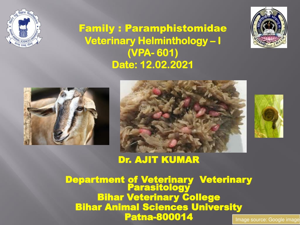
Final host:
Sheep, goat cattle &
buffaloes
Intermediate host;
Aquatic snail
(
Indoplanorbis exustus
)
Life-cycle:
similar to
Fasciola
sp.
Infective stage :
Metacercaria
E
g
g
M
i
r
a
c
i
d
i
u
m
S
p
o
r
o
c
y
s
t
Redia
C
e
r
c
a
r
i
a
cercaria emerge
from the aquatic snail and encyst on aquatic
vegetation
and
form
Metacercaria
.
I
N
S
I
D
E
T
H
E
S
N
A
I
L
Image source: Google image
Mature cercariae are dark brown in colour,
possess two distinct sensory eye spots and
anterior and posterior suckers
(Cercaria
pigmentata).
Final hosts get infection by the
ingestion of
metacercaria infected aquatic vegetation.
T
h
e
a
d
u
l
t
p
a
r
a
m
p
h
i
s
t
o
m
e
s
a
r
e
n
o
n
-
p
a
t
h
o
g
e
n
i
c
i
n
t
h
e
f
o
r
e
s
t
o
m
a
c
h
(
r
u
m
e
n
)
e
v
e
n
w
h
e
n
p
r
e
s
e
n
t
i
n
l
a
r
g
e
n
u
m
b
e
r
s
w
h
e
r
e
a
s
i
m
m
a
t
u
r
e
(
j
u
v
e
n
i
l
e
)
s
t
a
g
e
s
o
f
t
h
e
p
a
r
a
m
p
h
i
s
t
o
m
e
s
i
n
t
h
e
d
u
o
d
e
n
u
m
a
r
e
p
r
o
d
u
c
e
d
s
e
v
e
r
e
p
a
t
h
o
g
e
n
i
c
c
h
a
n
g
e
s
k
n
o
w
n
a
s
i
m
m
a
t
u
r
e
a
m
p
h
i
s
t
o
m
o
s
i
s
(
l
o
c
a
l
l
y
c
a
l
l
e
d
p
i
t
t
u
g
i
l
l
a
r
o
r
b
i
s
s
i
r
o
g
o
r
C
h
h
e
r
a
r
o
g
)
By the
excystation
process in duodenum,
immature paramphistomes liberated which
are
plug feeders
lead to
severe erosions of
duodenal mucosa.
Plug feeders
( Drawing pieces of the mucosa
into the suckers which pinch them causing
necrosis and haemorrhage)
o
L
o
s
s
o
f
l
a
r
g
e
a
m
o
u
n
t
o
f
p
r
o
t
e
i
n
l
e
a
d
s
t
o
h
y
p
o
p
r
o
t
e
i
n
e
m
i
a
(
a
c
u
t
e
s
t
a
g
e
)
u
n
l
i
k
e
F
a
s
c
i
o
l
a
s
p
.
(
c
h
r
o
n
i
c
s
t
a
g
e
)
o
A
l
t
e
r
a
t
i
o
n
o
f
t
h
e
s
e
l
e
c
t
i
v
e
p
e
r
m
e
a
b
i
l
i
t
y
o
f
t
h
e
i
n
t
e
s
t
i
n
a
l
w
a
l
l
(
d
u
e
t
o
m
u
c
o
s
a
l
r
u
p
t
u
r
e
)
l
e
a
d
s
h
y
p
o
p
r
o
t
e
i
n
e
m
i
a
,
t
h
e
n
s
u
b
m
a
n
d
i
b
u
l
a
r
o
e
d
e
m
a
(
b
o
t
t
l
e
j
a
w
)
o
N
e
c
r
o
s
i
s
a
n
d
h
a
e
m
o
r
r
h
a
g
e
i
n
i
n
t
e
s
t
i
n
e
The most common symptoms of immature
amphistomosis are -
Profuse fluid foetid diarrhoea
(shooting
diarrhoea),
Foetid (smell)
of the diarrhoeic faecs is due
to presence of the
decomposed plasma
protein in the G.I. tract
Necrosis
is indicated
by
haemorrhage
in
faeces
, which in turn is a
sign of severe enteritis.
The most common symptoms of
immature amphistomosis are
Anorexia
Hypoproteinaemia
Submandibular oedema
( bottle jaw)
Anaemia and
Marked weakness.
Soiling of hind quarters and tail
with the faeces.
The animals are thirsty and drink
frequently
(
P
o
l
y
d
i
p
s
i
a
)
.
Image source: Google image
Submandibular oedema ( bottle jaw)
.
Image source: Google image
On the basis of
Clinical signs
( Profuse fluid foetid
diarrhoea or shooting diarrhoea)
Microscopic examination of faecal
sample which reveals the presence
of characteristic eggs of
amphistomes
E
g
g
s
a
r
e
o
v
a
l
,
t
r
a
n
s
p
a
r
e
n
t
s
h
e
l
l
,
o
p
e
r
c
u
l
u
m
a
n
d
e
m
b
r
y
o
n
i
c
c
e
l
l
s
a
r
e
d
i
s
t
i
n
c
t
a
n
d
t
h
e
r
e
i
s
a
s
m
a
l
l
k
n
o
b
a
t
t
h
e
p
o
s
t
e
r
i
o
r
p
o
l
e
.
.
Image source: Google image
For the diagnosis of
immature amphistomosis-
Gross examination of fluid
foetid diarrhoea may
reveal the presence of
pink coloured pear shaped
immature flukes.
Image source: Google image
Specific drugs /Anthelmintics:
Oxyclozanide @ 15 mg/kg b.wt orally
( 100 % effective against both
mature and immature flukes)
Niclosamide @ 100 mg/kg. b. wt. orally.
Rafoxanide- 7.5 mg/kg body weight orally
Supportive drugs :
Astringent-
Neblon
powder
Liver tonic:
Pepsid, Broton syrup, Liv-52
Fluid therapy
Image source: Google image
This content delves into Paramphistomidae, focusing on species, final hosts, intermediate hosts, and anatomical characteristics such as lobed testes and suckers. The text describes common species like Paramphistomum cervi and Paramphistomum sp., detailing their physical attributes and distinguishing features. Images and detailed information provide insights into the world of veterinary helminthology at Bihar Animal Sciences University.
- Veterinary Helminthology
- Paramphistomidae
- Bihar Animal Sciences University
- Parasitology
- Veterinary Science

Download Presentation

Please find below an Image/Link to download the presentation.
The content on the website is provided AS IS for your information and personal use only. It may not be sold, licensed, or shared on other websites without obtaining consent from the author. Download presentation by click this link. If you encounter any issues during the download, it is possible that the publisher has removed the file from their server.
E N D
Presentation Transcript
Bihar Animal Sciences University | Family : Paramphistomidae Veterinary Helminthology Veterinary Helminthology I I (VPA (VPA- - 601) Date: 12.02.2021 Date: 12.02.2021 601) Dr. AJIT KUMAR Dr. AJIT KUMAR Department of Veterinary Veterinary Department of Veterinary Veterinary Parasitology Parasitology Bihar Veterinary College Bihar Veterinary College Bihar Animal Sciences University Bihar Animal Sciences University Patna Patna- -800014 800014 Image source: Google image
Thick fleshy flukes commonly called amphistomes Oral sucker is visible at the tip of cone and the situated at the posterior end called acetabulum. Due location of posterior sucker (acetabulum) at posterior end called as amphistomes. Pharynx absent and tegument is spineless. Intestinal caeca are simple. Testes are frequently lobed and usually anterior to the small ovary ventral subterminally sucker
Species Species Final host Final host Intermediate Intermediate host host Location Location Paramphistomum cervi Sheep, goat, cattle & buffalo Indoplanorbis exustus Adult in Rumen & reticulum Immature fluke in duodenum Cotylophoron cotylophorum -do- -do- -do- Gastrothylax crumenifer -do- Gyraulus convexiusculus -do- Gigantocotyle explanatum Buffaloes (mainly) do- Bile duct, gall bladder & duodenum Fischoederius elongates Sheep, goat, cattle & buffalo Lymnaea luteola Rumen & reticulum
Species Final host Intermediate host Cleopatra spp. of snail Location Gastrodiscus aegyptiacus Equines & pig Large & Small intestine Gastrodiscus secundus Horse & elephant Indoplanorbis exustus Colon Gastrodiscoides hominis Man & Pig Helicorbis coenosus Caecum of man & colon in pig Pseudodiscus collinsi Horse & elephant Planorbis, Bulinus spp. Colon
Paramphistomum sp. commonest species The live adult specimens are light red /pink in colour. Slightly concave ventrally and converse dorsally Slightly lobed testes are tandem in position and lie anterior to the ovary. Oral sucker is visible at the tip of cone and the ventral sucker situated at the sub-terminally posterior end called acetabulum. is the Cross-sections of the flukes are almost circular.
It is closely resembles to P. cervi but there is a Genital surrounding genital pore. sucker the Genital sucker
Other Other Name explanatum explanatum Body is conical with dorsal side strongly convex. Oral sucker small but Posterior sucker is very large, deep and located ventrally. Name : : Paramphistomum Paramphistomum Testes Testes Testes are lobed and diagonally placed. Rests of morphological characters are similar to Paramphistomum sp. Eggs are yellowish in colour. o o v a r y A series of superficial haemorrhage may be found in bile ducts and gall-bladder. Immature and mature fluke caused biliary amphistomosis in buffaloes. Large deep ventral sucker biliary
Two Two Posterolateral Posterolateral pouches pouches Body aegyptiacus is divided into two parts- cylindrical anterior part and posterior part. Posterior sucker is small and subterminal. Oral sucker posterolateral pouches. Testes Testes are are diagonal diagonal and and ovary ovary Papillae Papillae at at surface surface of Gastrodiscus testes saucer-shaped has two branched, branched, anterior anterior to to Ovary the the ventral ventral
It measures 9-18 mm in length and about 5 mm in thickness. The anterior end is covered with papillae. It has a very large ventral pouch which opens near the anterior end up to the ventral sucker Genital pore opens into the ventral pouch. Testes are lobed and horizontal in position Uterus has a typical direction which crosses from right to left at about the middle of the body Intestinal Intestinal caeca caeca reach reach up testes testes ventral pouch up to to the the level level of of
Body is tapering anteriorly and rounded posteriorly Oral sucker has two pairs oral (pharyngeal) pouches and acetabulum sub-terminal. Intestinal caeca are wavy Testes are highly branched and horizontal in position. Oral Oral pouches pouches Intestinal caeca Testes
Gastridiscoides Gastridiscoides hominis and and colon colon of of pig Unlike Unlike Gastrodiscus Gastrodiscus, , anterior Papillae Papillae absent absent on Two Two postero postero- -lateral lateral oral Testes Testes are are branched, branched, diagonal Presence Presence of of anal anal cleft hominis : : caecum caecum of of man man pig anterior part on ventral ventral surface oral pouches pouches diagonal and cleft at at the the posterior part is surface. . is rounded rounded and anterior anterior to posterior end to ovary ovary end. . Adult Gastrodiscoides hominis Helicorbis coenosus snail
Common Name: Conical fluke/ Stomach fluke/Rumen fluke Final host: Sheep, goat cattle & buffaloes Intermediate host; Aquatic snail ( Indoplanorbis exustus) Life-cycle: similar to Fasciola sp. Infective stage : Metacercaria
Egg Miracidium Miracidium INSIDE INSIDE Sporocyst Sporocyst Redia THE THE Cercaria Cercaria SNAIL SNAIL cercaria emerge from the aquatic snail and encyst on aquatic vegetation and form Metacercaria.
Mature cercariae are dark brown in colour, possess two distinct sensory eye spots and anterior and posterior pigmentata). suckers (Cercaria Final hosts get infection by the ingestion of metacercaria infected aquatic vegetation.
The adult paramphistomes are non- pathogenic in the fore stomach (rumen) even when present in large numbers whereas immature immature (juvenile) (juvenile) stages stages of the paramphistomes in the duodenum are produced severe pathogenic changes
By the excystation process in duodenum, immature paramphistomes liberated which are plug feeders lead to severe erosions of duodenal mucosa. Plug feeders ( Drawing pieces of the mucosa into the suckers which pinch them causing necrosis and haemorrhage)
oLoss Loss of hypoproteinemia hypoproteinemia Fasciola Fasciola sp of large large amount amount of chronic stage) of protein protein leads (acute (acute stage) stage) stage) leads to unlike unlike to sp. . ( ( chronic oAlteration Alteration of the the intestinal intestinal wall leads leads hypoproteinemia hypoproteinemia, , then oedema oedema ( ( bottle bottle jaw) of the wall ( ( due the selective selective permeability due to to mucosal mucosal rupture) then submandibular jaw) permeability of rupture) submandibular of oNecrosis Necrosis and and haemorrhage haemorrhage in in intestine intestine
The most common symptoms of immature amphistomosis are - Profuse fluid foetid diarrhoea (shooting diarrhoea), Foetid (smell) of the diarrhoeic faecs is due to presence of the decomposed plasma protein in the G.I. tract Necrosis by haemorrhage in faeces, which in turn is a sign of severe enteritis. is indicated
The most common symptoms of immature amphistomosis are Anorexia Hypoproteinaemia Submandibular oedema ( bottle jaw) Anaemia and Marked weakness. Soiling of hind quarters and tail with the faeces. The animals are thirsty and drink frequently (Polydipsia) . . Image source: Google image
Submandibular oedema ( bottle jaw) . . Image source: Google image
On the basis of Clinical signs ( Profuse fluid foetid diarrhoea or shooting diarrhoea) Microscopic examination of faecal sample which reveals the presence of characteristic amphistomes eggs of Eggs are oval, transparent operculum and embryonic cells are distinct and there is a small knob at the posterior pole. transparent shell shell, Image source: Google image
For immature amphistomosis- the diagnosis of Gross examination of fluid foetid diarrhoea reveal the presence of pink coloured pear shaped immature flukes. may Image source: Google image
Specific drugs /Anthelmintics: Oxyclozanide @ 15 mg/kg b.wt orally ( 100 % effective against both mature and immature flukes) Niclosamide @ 100 mg/kg. b. wt. orally. Rafoxanide- 7.5 mg/kg body weight orally Supportive drugs : Astringent- Neblon powder Liver tonic: Pepsid, Broton syrup, Liv-52 Fluid therapy Image source: Google image


")