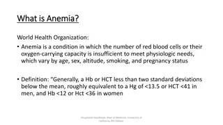
Iron Deficiency Anemia in a 78-Year-Old Man: A Case Study

A 78-year-old man presents with symptoms of fatigue, headache, and abdominal pain, leading to a diagnosis of iron deficiency anemia. His medical history includes hypertension, type II diabetes, and chronic shoulder pain. Investigations reveal hypochromic, microcytic anemia with low iron levels and high total iron-binding capacity. Management includes oral iron replacement therapy and addressing the underlying cause.

Download Presentation

Please find below an Image/Link to download the presentation.
The content on the website is provided AS IS for your information and personal use only. It may not be sold, licensed, or shared on other websites without obtaining consent from the author. Download presentation by click this link. If you encounter any issues during the download, it is possible that the publisher has removed the file from their server.
E N D
Presentation Transcript
Haematology RBC REVIEW DR DEA THOMAS
CASE 1 A 78-year-old man comes to the physician because of progressively worsening fatigue and headache for 6 months and occasional abdominal pain. He has not had any changes in his weight. Hemoglobin 10.3 g/dL 74 m3 Mean corpuscular volume 280,000/mm3 Platelet count He has a history of HTN, DM TII and chronic shoulder pain. 6,000/mm3 Leukocyte count Drug Hx: hydrochlorothiazide, metformin, and ibuprofen. He does not drink alcohol and does not smoke. O/E: P = 92/min, BP = 135/80 mm Hg. Conjunctival and mucosal pallor. Nil other examination findings
CASE 1 A 78-year-old man comes to the physician because of progressively worsening fatigue and headache for 6 months and occasional abdominal pain. He has not had any changes in his weight. Hemoglobin 10.3 g/dL (L) 74 m3 Mean corpuscular volume (L) 280,000/mm3(N) Platelet count He has a history of HTN, DM TII and chronic shoulder pain. 6,000/mm3 (N) Leukocyte count Drug Hx: hydrochlorothiazide, metformin, and ibuprofen. He does not drink alcohol and does not smoke. O/E: P = 92/min, BP = 135/80 mm Hg. Conjunctival and mucosal pallor. Nil other examination findings
IRON DEFICIENCY ANAEMIA
Causes of Iron Deficiency Anaemia 1. Low intake of bioavailable iron 2. Increased iron requirements rapid growth, pregnancy, menstruation 3. Chronic GI blood loss pathologic infections, such as hook worm and whipworm Malignancy Inflammatory bowel disease 4. Malabsorption Autoimmune gastritis Gastrectomy
Investigations HYPOCHROMIC, MICROCYTIC ANAEMIA Reticulocyte index: Hypo-proliferative Blood Film:
Investigations: iron studies Parameter Serum Iron Total Iron Binding Capacity Percentage transferrin saturation Serum ferritin Value Low High Low Low
Additional Investigations Faecal Occult Blood Colonoscopy Upper GI endoscopy
Management Treat the underlying cause! Oral iron replacement Ferrous Sulphate tablets Side effects: Nausea, abdominal pain, constipation or diarrhoea Can be given with Vitamin C to enhance absorption Oral therapy should continue until stores are replenished: 6 months Parenteral Iron IV or IM Non-compliant patients Cannot tolerate oral iron Severe, chronic bleeding: eg, severe menorrhagia, haemodialysis
Response Symptomatic improvement Reticulocytosis (7 10 days) Rise in Hb ~2g/dL every 3 weeks Normalisation of ferritin
CASE 2 MCV TIBC Serum Ferritin A 2-year-old boy presents with fatigue for several months. The parents report that their son is not as active as he used to be. He was solely breastfed until he was 14 months old. He currently eats some table foods and cow's milk daily. A Decreased Decreased Increased B Normal Decreased Increased C Normal Increased Normal O/E D Decreased Decreased Increased Conjunctival pallor. Redness and fissures of bilateral corners of the mouth. E Increased Normal Normal F Decreased Increased Decreased Laboratory studies are most likely to show which of the following?
CASE 2 MCV TIBC Serum Ferritin A 2-year-old boy presents with fatigue for several months. The parents report that their son is not as active as he used to be. He was breastfed until he was 14 months old. He currently eats some table foods and cow's milk daily. A Decreased Decreased Increased B Normal Decreased Increased C Normal Increased Normal O/E D Decreased Decreased Increased Conjunctival pallor. Redness and fissures of bilateral corners of the mouth. E Increased Normal Normal F Decreased Increased Decreased Laboratory studies are most likely to show which of the following?
CASE 3 A 22-year-old primigravid woman comes to the physician for her initial prenatal visit at 12 weeks' gestation. She has had generalized fatigue and shortness of breath over the past 2 months. She has also had a tingling sensation in her toes for the past month. She follows a strict vegan diet since the age of 13 years. O/E: T = 37 C, P = 111/min, BP = 122/80 mmHg. Pale conjunctivae and a shiny tongue. Sensation to vibration and position is decreased over the upper and lower extremities. When asked to stand, hold her arms in front of her, and close her eyes, she loses her balance Hemoglobin 9.4 g/dL Mean corpuscular volume 112 fL 8,700 /mm3 Leukocyte count 150,000 /mm3 Platelet count
CASE 3 A 22-year-old primigravid woman comes to the physician for her initial prenatal visit at 12 weeks' gestation. She has had generalized fatigue and shortness of breath over the past 2 months. She has also had a tingling sensation in her toes for the past month. She follows a strict vegan diet since the age of 13 years. O/E: T = 37 C, P = 111/min, BP = 122/80 mmHg. Pale conjunctivae and a shiny tongue. Sensation to vibration and position is decreased over the upper and lower extremities. When asked to stand, hold her arms in front of her, and close her eyes, she loses her balance Hemoglobin 9.4 g/dL (L) Positive Romberg s Mean corpuscular volume 112 fL (H) 8,700 /mm3 Leukocyte count 150,000 /mm3 Platelet count
Megaloblastic Anaemia Megaloblasts: large cells with an arrest in nuclear maturation Asynchronous maturation of the nucleus and cytoplasm Defective DNA synthesis RNA and protein synthesis relatively unaffected Most apparent in rapidly dividing cells erythropoietic cells GI cells Megaloblasts in the BM Hyper segmented neutrophils and macrocytes in peripheral smear Affects all cell lines
Causes Vitamin B12 deficiency Folate deficiency Abnormalities of vitamin B12 or folate metabolism transcobalamin deficiency, nitrous oxide, antifolate drugs (eg. methotrexate) Alcohol Drugs: hydroxyurea, cytosine arabinoside
Investigations Complete blood count Macrocytic anaemia Pancytopenia Low reticulocyte index High LDH haemolysis Hyperbilirubinaemia Peripheal blood smear Oval macrocytes, hyper-segmented neutrophils anisocytosis, poikilocytosis Hypercellular bone marrow
Causes Diet veganism Pernicious anaemia Gastrectomy Decreased gastric acidity Atrophic gastritis Severe pancreatitis Ileal resection Increased demand Relative deficiency Pregnancy Causes of B12 Deficiency
Investigations Serum methylmalonic acid is a test for B12 deficiency Serum homocysteine is a test for folate or B12 deficiency Tests are not widely available IF and parietal cell antibodies
Management Oral B 12 therapy Need high doses for prolonged periods Just as effective as IM therapy IM B12 therapy Dosing depends on severity For severe: 1mg IM daily for I week, then weekly for 1 month Maintenance: q3monthly Treat underlying issue Given prophylactically in gastrectomy patients and increased demand conditions
Folate deficiency Inadequate dietary intake Malabsorption Small intestine resections Increased demand Pregnancy Periods of growth Impaired utilization B12 deficiency Drugs: eg methotrexate
Investigations Serum homocysteine is a test for folate or B12 deficiency Folate deficiency can be confirmed with a normal B12 and MMA level and elevated homocysteine levels Tests are not widely available
Management Exclude B12 deficiency first! Giving folate alone in the presence of B12 deficiency may improve haematological issues but worsens neurological defects If B12 status unknown, give both Never give folate alone when B12 status is not known Oral folic acid 5mg daily for ~4 months Given prophylactically in pregnancy, pre-maturity, haemolytic anaemias
Differentials of macrocytic anaemia Alcohol Reticulocytosis Liver disease Myxoedema Myelodysplastic syndromes
CASE 4 A 35-year-old presents with a 3-month history of worsening fatigue. She has difficulty concentrating at work despite sleeping well most nights. Three years ago, she was diagnosed with Crohn s disease. She has about 7 non-bloody, mildly painful bowel movements daily. Her current medications include 5-aminosalicylic acid and topical budesonide. She does not smoke or drink alcohol. O/E T = 37.9 C, P = 92/min, BP = 110/65 mmHg. She appears pale. The abdomen is diffusely tender to palpation No guarding.
CASE 4 A 35-year-old presents with a 3-month history of worsening fatigue. She has difficulty concentrating at work despite sleeping well most nights. Three years ago, she was diagnosed with Crohn s disease. She has about 7 non-bloody, mildly painful bowel movements daily. Her current medications include 5-aminosalicylic acid and topical budesonide. She does not smoke or drink alcohol. O/E T = 37.9 C, P = 92/min, BP = 110/65 mmHg. She appears pale. The abdomen is diffusely tender to palpation No guarding.
Anaemia of Chronic Disease Most common anaemia in hospitalized patients Pathophysiology Inflammatory stimuli increase hepcidin expression defective iron incorporation in erythropoiesis a decrease in erythropoietin production and impaired response to erythropoietin Cytokine induced shortening of red cell survival Causes: chronic illnesses, malignancies, inflammatory diseases Usually a mild anaemia but depends on severity of underlying disorder Mildly hypochromic or normochromic, mildly microcytic or normocytic Underlying cause, EPO in chronic renal failure patients
CASE 5 A 56-year-old female, known to have HTN and DM TII, presents with a 2-week history of fatigue and painless bruising over her arms and trunk. She has also had several episodes of nosebleeds that resolved with compression after a few minutes. She recently completed treatment for a urinary tract infection. She has had no changes in her weight. Her last menstrual cycle was 5 years ago. She does not smoke or drink alcohol. Her vital signs are within normal limits. Physical examination shows pale conjunctivae. There are ecchymoses and petechiae over the upper extremities, chest, and back. There is no lymphadenopathy. The remainder of the physical examination is unremarkable. Hemoglobin 8.7 mg/dL Leukocyte count 11 Platelet count 54 Reticulocyte count 0.1% 93 m3 MCV Total bilirubin 1.1 mg/dL LDH 80 U/L
CASE 5 A 56-year-old female, known to have HTN and DM TII, presents with a 2-week history of fatigue and painless bruising over her arms and trunk. She has also had several episodes of nosebleeds that resolved with compression after a few minutes. She recently completed treatment for a urinary tract infection. She has had no changes in her weight. Her last menstrual cycle was 5 years ago. She does not smoke or drink alcohol. Her vital signs are within normal limits. Physical examination shows pale conjunctivae. There are ecchymoses and petechiae over the upper extremities, chest, and back. There is no lymphadenopathy. The remainder of the physical examination is unremarkable. Hemoglobin 8.7 mg/dL Leukocyte count 11 Platelet count 54 Reticulocyte count 0.1% 93 m3 MCV Total bilirubin 1.1 mg/dL LDH 80 U/L
APLASTIC ANAEMIA Pancytopenia due to hypoplasia of the bone marrow Haemopoietic cells replaced by fat cells Defect maybe with the stem cell or with the bone marrow environment Causes Congenital Acquired Idiopathic Secondary
Congenital Aplastic Anaemia Fanconi Anaemia Autosomal recessive Multiple genes involved but they ultimately damage a protein needed to protect against genetic damage Growth retardation, developmental delay, renal tract abnormalities Presentation in age 3 14 May develop AML Mx: SCT Dyskeratosis Congenita Nail and skin abnormalities, pulm fibrosis, cirrhosis Diamond-Blackfan Anaemia
Acquired Aplastic Anaemia Idiopathic 67% of acquired cases Need distinguish from late onset congenital aplastic anaemia Secondary Drugs eg Bactrim, carbamazepine, methimazole, NSAIDs, propylthiouracil, chloramphenicol Viral infections eg. Few months after hepatitis, EBV Ionising radiation Paroxysmal Nocturnal Haematuria
Presentation Symptoms of anaemia Increased risk of infections mouth and throat especially Frequently severe No lymphadenopathy Bleeding tendencies Petechiae Retinal haemorrhages
Investigations Parameter Aplastic Severe Very Severe Hb <10 g/dL Neutrophil <1.5 * 10^9 /L <0.5 <0.2 Platelets <50 <20 Reticulocytes Low <1% Bone marrow Hypoplastic <25% cellularity or 25 50% cellularity with <30% haemopoietic cells Normal cells on peripheral smear.
Investigations To find out underlying cause LFTs Serology: hepatitis, EBV, HIV Congenital studies Flow cytometry for PNH Check for haemosiderinuria
Management Stop offending agent Supportive Transfusions Treat and prevent infections Specific Stem Cell Transplant Immunosuppressive Therapy antithymocyte globulin (ATG) and cyclosporine Androgens in FA Haemopoietic growth factors
CASE 6 INVESTIGATIONS A 30-year-old woman presents with weakness and fatigue for 2 days. She has also noticed that her urine is darker than usual. She has never had these symptoms before. WBC 8 Hb 7.1 Hct 0.21 For the past week, she has had a persistent non- productive cough and low-grade fever. She has seasonal allergies. Plt 160 MCV 94 Bili TB: 4.3, DB = 1.1, IB = 3.2 She drinks one to two glasses of wine on social occasions and does not smoke. AST 15 ALT 17 O/E: T = 37.9 C, P = 88, R = 18, BP = 110/76 mm Hg. Mucous membranes are pale and jaundiced. Bilateral lower zone creps on Resp exam LDH 1, 251 DCT Positive CXR Bilateral reticular infiltrates
CASE 6 INVESTIGATIONS A 30-year-old woman presents with weakness and fatigue for 2 days. She has also noticed that her urine is darker than usual. She has never had these symptoms before. WBC 8 N Hb 7.1 Hct 0.21 For the past week, she has had a persistent non- productive cough and low-grade fever. She has seasonal allergies. Plt 160 N MCV 94 N Bili TB: 4.3, DB = 1.1, IB = 3.2 She drinks one to two glasses of wine on social occasions and does not smoke. AST 15 N ALT 17 N O/E: T = 37.9 C, P = 88, R = 18, BP = 110/76 mm Hg. Mucous membranes are pale and jaundiced. Bilateral lower zone creps on Resp exam LDH 1, 251 DCT Positive CXR Bilateral reticular infiltrates
AUTOIMMUNE HAEMOLYTIC ANAEMIA Caused by antibodies against body s own RBCs Haemolysis is extravascular Characterised by a positive Coombs test
Coombs Test Coombs Test Coombs serum: contains anti- human globulins sample is analyzed for erythrocyte agglutination No agglutination = negative test Direct Coombs Abs bound to RBCs Indirect Coombs Free Abs in serum
Warm AIHA Heat-sensitive antibodies binding to RBC IgG Complement Cells become progressively spherical Destroyed mainly in the spleen Triggered by increased body temperature (> 37 C) Causes Idiopathic Secondary: SLE, CLL, drugs eg. methyldopa
Warm AIHA: Presentation Gradual onset Chronic course Mild splenomegaly Varying severity Evan s syndrome: Warm AIHA + idiopathic thrombocytopenic purpura
Warm AIHA: Investigations Blood film: polychromasia Micro-spherocytes Coombs test best at 37 C
Management Treat underlying cause Folate supplementation in severe cases First line therapy: corticosteroids 1mg/kg/day and then tapered down Rituximab: Anti CD20, emerging first line Failure to control in appropriate doses: Splenectomy Immunosuppression may be tried: azathioprine, cyclophosphamide Intravenous immunoglobulin not very effective
Cold AIHA cold-sensitive antibodies binding to RBC IgM Directed mainly to I antigen on RBCs Optimal at 4 C Effective at binding complement contributing intravascular Agglutination occurs mainly in the peripheries, especially in cold temperatures Causes Idiopathic - Primary cold agglutinin disease (monoclonal) Secondary Lymphoproliferative disorder (monoclonal), Waldenstrom macroglobulinemia Post infectioys (polyclonal): eg. Mycoplasma pneumonia, infectious mononucleosis (anti-I antibodies) Paroxysmal cold haemoglobinuria : anti-P aka Donath Landsteiner antibody


![Enhancing Patient Blood Management in Pregnancy at [INSERT LOCAL HOSPITAL NAME]](/thumb/143207/enhancing-patient-blood-management-in-pregnancy-at-insert-local-hospital-name.jpg)