Understanding Red Blood Cell Abnormalities in Anemia: A Detailed Overview

This in-depth article delves into the various types of red blood cell abnormalities seen in anemia, such as anisocytosis, poikilocytosis, spherocytes, ovalocytes, acanthocytes, codocytes, drepanocytes, and stomatocytes. It explains the characteristics of each abnormality, their significance in different conditions, and their implications in diagnosing and managing patients with anemia and related disorders.

Download Presentation

Please find below an Image/Link to download the presentation.
The content on the website is provided AS IS for your information and personal use only. It may not be sold, licensed, or shared on other websites without obtaining consent from the author. Download presentation by click this link. If you encounter any issues during the download, it is possible that the publisher has removed the file from their server.
E N D
Presentation Transcript
Odesa National Medical University Department of internal medicine 1 with course of cardio-vascular pathology Anemia
Complete blood count (CBC) RED BLOOD CELL COUNT M 4,5-5,5 *1012; F 3,5-4,5 *1012 HEMOGLOBIN M 130-160 g/l (13-16 mg/dl); F 120-140 f/l (12-14 mg/dl) HEMATOCRIT (HCT) 34-46% PLATELET COUNT 180-400 *109 MCV 80 100 fl MCH 26 34 pg MCHC 30 370 g/l COLOR INDEX 0,85 1,05 RETICULOCYTES 0,2 1,2% ESR M 2-10 mm/hour; W 5-15 mm/hour 11.5-14.5% RDWc
MCV (Mean corpuscular volume) size of the RBC RDW (Red Cell Distribution Width) - this indicator tells how much red cells differ in size MCH (Mean corpuscular hemoglobin) shows how much hemoglobin is contained in one erythrocyte MCHC (Mean corpuscular hemoglobin concentration) shows as erythrocyte is saturated with hemoglobin
Anisocytosis- the content of macro- and microcytes is more than 15% of all red blood cells Poikilocytosis - different forms of red blood cells o Spherocytes- erythrocytes have a spherical shape, a greater thickness, they have no central illumination. Spherocytes - cells, ready to hemolysis. Spherocytosis observed in hemolytic anemia, septicemia, blood incompatibility AB0 system, DIC syndrome, with implantation of artificial blood vessels, heart valves, burns, autoimmune disease. Microspherocytosis is a pathognomonic sign for anemia Minkowski Chauffard(hereditary microspherocytosis). In a small number microspherocytes may occur in other hemolytic anemias. o Ovalocytes - cell oval or elongated form due to the anomaly of the membrane or hemoglobin. It occurs in hereditary elliptocytosis, thalassemia, megaloblastic anemia, iron deficiency anemia, liver cirrhosis, anemia associated with deficiency of G-6-PDG, glutathione, sickle-cell anemia. It may occur as an artifact (in the thickest part of the drug).
o acanthocytes (sheet-like cell, spur-like cell) RBC with protrusions of various sizes, arranged at different distances from each other. Occur in severe liver disease (chronic hepatitis, cirrhosis, alcoholic liver disease), hereditary deficiency of pyruvatekinase, hereditary spherocytosis (severe), in violation of lipid metabolism, treatment with heparin. A small number of acanthocytes can be found in patients after splenectomy. o codocytes (target cells) - flat pale colored periphery of RBC with accumulation of hemoglobin in a center as a target. Codocytes have an increased surface area due to excess of cholesterol. Typical for thalassemia, hemoglobinopathies S, C, D and E, iron deficiency anemia, lead poisoning, liver disease, especially accompanied by obstructive jaundice, after removal of the spleen. o drepanocytes (sickle cell) - the cells like a sickle or holly leaves. Typical for sickle cell anemia and other hemoglobynopathies o stomatocytes (mouth cells) remind the mouth. Typical for hereditary spherocytosis and stomatocytosis, tumors, alcoholism, cirrhosis and obstructive liver disease, cardiovascular diseases, after transfusions, when taking certain medications. Can be revealing as artifacts.
o Echinocytes (Burr cells) - cell has spikes of the same size and symetrical localization. It occurs in uremia, a transfusion of blood containing the old RBC, gastric cancer, peptic ulcer, complicated by bleeding, hypophosphatemia, hypomagnesemia, in hereditary deficiency pyruvate kinase, phosphoglycerate. It often occurs as an artifact. o Degmacytes ("bitten cell") - a cell looks like it took a bite. Occurs in G- 6-PDG deficiency, unstable hemoglobin o Bubble cells - cell looks as though on the surface there is a bubble or blister. The mechanism of formation is not clear. It occurs at a the immune hemolytic anemia. o Schistocytes - cells similar to the helmet, cocked hat, splinters. Observed in a microangiopathy, hemolytic anemia by physical factors, malignant hypertension, uremia, complications with prosthetic valves and vessels, DIC, sepsis, tumor, when taking certain medications, exposure to toxins. o Dacrocytes (Teardrop Cells) - cells remind drop or a tadpole. Observed in a myelofibrosis, myeloid metaplasia, hypoplastic anemia due to tumor, granuloma, lymphoma and fibrosis, thalassemia, severe iron deficiency, toxic hepatitis.
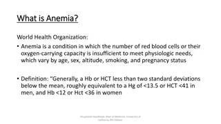
Anemia Definition: Low oxygen carrying capacity in the blood due to low erythrocyte mass. We use the hematocrit or hemoglobin as surrogate markers for the erythrocyte mass because that it too difficult to measure. Hemoglobin of < 12 g/dL in women or a Hemoglobin of <13 g/dL in men is considered abnormal (WHO criteria). The hematocrit is approximately 3 x the hemoglobin.
Classifications of anemia (1) 1. Posthemorrhagic (acute / chronic) 2. Due to the formation of RBC disorders (iron deficiency, deficiency of vitamin B12 and folic acid, hypo/aplastic) 3. Hemolytic (congenital/ acquired) 4. Mixed
Classifications of anemia (2) Macrocytic (MCV>100 fl) Microcytic (MCV<80 fl) Normocytic (MCV 81-99 fl)
Classifications of anemia (3) Hypochromic (color index <0.8) Normochromic (color index 0.85-1.05) Hyperchromic (color index >1.05)
Classifications of anemia (4) Severity of anemia Mild (Hb >90 g/L or 9.0 mg/dl) Moderate (Hb 90-70 g/L or 9.0-7.0 mg/dl) Severe (Hb <70 g/L or 7.0 mg/dl)
Acute posthemorrhagic anemia Etiology bleeding Symptoms (depends on the duration and volume of blood loss) severe weakness, shortness of breath, dizziness, syncope, ringing in the ears, darkening of the eyes, dry mouth, cold sweat, paleness of skin and mucose membranes, tachycardia, arrhythmias, hypotension, tachypnea
Lab diagnostics Reflex-vascular phase (1-2 days) normal level of RBC, Hb; possible WBC, Plt; Hydremic phase (2-3 days) - RBC, Hb; N color index; Bone marrow phase (5-6 days) - reticulocytes; presence of young WBC Normalization in 2-3 weeks Treatment - Stop the bleeding - Replenishment of circulating blood volume - If needed - the transfusion of blood or its components
Chronic posthemorrhagic anemia Etiology Repeated and prolonged bleeding Symptoms, diagnosis is similar to iron- deficiency anemia
Anemia due to RBC and Hb formation disorders
Iron is a part of hemoglobin, myoglobin, cytochrome, catalase enzymes , lactateperoxidase; ferritin, hemosiderin, transferrin and other enzymes.
Iron-deficiency anemia (sideropenic anemia) Etiology 1. Chronic bleedings (hyperpolymenorrhea, GIT diseases, hematuria, epistaxis, lung diseases, iatrogenic blood loss) 2. Increased need for iron (pregnancy, childbirth, lactation, puberty, intense exercise) 3. Insufficient intake of iron from food 4. Malabsorption syndrome 5. Violation of transport (reducing amount of transferrin) Congenital Hypoproteinemia Presence of antibodies to transferrin and its receptors
Symptoms and sings Anemic syndrome Sideropenic syndrome Taste perversion Addicted to spicy, salty, sour, spicy food Smell perversion Severe muscle weakness and fatigue, muscle atrophy Degenerative changes in the skin and its appendages (skin is dry, flaking; hair is thin, faint, rapidly fall and turning gray; koilonychia) Angular stomatitis Glossitis Atrophic gastritis and enteritis a symptom of the "blue sclera sideropenic subfebrilitet moderate immunosuppression reduction of reparative processes of the skin, mucous membranes 1. 2.
Lab diagnostic 1. CBC ( RBC and Hb, MCH, color index, MCV, anisocytosis, poikilocytosis, N reticulocytes, moderate ESR) 2. Checking iron ( ferritin, serum iron (FE), total iron-binding capacity (TIBC), iron saturation, test with desferal) 3. Puncture of the bone marrow ( sideroblast cells)
Treatment 1. Treatment of etiological factors 2. Diet 3. Treatment with iron-consist drugs Elimination of iron deficiency(100-300 mg BID/TID per os, rarely IM or IV) 6-8 weeks Restocking (1/2-1/3 from main dose) 3 month Preventive treatment (if necessary) a. b. c.
Iron redistribution anemias (Anemia in impaired iron reutilization, Anemia of chronic disease) anemia caused by disturbance of movement of iron into the plasma and into bone marrow from the depot.
Etiology Acute and chronic infection diseases Sepsis Tuberculosis Infective endocarditis Chronic leukemia Sarcoidosis Rheumatoid arthritis Chronic active hepatitis Oncology Alcoholic damaging of liver IHD
Lab diagnostic 1. CBC ( RBC and Hb, MCH, color index) 2. Checking iron ( ferritin, N/ serum iron, /N total iron-binding capacity (TIBC)) 3. Puncture of the bone marrow ( sideroblast cells) 4. Clinical and laboratory sings of inflammation Treatment mainly its treatment of main disease
Megaloblast anemia/ macrocytic anemia
Folate deficiency Found in: Fruits (e.g. citrus, melon, bananas), leafy green vegetables, and fortified grain products Causes include: alcohol due to both a decreased ability to absorb folate and, frequently, decreased intake of folate rich foods Malabsorption (celiac disease, IBD) Diseases/conditions associated with rapid cell turnover such as sickle cell disease, psoriasis, pregnancy Medications: trimethoprim, phenytoin, methotrexate
B12 deficiency anemia Etiology: 1. Problems with absorption 2. Competitive consumption of vitamin B12 3. Increased consumption (multiple pregnancy, ch hemolytic anemia, myeloma, thyrotoxicosis) 4. Violation of dietary intake 5. Decreasing of storage (liver cirrhosis) 6. Violation of vitamin B12 transport
Symptoms 1. Damaging of the digestive system (lack or loss of appetite, belching, pain and burning in the tongue. Development of glossitis, atrophic gastritis, atrophic changes of the intestinal mucosa) 2. Damage of the hematopoietic system/ anemic syndrome (specificity - yellowness of the skin and sclera) 3. Nervous system damage / funicular myelosis (violation of proprioceptive sensitivity, reduced reflexes, muscle atrophy of the lower extremities, pelvic organ dysfunction OR lower spastic paraparesis)
Lab diagnostic CBC - Hb/RBC, MCV, color index, anisocytosis, poikilocytosis, Howell-Jolly bodies, Cabot rings, reticulocytes, WBC, neutrophils, eosinophils, lymphocytes, platelets , hypersegmented neutrophils Myelogram - irritation red hematopoietic, appearance of the megaloblast type of hematopoesis, changing in myeloid cells The amount of folate, vitamin B-12 and vitamin C in blood antibodies to intrinsic factor Methylmalonic acid test Plasma Total Homocysteine
Vitamin B12 If levels are at the lower limits of normal (200-300), then consider checking methylmalonic acid and homocysteine levels, these will be elevated in the case of B12 deficiency Should be given orally, IM in rare situations Oral dosing is 1000 2000 mcg po q day IM dosing is 1000 mcg IM q day x 7 days, then q wk x 4 wks then q month
Criteria for of treatment effectiveness - Subjective improvement - Reticulocytosis in 5-7 days of treatment (20%) - Increase in hemoglobin and RBC from 2 week of treatment - Normalization of the CBC in 3-4 weeks
Hemolytic anemia Labs to order: reticulotye count, peripheral smear, haptoglobin, indirect bilirubin and LDH The peripheral smear will help you differentiate between causes of hemolysis
Extravascular Hemolytic Anemia Etiologies of extravascular hemolysis include: Congenital hemoglobin abnormalities: hemoglobinopathies Erythrocyte membrane abnormalities: hereditary spherocytosis Erythrocyte metabolic abnormalities: G6PD deficiency Auto-immune process: idiopathic, underlying malignancy, CVD, lymphoproliferative disorders, medications (PCN, cephalosporins, NSIADs)
Glucose 6-phospate dehydrogenase deficiency X-linked enzyme deficiency that results in decreased glutathione levels Most common form results in severe hemolysis with medications (primaquine, sulfa, dapsone), fava beans and some infxn. Dx: enzyme activity testing, may have false negative test in the setting of acute hemolysis, re-test at 2-3 months
Intravascular hemolytic anemia Etiologies include: Microangiopathic hemolytic anemia TTP, HUS, DIC, HELLP Shearing due to malfunctioning mechanical heart valves
Transfusions When to transfuse? General rule: Hct <21%, consider at <25% in pt s with multiple medical problems Other times to consider: on-going rapid blood loss, highly symptomatic patients, patient with low Hct and severe cardiovascular disease You expect the Hct to increase 3% for each unit of blood transfused
Anemia of Chronic Kidney Disease Hypoproliferative (low reticulocyte count), normocytic anemia For erythropoietin therapy to be effective need adequate iron stores ferritin >100 and transferrin saturation >20% Treatment goal is hemoglobin of 11-12 (Hct 33-36%), higher levels are associated with increased morbidity and mortality carries a Black Box warning Can use darbopoetin or epoetin
Thalassemia Thalassemia trait: mild anemia with pronounced microcytosis (MCV <70) Ethnicity: Mediterranean, Asian, African Pt are asymptomatic FamHx is often negative Dx thal trait: Hemoglobin electrophoresis Thalassemia intermedia: Ineffective erythropoiesis, more severe anemia, often require frequent transfusions, at risk of development of iron overload Iron deficiency anemia: usually more pronounced microcytic anemia than thal trait

")
 – size of the")

 –RBC with")
 - cell has spikes of the same")

")
")
")
")