Understanding Bronchiectasis: Causes, Symptoms, and Diagnosis
Bronchiectasis is a condition characterized by abnormal and permanently dilated airways, often resulting in chronic cough, copious sputum production, and frequent bacterial infections. The disease can be congenital or acquired, with causes ranging from cystic fibrosis to inhaled toxins. Common symptoms include pleuritic pain, haemoptysis, and general debility. Physical signs may include crackles over affected areas, diminished breath sounds, and clubbing of fingers in advanced cases.

Uploaded on Oct 01, 2024 | 0 Views
Download Presentation

Please find below an Image/Link to download the presentation.
The content on the website is provided AS IS for your information and personal use only. It may not be sold, licensed, or shared on other websites without obtaining consent from the author. Download presentation by click this link. If you encounter any issues during the download, it is possible that the publisher has removed the file from their server.
E N D
Presentation Transcript
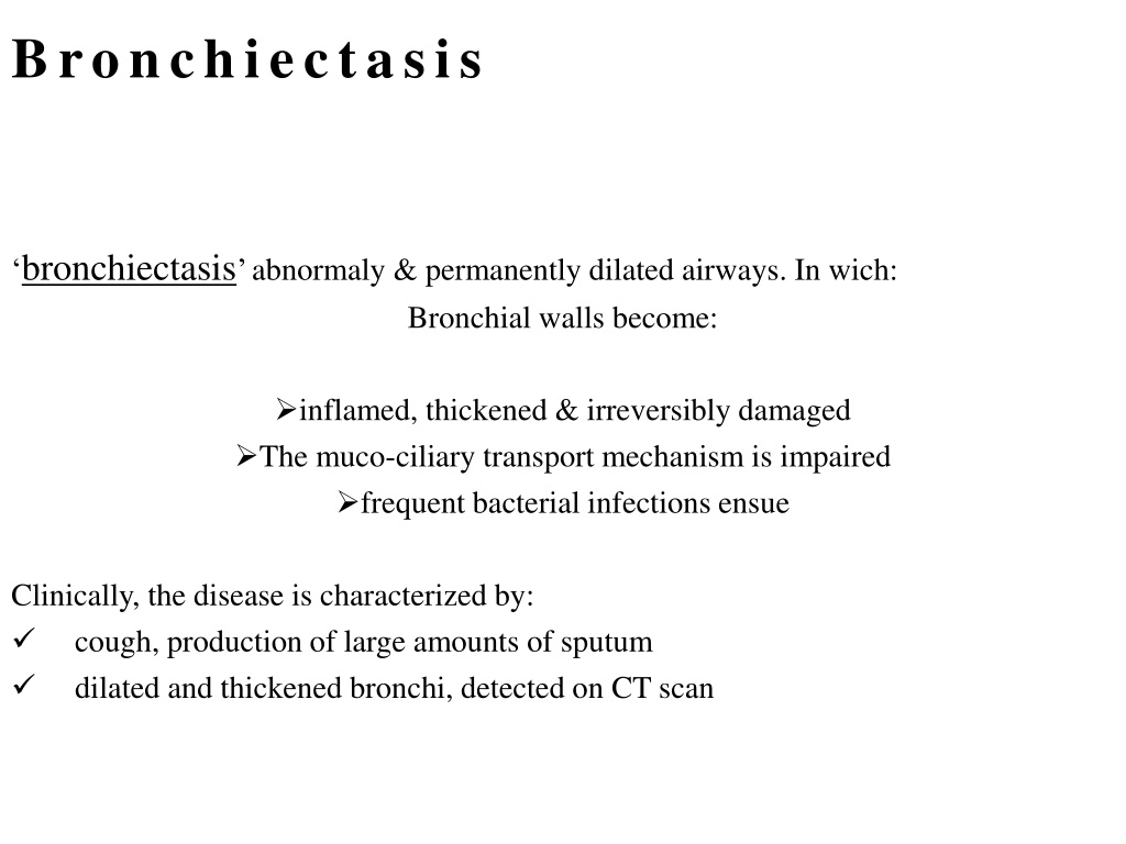
Bronchiectasis bronchiectasis abnormaly & permanently dilated airways. In wich: Bronchial walls become: inflamed, thickened & irreversibly damaged The muco-ciliary transport mechanism is impaired frequent bacterial infections ensue Clinically, the disease is characterized by: cough, production of large amounts of sputum dilated and thickened bronchi, detected on CT scan
Aetiology and pathology Generalized Congenital defect affecting airway ion transport or ciliary function (cystic fibrosis) Acquired secondary to damage airways by: destructive infection inhaled toxin foreign body. The result is chronic inflammation & infection in airways. Localized accumulation of pus beyond an obstructing bronchial lesion bronchiectatic cavities inflammatory changes in the deeper layers of the bronchial wall, hypertrophy of the bronchial arteries Chronic inflammatory and fibrotic changes in the surrounding lung tissue resulting in: progressive destruction of the normal lung architecture in advanced cases.
Causes of bronchiectasis Congenital Cystic fibrosis Ciliary dysfunction syndromes Primary ciliary dyskinesia (immotile cilia syndrome) Kartagener s syndrome (sinusitis & transposition of the viscera) Primary hypogammaglobulinaemia Acquired: children Pneumonia Primary TB & Inhaled foreign body Acquired: adults Suppurative pneumonia Pulmonary TB Allergic broncho-pulmonary aspergillosis complicating asthma Bronchial tumours
Clinical features Symptoms : Cough: chronic, daily, persistent Sputum: copious, continuously purulent Pleuritic pain: when infection spreads to involve pleura, or with segmental collapse due to retained secretions Haemoptysis: Streaks of blood common, larger volumes with exacerbations of infection, Massive haemoptysis Infective exacerbation: increased sputum volume with fever, malaise, anorexia Halitosis: frequently accompanies purulent sputum General debility: weight loss, anorexia, exertional breathlessness
Physical signs may be unilateral or bilateral If bronchiectatic lesion not contain secretions & no associated lobar collapse, there are no abnormal physical signs When there are large amounts of sputum in the bronchiectatic spaces, numerous coarse crackles may be heard over the affected areas. Collapse with retained secretions blocking a proximal bronchus may lead to locally diminished breath sounds In advanced disease may cause scarring & overlying bronchial breathing Clubbing of fingers
Clubbing: 1.loss of angle 2.increase curvature 3.flactuation 4.drum stick
Investigations Investigations Sputum examination with culture & sensitivity is essential for adequate treatment. The major pathogens are: Staph. Aureus Pseudomonas aeruginosa H. influenzae and anaerobes Strep. pneumoniae & Klebsiella pneumoniae. Aspergillus fumigatus 10% of sputum (cystic fibrosis) Mycobacterium avium-intracellulare complex (MAI) is being increasingly found. Chest X-ray: normal dilated bronchi with thickened bronchial walls multiple cysts containing fluid. High-resolution CT scanning (sensitivity of 97%) bronchial dilatation, bronchial wall thickening
0ther investigations Sinus X-rays 30% have concomitant rhino sinusitis Serum immunoglobulins 10% of adults antibody deficiency ( IgA) Sweat electrolytes test if cystic fibrosis is suspected Mucociliary clearance (nasal clearance of saccharin) A 1 mm cube of saccharin is placed on the inferior turbinate and the time to taste measured (normally less than 20 minutes).
Management: to enhance airway patency Inhaled bronchodilators & Corticosteroids Physiotherapy regular daily physiotherapy to assist the drainage of excess bronchial secretions, TO reducing the amount of sputum preventing recurrent episodes of broncho-pulmonary infection
Antibiotic therapy Antibiotic therapy For most patients larger doses & longer courses are required resolution of symptoms is often incomplete When secondary infection with staphylococci & Gram-negative bacilli, Pseudomonas , antibiotic therapy should be guided by the microbiological sensitivities. For Pseudomonas oral ciprofloxacin (500 750 mg twice daily) or ceftazidime by IV injection or infusion (1 2 g 3 times daily) Haemoptysis often responds to treatment of the underlying infection in severe cases, percutaneous embolisation of the bronchial circulation
Surgical treatment only indicated in a small no. of cases. if the bronchiectasis is confined to a single lobe or segment on CT many of the patients in whom medical treatment proves unsuccessful are also unsuitable for surgery because of either extensive bilateral bronchiectasis or coexisting severe airflow obstruction. In progressive forms of bronchiectasis, resection of destroyed areas of lung that are acting as a reservoir of infection should only be considered as a last resort.
Prognosis The disease is progressive when associated with ciliary dysfunction and cystic fibrosis, and eventually causes respiratory failure. In other, the prognosis can be relatively good if physiotherapy is performed regularly and antibiotics are used aggressively. Prevention adequate prophylaxis for and treatment of : childhood measles whooping cough primary tuberculous infection are essential The early recognition and treatment of bronchial obstruction .
Occupational Lung Disease Exposure to dusts, gases, vapours and fumes at work can cause several different types of lung disease: acute bronchitis, pulmonary oedema from irritants (sulphur dioxide) extrinsic allergic alveolitis (organic dust) bronchial carcinoma (e.g. asbestos ). pulmonary fibrosis mineral dust occupational asthma (developed world) mesotheloma
Extrinsic allergic alveolitis Hypersensitivity pneumonitis (HP) HP; results from the inhalation of a wide variety of organic antigens, Lead to a diffuse immune complex reaction in the alveoli and bronchioles. Common causes : farmer s lung and bird fancier s lung. HP is not exclusively occupational or environmental, & other important causes include medications
Extrinsic Allergic Extrinsic Allergic Alveolitis Alveolitis (HP) (HP) In this disease there is a widespread diffuse inflammatory reaction in both the small airways of the lung and the alveoli It is due to the inhalation of a number of different antigens, the most common being microbial spores contaminating vegetable matter (e.g. straw, hay, mushroom compost)
By far the most common of these diseases world-wide is farmer's lung. farmer s lung, which affects up to 1 in 10 of the farming community around the world. Cigarette smokers have a lower risk of developing the disease due to decreased antibody reaction to the antigen.
Clinical features Typically fever, malaise, cough, shortness of breath, several hours after exposure to the causative antigen(farmer forking hay in the morning) may notice symptoms during the late afternoon and evening with resolution by the following morning. On examination, the patient may have a: fever, tachypnea. coarse end-inspiratory crackles wheezes throughout the chest Cyanosis caused by V/P mismatch may be severe even at rest Continued exposure leads to a chronic illness characterized by severe weight loss, effort dyspnoea and cough as well as the features of idiopathic pulmonary fibrosis .
Investigations Chest X-rayshows fluffy nodular shadowing with the subsequent development of streaky shadows, particularly in the upper zones. In very advanced cases, honeycomb lung occurs. High-resolution CT shows reticular and nodular changes with ground glass opacity, Lung function tests show a restrictive ventilatory defect. Polymorphonuclear leucocyte count is raised in acute cases. Eosinophilia is not a feature. Precipitating antibodies are present in the serum. Precipitating antibodies are evidence of exposure, not disease. Bronchoalveolar lavage shows increased T lymphocytes and granulocytes.
Managements Prevention is achieved by: changes in work practice, with the use of silage for animal fodder and the drier storage of hay & grain. Pigeon fancier s lung is more difficult to control (hobby). Prednisolone, initially in large doses of 30 60 mg daily, may achieve regression during the early stages of the disease. Established fibrosis will not resolve and in some patients the disease may progress to respiratory failure in spite of intensive therapy. .
Management If practical, the patient should: cease exposure to the inciting agent. Dust masks with appropriate filters may minimise exposure methods of reducing levels of antigen (e.g. drying hay before storage). In acute cases : prednisolone should be starting with an oral dose of 40 mg/day given for 3 4 weeks Severely hypoxemic require high concentration oxygen If fibrosis may progress to cause severe: respiratory disability, hypoxaemia, pulmonary hypertension cor pulmonale and eventually death