Abdominal Wall Defects: Omphalocele and Gastroschisis Overview
Abdominal wall defects such as omphalocele and gastroschisis are congenital conditions where abdominal organs protrude through an unusual opening in the abdomen. These defects result from disruptions during embryonic development, leading to serious implications for affected individuals. Different types, management strategies, and outcomes are discussed in detail.

Download Presentation

Please find below an Image/Link to download the presentation.
The content on the website is provided AS IS for your information and personal use only. It may not be sold, licensed, or shared on other websites without obtaining consent from the author. Download presentation by click this link. If you encounter any issues during the download, it is possible that the publisher has removed the file from their server.
E N D
Presentation Transcript
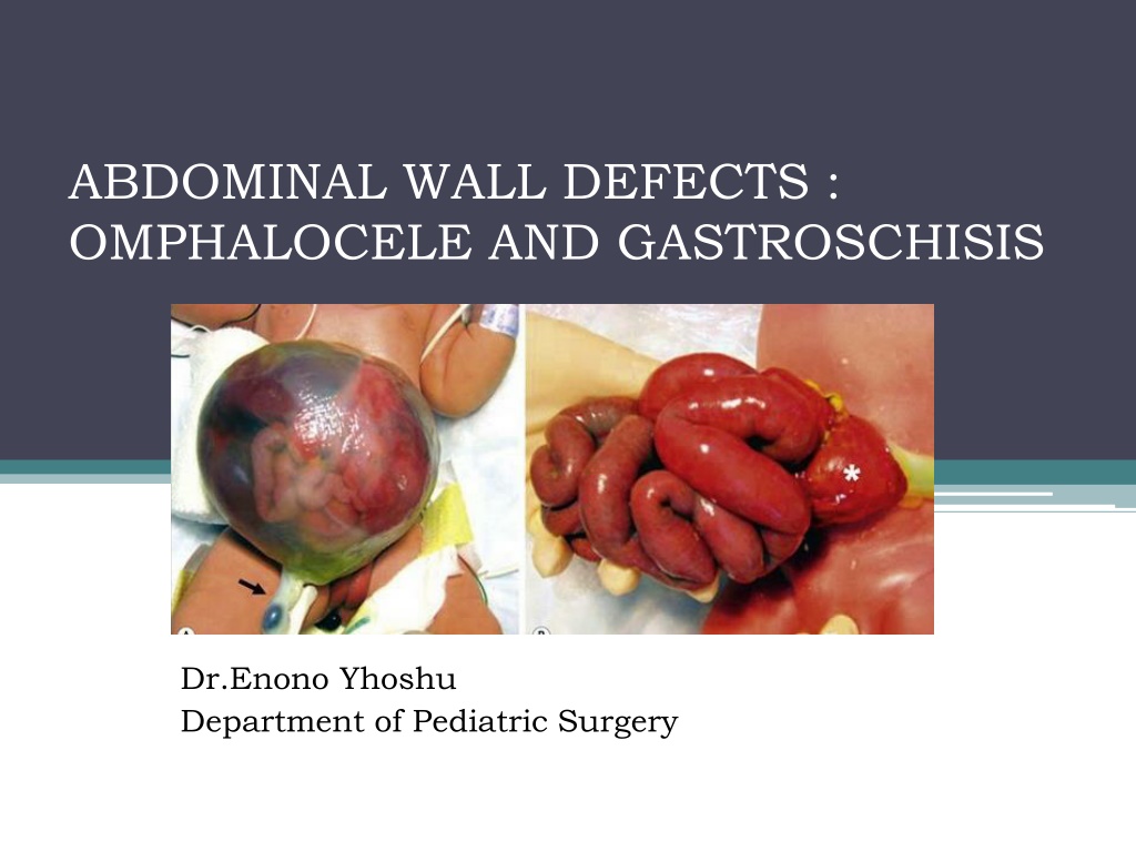
ABDOMINAL WALL DEFECTS : OMPHALOCELE AND GASTROSCHISIS Dr.Enono Yhoshu Department of Pediatric Surgery
ABDOMINAL WALL DEFECTS A type of congenital defect that allows the abdominal organs to protrude through an unusual opening (blue arrows)that forms on the abdomen.
CONTENTS Embryology Types Gastroschisis Omphalocele Management Outcome Differences
EMBRYOLOGY Closure of the body wall begins at 3 weeks gestation and results from growth and longitudinal infolding of the embryonic disks.
The cephalic fold forms the thoracic and epigastric wall. The lateral folds form the lateral abdominal walls. The caudal fold contributes the hindgut, bladder, and hypogastric wall. These four folds meet in the midline to form the umbilical ring.
During 6th week of gestation, rapid growth of intestines causes herniation of the midgut into the umbilical cord. Week 10, the midgut is returned to the abdominal cavity and the small bowel and colon assumes a fixed position. Any disruption in process may result in an abdominal wall defect.
TYPES 1. Ectopia cordis thoracis cephalic fold defect. 2. Pentalogy of Cantrell- cephalic fold defect. 3. Omphalocele Failure of folding. 4. Umbilical cord Hernia Small defect and normal abdominal wall. 5. Gastroschisis 6. Cloacal exstrophy caudal fold defect.
GASTROSCHISIS- Most common Incidence : 2 to 4.9 per 10,000 live births. Herniation of intestinal loops through full-thickness defect in anterior abdominal wall. Defect lateral to the umbilicus (right>left), usually less than 4cm in size. No sac covers the extruded viscera (usu. only intestines). Preterm babies (28%). Young mothers (<25years).
Etiology: In-utero vascular accident. 2 theories 1. Involution of the right umbilical vein causes necrosis in the abdominal wall leading to a right-sided defect. 2.Right omphalomesenteric artery prematurely involutes Other theories: In-utero rupture of omphalocele. Abnormal midline fusion of the abdominal folds.
ASSOCIATED ANOMALIES 10-20% - intestinal stenosis or atresia that results from vascular insufficiency to the bowel. Vanishing bowel - very small defect strangulates bowel development.
ANTENATAL CONSIDERATIONS Diagnosis can often be made < 20 weeks of pregnancy by ultrasound. Amniotic fluid and serum tests of AFP and amniotic fluid acetylcholinesterase (AChE)- raised in abdominal wall defects. Opportunity to counsel the family (Increased risk : - Intrauterine growth retardation (IUGR), - Fetal death, and - Premature delivery). Prepare for optimal postnatal care.
Mode of delivery. Optimal mode- debated. - Proponents of LSCS: Vaginal delivery may damage bowel. - Studies have failed to show difference in outcome between Caesarean and vaginal delivery. - The delivery method should be at the discretion of the obstetrician and the mother
Timing of delivery Considerations : 1. Because bowel edema and peel formation increase as pregnancy progresses. 2. LBW and preterm negatively influences outcome, with neonates weighing <2 kg having - increased time to full enteral feeding, - ventilated days, and - duration of parenteral nutrition. The presumption is that earlier delivery based on serial measurements of the bowel may decrease the incidence of intestinal complications.
PERINATAL CARE Outcome depends on - amount of intestinal damage that occurs during fetal life. Combination of exposure to amniotic fluid and constriction of the bowel at the abdominal wall defect. Intestinal damage impaired motility and mucosal absorptive function prolonged need for total parenteral nutrition and severe irreversible intestinal failure.
Prenatal diagnosis provides a potential opportunity to modulate mode, location, and timing of delivery in order to minimize these complications.
Neonatal resuscitation and management Gastroschisis causes significant evaporative water losses from the exposed bowel. 1. Warm saline-soaked gauze, placed in a central position on the abdominal wall and wrapped with plastic wrap. 2. IV Fluid resuscitation. 3. Gastric decompression. 4. Baby right side down- prevent mesenteric pedicle kinking. 5. IV antibiotics.
SURGICAL MANAGEMENT The primary goal of every surgical repair is to return the viscera to the abdominal cavity while minimizing the risk of damage to the viscera. Options include: (i) Primary reduction with operative closure of the fascia; (ii) silo placement, serial reductions, and delayed fascial closure;
Primary closure with fascial closure In neonates considered to possess sufficient intraabdominal domain to permit full reduction of the herniated viscera. Warm bowel and clean the peel; check quickly for intestinal anomalies.
Primary closure- without fascial closure Umbilicus as an allograft. Prosthetic non absorbable mesh. Prosthetic biosynthetic absorbable options dura or porcine small intestinal submucosa.
Staged closure Bowel placed into Spring loaded silo - Silastic sheet silo Delivery room or OT. Bowel is reduced once or twice daily into the abdominal cavity as the silo is shortened by sequential ligation. Once contents entirely reduced, definitive closure. Usually takes 1-14 days.
Intra-abdominal pressure Either as intravesical or intragastric pressure, can be used to guide the surgeon during reduction. Pressures >20 mmHg are correlated with decreased perfusion to the kidneys and bowel. Following reduction, monitor: - Physical examination, - Urine output, and - lower limb perfusion With a low threshold to reopen a closed abdomen for signs of abdominal compartment syndrome
Management of associated intestinal atresia or perforation Upto 10 % cases associated. Usually jejunal and ileal. Options - Resection and end to end anastomosis - Stoma - Initial gastroschisis repair and 4-5 weeks later, atresia surgery.
Postoperative Course Abnormal intestinal motility. Abnormal nutrient absorption. Delayed enteral feeding. Prokinetics. Parenteral nutrition.
OMPHALOCELE- 2ndMost common Incidence is 1.5 to 3 per 10,000 live births. Omphalocele represents a failure of the body folds to complete their journey. Herniated viscera covered by a membrane consisting of peritoneum on the inner surface, amnion on the outer surface, and Wharton s jelly between the layers.
OMPHALOCELE (EXOMPHALOS) The umbilical vessels insert into the membrane and not the body wall. The hernia contents include a variable amount of intestine, often parts of the liver, and occasionally other organs.
OMPHALOCELE (EXOMPHALOS) Whatever the insult may be that causes it, this aberration occurs early in embryogenesis- more associated anomalies.
ANTENATAL CONSIDERATIONS Distinguished by presence of sac and presence of liver. Other associated anomalies- ultrasound especially for cardiac and chromosomal studies. Increased levels of AFP and AChE Risks of : - IUGR (5-35%) - Fetal death - Premature labour (5-60%)
PERINATAL CARE Neither caesarean nor vaginal delivery superior. Most practitioners choose to deliver neonates with large omphaloceles by cesarean section because of the fear of liver injury or sac rupture during vaginal delivery. Delivery at tertiary perinatal centre- immediate access to expert care. No advantage of preterm delivery.
NEONATAL RESUSCITATION AND MANAGEMENT Careful attention to cardiopulmonary status- unsuspected pulmonary hypoplasia- requires immediate intubation and ventilation. Directed cardiac evaluation: - auscultation, - four-limb blood pressures, and - peripheral pulse examination. Dressed with saline soaked gauze and an impervious dressing to minimize fluid and temperature losses. If sac ruptured, then treat as gastroschisis. IV fluids and nasogastric tube.
SURGICAL MANAGEMENT Treatment options in infants with omphalocele depend on: - The size of the defect, - gestational age, and - the presence of associated anomalies. Options: 1. Primary closure 2. Staged closure
PRIMARY CLOSURE Only when the baby is stable and defect is small. Steps: - Excising the omphalocele membrane, - reducing the herniated viscera, and - closing the fascia and skin.
STAGED CLOSURE If the covering sac is intact, then there is no urgency to perform operative closure. Escharotic therapy , which results in gradual epithelialization of the omphalocele sac. Usually takes many months for the sac to granulate and epithelialize. Options: 1. Silver sulfadiazine 2. Mercurochrome 3. Povidone iodine 4. Gentian violet
Mercurochrome - scarificant and disinfectant. - reports of mercury poisoning. Povidone iodine - systemic absorption of the iodine- transient hypothyroidism. Gentian violet Antibacterial and antifungal.
STAGED CLOSURE Sac is epithelialized or sturdy enough to withstand external pressure Compression is done with elastic bandages and serially increased until the abdominal contents are reduced. VENTRAL HERNIA REPAIR
VENTRAL HERNIA REPAIR 1. Flaps that mobilize the muscle, fascia, and skin of the abdominal wall toward the midline and allow midline fascial closure. 2. Tissue expanders-to create an abdominal cavity big enough to house the viscera. 3. Prosthetic patches in abdominal wall.
Long-term outcomes GASTROSCHISIS Generally excellent. Many patients with atresia do very well as long as the bowel is not irreversibly damaged during fetal life. Majority - will achieve normal growth and development after an initial catch-up period in early childhood.
Long-term outcomes OMPHALOCELE Most infants recover well with no long term issues, provided that there are no significant structural or chromosomal abnormalities. Long term medical problems occur in patients with large omphaloceles: - gastroesophageal reflux, - pulmonary insufficiency, - recurrent lung infections or asthma, and - feeding difficulty with failure to thrive, reported in up to 60% of infants with a giant omphalocele.
OMPHALOCELE GASTROSCHISIS INCIDENCE 1.5-3: 10,000 2 -4.9: 10,000 SAC Present Absent ASSOCIATED ANOMALIES Common Uncommon DEFECT At umbilicus; 1-15 cm Right of umbilicus; <4cm MATERNAL AGE Average Younger SURGICAL MANAGEMENT Non urgent Urgent PROGNOSTIC FACTORS Associated anomalies Bowel condition MORTALITY <5% ~ 25%
THANK YOU FOR YOUR PATIENT LISTENING


")
")
")