Understanding Dairy Farming: Udder Anatomy and Milk Production


DAIRY FARMING
ABC01(0-4-0)
Session: 11
Skill of milking procedure- hand milking as well as
machine milking, knowledge of retention of milk in
the udder, letting down procedure etc.


External
Anatomy
The
udder
consists
of
4
separate
glands
Located
in
the
inguinal
region
of
the
ventral
aspect.
Each
gland
has
one
teat
Each
teat
has
one
opening
The
glands
are
covered
with
hair
Teats
do
not
have
hair.
The
right
and
left
halves
are
entirely
separate
externally
indicated
by
intermammary
groove.
The
rear
quarters
account
for
55-60%
of
the
milk
produced
and
55-60%
of
udder
weight.
Rear
teats
are
usually
shorter
than the
front
teats.
The
Teats
-
(papilla
mammae)
Functions
as
the
only
exit
for
the
mammary
secretion
The
only
means
for
the calf
to
receive
milk.
Usually,
only
one
teat
drains
one
gland.
No
hair,
sweat
glands
or
sebacious
glands
on
the
teats.
Supernumerary
Teats
About
50%
of
all
cows
have
extra
teats- supernumerary
teats.
Some
of
these
extra
teats
open
into
a
"normal"
gland,
but
many
do
not.
Streak
canal (ductus
papillaris)
Functions
as
the
only
orifice
of
the
gland
between
the internal
milk
secretory
system
and
the
external
environment.
The
streak
canal
is
the
main barrier
against
infection.
Lined
with
a
skin-like
epidermis.
Closed
by
sphincter
muscles
around
the
streak
canal.
streak
canal
length
increases
with
increasing
lactation
number.
The
interior
of the
gland
is
made
up
of:
Connective
tissue
-
fibrous tissue
(collagen)
and
fatty
tissue
(adipose
cells).
Secretory
tissue
-
secretory
epithelial
cells-
produce
the
milk.
The
relative
amount
of
connective
vs.
secretory
tissue
varies
from
animal
to
animal,
by
stage
of
mammary
development
Gland
Cistern
-
(sinus
lactiferus)
Also
called
the
udder
cistern
or
milk
cistern
opens
directly
into
the
teat
cistern.
The
cisterns
function
for
milk
storage
(holds
~100-
400
ml).
The
gland
cistern
varies
greatly
in
size
and
shape.
There
are
often
pockets
formed
in
the
cistern
at
the
end
of the
larger
ducts.
The
major
ducts
which
empty
into
the
gland
cistern
sometimes are
called
cisternal
ducts.
Secretory
tissue
in
the
udder
is
organized
into
lobes-
many
lobules-lobule
contains
150-220
microscopic
alveoli.
Alveoli
-
(acini)
Sack-like
structures
where
milk
is
synthesized
and
secreted.
A
single
layer
of
secretory
epithelial
cells
lines
the
lumen.
Contractile myoepithelial
cells
surround
the epithelial
lining.
Myoepithelial cells
contract
in
response
to
the
hormone
oxytocin-milk
being
squeezed
out of
the
alveolar
lumen
and
into
the
small
ducts.
Outside
of
the
myoepithelial
cells
the
alveolus
is
surrounded
by
a
connective
tissue
basement
membrane.
The
capillary
bed on
the
outside
of
the
alveolus
is
part
of
the
stromal
connective
tissue
between
alveoli.
A
group
of
alveoli
can
be
visualized
as
a
clump
of
grapes,
with
the
stems
acting
as
the
small
ducts
leading
from
the
alveoli.
Lobules
-
Clusters
of
150-220
alveoli
are
encapsulated
by
a
connective
tissue
sheath
and
are
organized
as
a
lobule
(~.7-.8
mm
dia.).
Lobes
-
Groups
of
lobules
are
surrounded
by
a
connective
tissue
sheath
and
comprise
a
lobe.
Each
mammary
gland
is
made
of
numerous
lobes.
Ducts
-tubules
by
which
milk
drains from
the
alveoli
down
to
the
gland
cistern.
Interlobar
or
primary
ducts
drain
multiple
lobes.
Intralobar
ducts
or
secondary
ducts
are
within
a
lobe
-drain
several
regions
of
the
lobe.
Intercalary or
tertiary
ducts
-small
ducts
which
exit
from
the
alveolus.
A
strong
suspensory
system
required-high
producers.
Mammary
gland
is
a
skin
gland,
and
is
therefore
external
to
the
body
cavity.
The
tissues,
which
provide
some
degree
of
support
for
the
udder:
Skin
-minor
support..
Superficial
fascia
or
Areolar
subcutaneous
tissue
-attaches
the
skin
to
the
underlying
tissues.
Suspensory
ligaments-main
suspensory
structures.
Suspensory
ligaments
are
three:
1.
Superficial
Lateral
suspensory
ligament
2.
Deep
Lateral
suspensory
ligament
3.
Median
suspensory
ligaments
Blood supply
to
the
mammary
gland
-
extremely
important
for
mammary
function.
All
of
the
milk
precursors
come
from
blood.
On
average
400
-
500
units
of
blood
passes
through
the
udder
for
each
unit
of
milk
synthesized
Total
udder
blood
volume
for
lactating
cows
is
about
8%
of
total
body
blood
volume,
non-lactating
cow
-about
7.4%.
There
is
a
2-6
fold
increase
in
blood
flow
in
the
mammary
gland
starting 2-3
days
prepartum.
Arterial
System
Blood
leaves
the
heart
and
flows
towards
the
rear
of
the
cow
-abdominal
aorta
.When
it
reaches
the
pubic
area
-
called
the
common
iliac
arteries.
These
divide
into
the internal
and
external
iliac
arteries.
The
external
iliac
artery
becomes
the
femoral
artery
(supplies
the
leg
muscles)
A
branch
off
of the
femoral
artery
forms
the
prepubic
artery from
which
branches
the
posterior
abdominal
artery
and
the
external
pudic
(or
external
pudental)
artery.
The
external
pudic
artery
passes
through
the
inguinal
canal
and
out of
the
body
cavity.
The
inguinal
canal -orifice
in
the
body
cavity
in
the
inguinal
region
where
blood
vessels,
lymph
vessels
and
nerves
enter
and
leave
the
body
wall
to
supply
the
skin
in
the
posterior
part
of
the
animal.
As
the
external
pudic artery
passes
out of
the
body
cavity
it
becomes
the
mammary
artery.
Once
it
enters
the
gland,
the
mammary
artery
then
divides
into
the
anterior
(or
cranial)
and
posterior
(or
caudal)
mammary
arteries,
which
then
it
further
branches
as
it
descends
down
into
the
gland
A
small amount
of
blood
also
reaches
the
mammary
gland
by
the
perineal artery
(from
the
internal iliac
artery),
but
this
only
supplies
the
upper
rear
portion of
gland.
Venous
System
Veins
leave
the
mammary
gland
anti-
parallel
to
the
arteries.
There
are
three
veins
on
each
side
that
carry
blood
away
from
the
gland:
1.
External
pudic
vein
leaves
the
udder
anti-parallel
to
the
external
pudic
artery
2.
Subcutaneous
abdominal
vein
(milk
vein)
exits
the
gland
at
the
anterior
end
of
the
front
quarters
and
passes
along
the
abdominal
wall-large
vein
visible
under
the
skin
on
the
belly
of the
cow.
Nervous
System
Innervation
of the
udder
is
sparse compared
with
other
tissues.
Sensory
nerves
are
found
in
the
teats
and
skin;
these
are
involved
in
the
afferent
pathway
of
the
milk
ejection
reflex.
There
is
no
parasympathetic
innervation
to
the
gland;
this
is
similar to
other
skin
glands.
There
is
no
innervation
of the
secretory
system:
myoepithelial
cells
are
not
innervated;
they
do
not
contract
in
response
to direct
innervation,
but
rather
they
contract
in
response
to
the
blood-
borne
hormone,
oxytocin.
The
lymphatic
network
There
is
extensive
lymph
drainage
from
the
teats.
originates
in
tissue
spaces as
lymphatic
capillaries.
Lymph
capillaries
converge
to
form
larger
vessels.
Lymph
flow
is
undirectional
from
the
udder
through
lymphatic
vessels,
eventually
dumping
lymph
into
the
vena
cava.
Lymph
is
a
clear,
colorless
liquid
with
a
composition
similar to
blood
plasma.
Valves
in
the
lymphatic
vessels
prevent retrograde
flow.
In
the
udder,
the
lymph
system
flows
through
the
supramammary
lymph
nodes.
Mastitis
Inflammation
of
udder
due
to
bacterial
or
fungal
infection.
Physical
and
chemical
composition
of the
milk
will
be
changed.
In
our
condition
one
of
the
reasons
for
poor
production
from
local
cattle
could
be
due
to
subclinical
mastitis
that
go
on
undetected
in
the field.
Mammary
gland
abalation:
Surgical
removal
of
mammary
glands
as
a
remedy
to
disease
process.
Carried
out
only
in
pets
for
saving
their
life.
Thank you
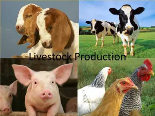
Exploring the external and internal anatomy of the udder in dairy cattle, including the functions of teats, streak canal, gland cistern, and supernumerary teats. Learn about milk retention, milking procedures, secretory tissue, and more crucial aspects of dairy farming.

Download Presentation

Please find below an Image/Link to download the presentation.
The content on the website is provided AS IS for your information and personal use only. It may not be sold, licensed, or shared on other websites without obtaining consent from the author. Download presentation by click this link. If you encounter any issues during the download, it is possible that the publisher has removed the file from their server.
E N D
Presentation Transcript
DAIRY FARMING ABC01(0-4-0) Session: 11 Skill of milking procedure- hand milking as well as machine milking, knowledge of retention of milk in the udder, letting down procedure etc.
ExternalAnatomy The udder consists of 4 separateglands Located in the inguinal region of the ventral aspect. Each gland has one teat Each teat has one opening The glands are covered with hair Teats do not havehair.
The right and left halves are entirely separate externally indicated by intermammary groove. The rear quarters account for 55-60% of the milk produced and 55-60% of udderweight. Rear teats are usually shorter than the frontteats.
The Teats -(papilla mammae) Functions as the only exit for the mammary secretion The only means for the calf to receive milk. Usually, only one teat drains onegland. No hair, sweat glands or sebacious glands on the teats.
Supernumerary Teats About 50% of all cows have extra teats- supernumerary teats. Some of these extra teats open into a "normal" gland, but many do not.
Streak canal (ductus papillaris) Functions as the only orifice of the gland between the internal milk secretory system and theexternal environment. The streak canal is the main barrier against infection. Lined with a skin-like epidermis. Closed by sphincter muscles around the streak canal. streak canal length increases with increasing lactation number.
The interior of the gland is made up of: Connective tissue -fibrous tissue (collagen) and fatty tissue (adipose cells). Secretory tissue -secretory epithelial cells- produce the milk. The relative amount of connective vs. secretory tissue varies from animal to animal, by stage of mammary development
Gland Cistern -(sinus lactiferus) Also called the udder cistern or milk cisternopens directly into the teatcistern. The cisterns function for milk storage (holds~100- 400 ml). The gland cistern varies greatly in size and shape. There are often pockets formed in the cistern at the end of the larger ducts. The major ductswhich empty into the gland cistern sometimes are called cisternal ducts.
Secretory tissue in the udder is organized intolobes- many lobules-lobule contains 150-220 microscopic alveoli. Alveoli -(acini) Sack-like structures where milk is synthesizedand secreted. A single layer of secretory epithelial cells lines the lumen. Contractile myoepithelial cells surround the epithelial lining.
Myoepithelial cells contract in response to the hormone oxytocin-milk being squeezed out of the alveolar lumen and into the smallducts. Outside of the myoepithelial cells the alveolusis surrounded by a connective tissue basement membrane.
The capillary bed on the outside of the alveolusis part of the stromal connective tissue between alveoli. A group of alveoli can be visualized as a clump of grapes, with the stems acting as the small ducts leading from the alveoli.
Lobules -Clusters of 150-220 alveoli are encapsulated by a connective tissue sheath and are organized as a lobule (~.7-.8 mm dia.). Lobes -Groups of lobules are surrounded by a connective tissue sheath and comprise alobe. Each mammary gland is made of numerous lobes.
Ducts -tubules by which milk drains from the alveoli down to the gland cistern. Interlobar or primary ducts drain multiple lobes. Intralobar ducts or secondary ducts are within a lobe -drain several regions of the lobe. Intercalary or tertiary ducts -small ducts whichexit from the alveolus.
A strong suspensory system required-high producers. Mammary gland is a skin gland, and is therefore external to the bodycavity.
The tissues, which provide some degree of support for the udder: Skin -minor support.. Superficial fascia or Areolar subcutaneoustissue -attaches the skin to the underlying tissues. Suspensory ligaments-main suspensory structures. Suspensory ligaments are three: 1. Superficial Lateral suspensoryligament 2. Deep Lateral suspensory ligament 3. Median suspensory ligaments
Blood supply to the mammary gland -extremely important for mammary function. All of the milk precursors come from blood. On average 400 -500 units of bloodpasses through the udder for each unit of milk synthesized Total udder blood volume for lactating cowsis about 8% of total body blood volume, non-lactating cow -about 7.4%. There is a 2-6 fold increase in blood flow in the mammary gland starting 2-3 days prepartum.
Arterial System Blood leaves the heart and flows towards therear of the cow -abdominalaorta .When it reaches the pubic area - calledthe common iliac arteries. These divide into the internal and externaliliac arteries. The external iliac artery becomes the femoral artery (supplies the leg muscles)
A branch off of the femoral artery forms the prepubic artery from which branches the posterior abdominal artery and the external pudic (or external pudental) artery. The external pudic artery passes through the inguinal canal and out of the bodycavity.
The inguinal canal -orifice in the body cavity in the inguinal region where blood vessels, lymph vessels and nerves enter and leave the body wall to supply the skin in the posterior part of theanimal. As the external pudic artery passes out of the body cavity it becomes the mammary artery. Once it enters the gland, the mammary artery then divides into the anterior (or cranial) and posterior (or caudal) mammary arteries, which then it further branches as it descends down into the gland
A small amount of blood also reaches the mammary gland by the perineal artery (from the internal iliac artery), but this only supplies the upper rear portion of gland.
VenousSystem Veins leave the mammary gland anti- parallel to the arteries. There are three veins on each side that carry blood away from the gland: 1. External pudic vein leaves the udder anti-parallel to the external pudicartery 2.Subcutaneous abdominal vein (milk vein) exits the gland at the anterior end of the front quarters and passes along the abdominal wall-large vein visible under the skin on the belly of thecow.
Nervous System Innervation of the udder is sparse comparedwith other tissues. Sensory nerves are found in the teats and skin; these are involved in the afferent pathway ofthe milk ejectionreflex. There is no parasympathetic innervation to the gland; this is similar to other skinglands. There is no innervation of the secretory system: myoepithelial cells are not innervated; they donot contract in response to direct innervation, but rather they contract in response to the blood- borne hormone, oxytocin.
The lymphatic network There is extensive lymph drainage from the teats. originates in tissue spaces as lymphatic capillaries. Lymph capillaries converge to form larger vessels. Lymph flow is undirectional from the udder through lymphatic vessels, eventually dumping lymph into the vena cava. Lymph is a clear, colorless liquid with a composition similar to bloodplasma.
Valves in the lymphatic vessels prevent retrograde flow. In the udder, the lymph system flows throughthe supramammary lymph nodes.
Mastitis Inflammation of udder due to bacterial orfungal infection. Physical and chemical composition of the milkwill be changed. In our condition one of the reasons for poor production from local cattle could be due to subclinical mastitis that go on undetected in the field.
Mammary gland abalation: Surgical removal of mammary glands as a remedy to disease process. Carried out only in pets for saving their life.