Understanding Uterine Leiomyosarcoma: A Case Presentation
A 58-year-old female with a history of fibroids presented with post-menopausal bleeding and was diagnosed with uterine leiomyosarcoma, a rare and aggressive malignancy of uterine smooth muscle cells. The case details the clinical course, diagnosis, and prognosis of this condition, emphasizing the importance of early detection and management.

Download Presentation

Please find below an Image/Link to download the presentation.
The content on the website is provided AS IS for your information and personal use only. It may not be sold, licensed, or shared on other websites without obtaining consent from the author. Download presentation by click this link. If you encounter any issues during the download, it is possible that the publisher has removed the file from their server.
E N D
Presentation Transcript
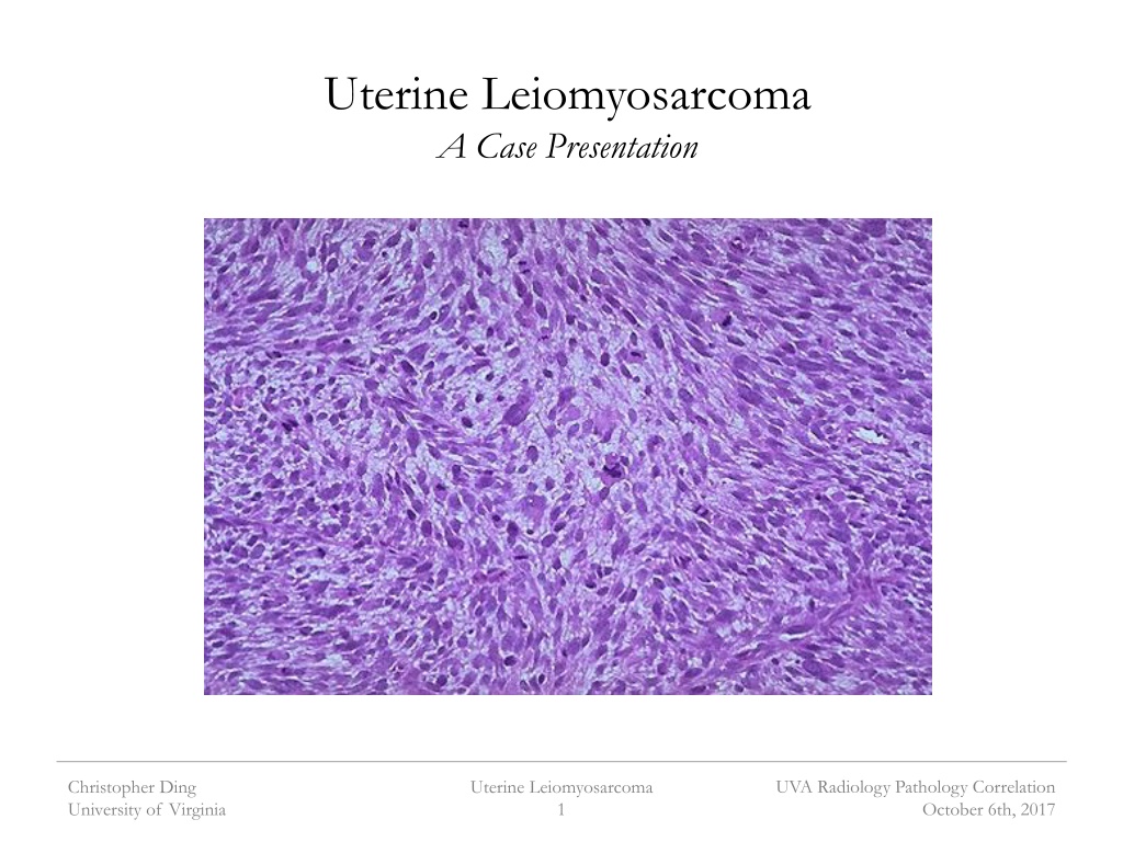
Uterine Leiomyosarcoma A Case Presentation UVA Radiology Pathology Correlation Christopher Ding University of Virginia Uterine Leiomyosarcoma 1 October 6th, 2017
Patient Presentation 58 year-old female with history of fibroids presented to UVA Ob/Gyn clinic by self-referral in June 2014 for 9 months of intermittent post-menopausal bleeding Endorsed progressive pelvic cramping, early satiety, and increasing abdominal girth Initially presented to primary care physician in September 2013 Pelvic ultrasound obtained at OSH in September 2013 demonstrated large fundal fibroid measuring 6.5 x 3.0 x 6.0 cm Endometrial biopsy obtained in December 2013 was inconclusive UVA Radiology Pathology Correlation Christopher Ding University of Virginia Uterine Leiomyosarcoma 2 October 6th, 2017
Clinical Course Repeat pelvic ultrasound performed June 2014 Uterine mass had doubled in size since last US in September Referred to UVA Cancer Clinic Scheduled for total abdominal hysterectomy with BSO for removal of large uterine fibroid CXR revealed multiple bilateral pulmonary nodules UVA Radiology Pathology Correlation Christopher Ding University of Virginia Uterine Leiomyosarcoma 3 October 6th, 2017
Clinical Course (cont.d) Intra-Op dx of uterine leiomyosarcoma on frozen section Multiple nodule visualized throughout and removed from the peritoneum, omentum, and bowel The fundus of the uterus is diffusely involved by a tan-yellow infiltrative mass measuring 7.5 x 5.5 x 10.0 cm.Grossly the mass has a tan-yellow cut surface with areas of increased vascularity and rare areas of necrosis UVA Radiology Pathology Correlation Christopher Ding University of Virginia Uterine Leiomyosarcoma 4 October 6th, 2017
Uterine Leiomyosarcoma Rare malignancy of uterine smooth muscle cells Comprises 1-2% of all uterine sarcomas Not derived from uterine fibroids Aggressive malignancy with poor prognosis and high rate of recurrence 5 year survival of 40%; minimal if outside the uterus Recurrence rate of 53-71% UVA Radiology Pathology Correlation Christopher Ding University of Virginia Uterine Leiomyosarcoma 5 October 6th, 2017
Uterine Leiomyosarcoma Gross description fleshy tan-yellow mass with areas of hemorrhage and/or necrosis Usually large (>6cm in diameter) and solitary UVA Radiology Pathology Correlation Christopher Ding University of Virginia Uterine Leiomyosarcoma 6 October 6th, 2017
Clinical Course (cont.d) Treatment with chemoradiation throughout subsequent years Four rounds of chemotherapy: gemcitabine/docetaxel, carboplatin/doxorubicin, trabectedin, temazolamide, gemcitabine/docetaxel Radiation therapy performed for control of hip lesions. Gamma knife for cerebellar lesions August 2017, chemotherapy stopped due to thrombocytopenia to 63, further tx deemed potentially harmful Palliative lower colostomy performed d/t concern for impending BO Patient expresses interest in XRT/pembrolizumab trial at UVA. Plan to irradiate painful RUQ abdominal wall lesion, neck mass, and perirectal masses. UVA Radiology Pathology Correlation Christopher Ding University of Virginia Uterine Leiomyosarcoma 7 October 6th, 2017
Sites of Metastasis UVA Radiology Pathology Correlation Christopher Ding University of Virginia Uterine Leiomyosarcoma 8 October 6th, 2017
Pembrolizumab Monoclonal antibody against PD-1 receptor Prevents suppression of immune activity against tumor cells mediated by PD-L1 and PD-L2 expression Currently approved for melanoma, NSCLC, H&N squamous CC, others UVA Radiology Pathology Correlation Christopher Ding University of Virginia Uterine Leiomyosarcoma 9 October 6th, 2017
Pembrolizumab (cont.d) UVA is undertaking Phase I safety trial of pembrolizumab with high-dose conformal radiation therapy Primary outcomes include adverse event profile at 30 and 90 days and tumor infiltration by T-cell through day 43 Notable inclusion criteria: Must be able to provide tissue from 2-3 separate biopsy procedures Patients must be resistant to at least 1 prior conventional chemotherapy regimen or other standard of care regimen Patient must have no remaining conventional treatment options proven to provide long-term disease control UVA Radiology Pathology Correlation Christopher Ding University of Virginia Uterine Leiomyosarcoma 10 October 6th, 2017
Most Recent Imaging Abdomen/pelvis CT Interval enlargement of all pre-existing peritoneal implants and right rectus abdominal implant New lesions of the liver and para-aortic lymph nodes Chest CT Interval enlargement of all pre-existing lung nodules and 8th rib lesion, development of new lung nodules Enlarged cervical lymph nodes MRI Brain No abnormal enhancement in area of previous treated cerebellar lesion UVA Radiology Pathology Correlation Christopher Ding University of Virginia Uterine Leiomyosarcoma 11 October 6th, 2017
UVA Radiology Pathology Correlation Christopher Ding University of Virginia Uterine Leiomyosarcoma 12 October 6th, 2017
Procedure Ultrasound-guided FNA and core biopsies of RUQ abdominal wall mas and left cervical lymph nodes 1 FNA and 4 core biopsies of rectus sheath mass 1 FNA and 1 core biopsy of left ventral cervical LN UVA Radiology Pathology Correlation Christopher Ding University of Virginia Uterine Leiomyosarcoma 13 October 6th, 2017
Cytology Findings Stanford criteria used in diagnosis Cellular atypia, abundant mitoses (>10 figures per 10 hpf) , & coagulative necrosis Stains used for confimation Smooth muscle markers: H-caldesmin, SMA, desmin Hormone receptors: ER, PR Others: Ki-67, EMA UVA Radiology Pathology Correlation Christopher Ding University of Virginia Uterine Leiomyosarcoma 14 October 6th, 2017
Stanford Criteria UVA Radiology Pathology Correlation Christopher Ding University of Virginia Uterine Leiomyosarcoma 15 October 6th, 2017
Hopeful News This patient exhibited complete pathologic response to pembrolizumab at all but one metastatic ULMS site. Genomic analysis of the resistant tumor revealed acquired bialllelic PTEN loss. UVA Radiology Pathology Correlation Christopher Ding University of Virginia Uterine Leiomyosarcoma 16 October 6th, 2017
References D Angelo E, Prat J. Uterine sarcomas: a review. Gynecol Oncol. 2010 Jan;116(1):131-9. Seagle BL, Sobecki-Rausch J, strohl AE, Shilpi A, Grace A, Shahabi S. prognosis and treatment of uterine leiomyosarcoma: A national Cancer Database study. Gynecol Oncol. 2017 Apr;145(1):61-70. Tirumani SH, Deaver P, Shinagare AB, et al. Metastatic pattern of uterine leiomyosarcoma: retrospective analysis of the predictors and outcome in 113 patients. Journal of Gynecologic Oncology. 2014;25(4):306-312. UpToDate. Treatment and prognosis of uterine leiomyosarcoma. Accessed October 2nd, 2017 UVA Radiology Pathology Correlation Christopher Ding University of Virginia Uterine Leiomyosarcoma 17 October 6th, 2017


")


")


")