Understanding Acidosis and Alkalosis in Acid-Base Disorders
Acid-base disorders, involving acidosis and alkalosis, are common in clinical practice. These conditions are characterized by changes in pH levels, with acidemia (pH < 7.35) and alkalemia (pH > 7.45) representing acidosis and alkalosis, respectively. The primary causes of these imbalances lie within the respiratory, metabolic, and renal systems. Various physiological mechanisms, such as the carbonic acid/bicarbonate buffer system, work to maintain the pH within a narrow range. Different types of acid-base disturbances, like metabolic acidosis and alkalosis, and respiratory acidosis and alkalosis, have specific pH and electrolyte profiles. Understanding these concepts is crucial for diagnosing and managing such conditions.

Download Presentation

Please find below an Image/Link to download the presentation.
The content on the website is provided AS IS for your information and personal use only. It may not be sold, licensed, or shared on other websites without obtaining consent from the author. Download presentation by click this link. If you encounter any issues during the download, it is possible that the publisher has removed the file from their server.
E N D
Presentation Transcript
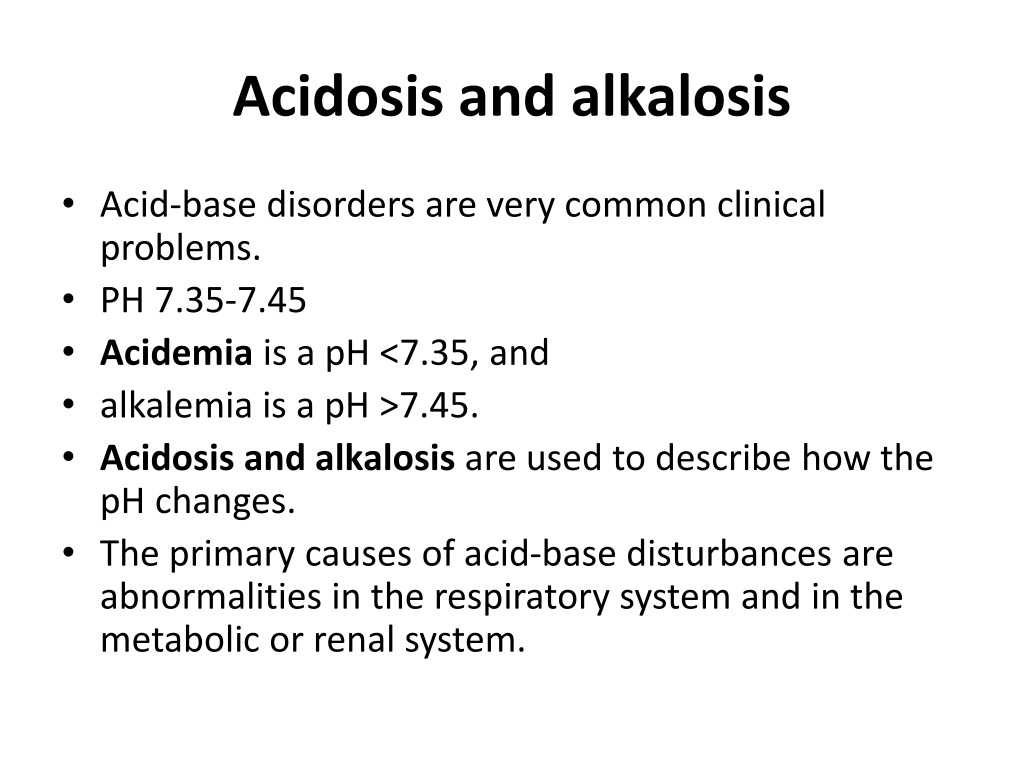
Acidosis and alkalosis Acid-base disorders are very common clinical problems. PH 7.35-7.45 Acidemia is a pH <7.35, and alkalemia is a pH >7.45. Acidosis and alkalosis are used to describe how the pH changes. The primary causes of acid-base disturbances are abnormalities in the respiratory system and in the metabolic or renal system.
DISORDERS OF ACID-BASE BALANCE The pH of the arterial plasma is normally The pH of the arterial plasma is normally 7.40 corresponding to a H corresponding to a H+ +concentration of concentration of 40 concentration corresponds to a decrease in concentration corresponds to a decrease in pH. this parameter is under tight homeostatic regulation, such that the H this parameter is under tight homeostatic regulation, such that the H+ + concentration does not vary outside the range concentration does not vary outside the range 36 under normal circumstances. under normal circumstances. IN acidosis there is IN acidosis there is anaccumulation anaccumulation of acid or loss of abase causing Ph . The converse Ph . The converse accure accure in alkalosis . in alkalosis . Abnormal acid Abnormal acid- -base balance occurs in a wide range of diseases base balance occurs in a wide range of diseases variety of physiological mechanisms act to prevent wide swings in the pH of the ECF. The first is the action of blood and tissue buffers, of which the most important is the carbonic acid/bicarbonate buffer system. This involves the reaction of H+ions with bicarbonate to form carbonic acid, which, under the influence of the enzyme carbonic anhydrase (c.a.), dissociates to form CO2and water, as follows: CO2+H2O---H2CO3----H2+HCO3 This buffer system is important because bicarbonate is present in relatively high concentration in the ECF (21-28 mmol/l), and two of its key components are under physiological control: the CO2by the lungs, and the bicarbonate, by the kidneys. 7.40, (pH , (pH 7.36 40 nmol nmol/l. An increase in H /l. An increase in H+ + pH. 7.36- -7.44 7.44) ) 36- -44 44 nmol nmol/l (pH /l (pH 7.44 7.44- -7.36 7.36) ) of acid or loss of abase causing afall afall in in
Metabolic acidosis :decrease HCO3<24mmol ,decrease CO2<5.33kpa ,PH>40 Metabolic alkalosis increase HCO3>24mmol ,increase CO2>5.33kpa ,PH<40 Respiratory acidosis: increase HCO3>24mmol ,increase CO2>5.33kpa ,PH>40 Respiratory alkalosis: increase HCO3<24mmol ,decrease CO2<5.33kpa ,PH<40
ACIDOSIS 1-METABOLIC ACIDOSIS Metabolic acidosis occurs when an acid other than carbonic acid (due to CO2retention) accumulates in the body, resulting in a fall in the plasma bicarbonate. The pH fall which would otherwise occur is blunted by hyperventilation, resulting in a reduced PCO2. If the kidneys are intact (i.e. not the cause of the initial disturbance), renal excretion of acid can be gradually increased over days to weeks, raising the plasma bicarbonate and hence the pH towards normal in the new steady state
Causes Base loss(Bicarbonate losses ) Extra renal = ( Renal = renal tubular acidosis ( Urinary loss of HCO3in proximal RTA Carbonic anhydrase inhibitors Failure of bicarbonate regeneration Distal renal tubular acidosis (impaired tubular acid secretion ) Aldosterone deficiency (Addison's disease Aldosterone insensitivity ( Interstitial renal disease, Aldosterone antagonists) Small bowel drainage Diarrhea ) Primary hyperparathyroidism Ureteroileostomy (ileal bladder) Acidifying salts ( Ammonium chloride , Lysine or arginine hydrochloride ) Diabetes mellitus Reduced excretion of acids ( Renal failure: Accumulation of organic acids Overproduction of acids Ketoacidosis 1- Diabetic(Accumulation of ketones1with hyperglycaemia) 2-Alcoholic 3-Starvation (Accumulation of ketones without hyperglycaemia) Lactic acidosis( Tissue hypoxia (e.g. shock) or liver disease) Toxin ingestion ( Methanol Ethylene glycol Salicylates)
Clinical features Rapid deep breathing ?stimulation of respiratory centre by reduction in PH of blood &to eleminat acid substance The urin strongly acidic
Management The first step in management of metabolic acidosis is identifying and correcting the cause when possible . This may involve control of diarrhoea, treatment of diabetes, correction of shock, cessation of drug administration, or dialysis to remove toxins. Since metabolic acidosis is frequently associated with sodium and water depletion, resuscitation with appropriate intravenous fluids will often be needed. Use of intravenous bicarbonate in this setting is controversial. Because rapid correction of acidosis has some inherent risks (e.g. induction of hypokalaemia or reduced plasma ionised calcium), use of bicarbonate infusions is best reserved for situations where the underlying disorder cannot be readily corrected and where the acidosis is severe (H+> 70 nmol/l, pH < 7.15) and associated with evidence of tissue dysfunction. The acidosis associated with RTA can sometimes be controlled by treating the underlying cause . Usually, however, supplements of sodium and potassium bicarbonate are necessary to achieve the target of a plasma bicarbonate level above 18 mmol/l with normokalaemia in types 1 and 2 RTA, while diuretics or corticosteroids (as appropriate) may be effective in reversing the underlying disturbance in type 4 RTA.
RESPIRATORY ACIDOSIS acondition where the PCO2 is above the normal range is caused by: 1-impairment in the rate of alveolar ventilation. 2-a sudden depression of the medullary respiratory center (narcotic overdose), 3-paralysis of the respiratory muscles(M.G,G.B ,toxines ,anasthesia 4-airway obstruction.(infection ,allergy ,tumoures ,F.B . 5-Chronic respiratory acidosis in chronic airway disease (emphysema), with extreme kyphoscoliosis, and with extreme obesity (pickwickian syndrome). The serum bicarbonate concentration is increased, the magnitude of which depends on the acuity and the severity of the respiratory disorder. Acute increases in the PCO2result in somnolence, confusion, and, ultimately, carbon dioxide narcosis. Asterixis may be present. Because carbon dioxide is a cerebral vasodilator, the blood vessels in the optic fundi are often dilated, engorged, and tortuous. Frank papilledema may be present in patients with severe hypercapnic states. treatment 1-The only practical therapy of acute respiratory acidosis involves treatment of the underlying disorder 2-O2 and 3- ventilatory support. I n patients with chronic hypercapnia who develop an acute increase in the PCO2, attention should be directed toward identifying factors that may have aggravated the chronic disorder. Alkalinizing salts should be avoided in patients with chronic
METABOLIC ALKALOSIS Aetiology and clinical assessment Metabolic alkalosis is characterised by an increase in the plasma bicarbonate concentration and the plasma pH . There is a compensatory rise in PCO2due to hypoventilation. CAUSES 1-Hypovolaemic metabolic alkalosis is the most common pattern, such as sustained vomiting where acid-rich fluid is lost directly from the body. during treatment with most diuretic drugs (other than carbonic anhydrase inhibitors and potassium-sparing drugs), since the diuretic action involves increased acid loss into the urine. 2- Normovolaemic (or hypervolaemic) metabolic alkalosis occurs in settings where both bicarbonate retention and volume expansion occur simultaneously. Classical causes include corticosteroid excess states such as primary hyperaldosteronism (Conn's syndrome, Cushing's syndrome and corticosteroid therapy. Occasionally, overuse of antacid salts for treatment of dyspepsia can produce a similar pattern.
. Clinically, apart from manifestations of the underlying cause, there may be few symptoms or signs related to alkalosis itself. When the rise in systemic pH is abrupt, plasma ionised calcium falls and signs of increased neuromuscular irritability such as tetany may develop
Management In metabolic alkalosis associated with hypovolaemia, treatment involves provision of adequate intravenous fluid, specifically isotonic sodium chloride, which interrupts the volume-conserving mechanisms and allows the kidney to excrete the excess alkali in the urine. Replacement of potassium helps correct the hypokalaemia and its consequences in the kidney. In metabolic alkalosis associated with normal or increased volume, treatment should focus on correcting the underlying cause, specifically the removal or blockade of excess mineralocorticoid activity. The approach depends on the underlying endocrinological diagnosis .
RESPIRATORY ALKALOSIS occurs when hyperventilation reduces the arterial PCO2(31-42)and consequently increases the arterial pH. Acute respiratory alkalosis is most commonly a result of the hyperventilation syndrome. It may also occur in damage to the respiratory centers ,hysterical acute salicylism, in fever and septic states association with various pulmonary processes (pneumonia, pulmonary emboli, or congestive heart failure). iatrogenically by injudicious mechanical ventilatory support. Chronic hyperventilation occurs in the acclimatization response to high altitudes (a low ambient oxygen tension), in advanced liver disease, and in pregnancy . Acute hyperventilation is characterized by lightheadedness, paresthesias, circumoral numbness, and tingling of the extremities. Tetany occurs in severe cases. When anxiety provokes hyperventilation, air rebreathing with a paper bag generally terminates the acute attack
Compensatory response Predicted compensation Blood H+ Disturbance Primary change > 401 HCO3< 24 mmol/l PCO2< 5.33 kPa2 Metabolic acidosis P CO2fall in kPa = 0.16 HCO3fall in mmol/l < 401 PCO2> 5.33 kPa2,3 Metabolic alkalosis HCO3> 24 mmol/l PCO2rise in kPa = 0.08 HCO3rise in mmol/l > 401 PCO2> 5.33 kPa2 Respiratory acidosis HCO3> 24 mmol/l Acute:HCO3rise in mmol/l = 0.75 PCO2rise in kPa Chronic: HCO3rise in mmol/l = 2.62 P CO2rise in kPa < 401 PCO2< 5.33 kPa2 Respiratory alkalosis HCO3< 24 mmol/l Acute:HCO3fall in mmol/l = 1.50 CO2fall in kPa Chronic: HCO3fall in mmol/l = 3.75 P CO2fall in kPa