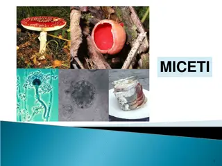
Zoonotic Diseases and Mycoses


UNIT-3
‘Zoonotic disease’
(Credit Hours-3+1)


 undefined


Superficial and Systemic
mycoses
Features of Fungi
•
Eukaryotic cell/ Prokaryotic cells?
•
Autophagy/hetrophagy ?
•
Unicelluler/Multicelluler?
•
Are they motile/non motile?
•
They multiply by mitotic/meiotic division?
Classification
- Depending on
cell morphology
1.
Yeasts
eg. Yeast,
Cryptococcus neoformans
2.
Yeast like fungi
eg.
Candida albicans
3.
Molds
eg
. Dermatophytes
4.
Dimorphic fungi
eg
.
Histoplasma capsulatum
Blastomyces dermatidis
Introduction:
Fungus are:
▫
Normal skin microbiota
▫
Opportunistic infection: Entry through a wound
Immunocompromised patients
▫
Causes a variety of disease conditions: Animals & man
▫
Infections, also K/ as
mycoses
▫
Most of mycoses: zoonotic in nature
▫
Effect all organs & structures of the body
▫
Moist environment: Promote growth & transmission
(sweaty shoes, communal showers, and locker rooms)
Types of Mycoses
Mycoses: 3 types
(Based on their invasiveness)
1. Cutaneous mycoses
Infections of the epidermis,
hair, & nail
Ex.
Tinea
2. Subcutaneous mycoses
Penetrates epidermis/dermis
to infect deeper tissues
Ex.
Sporotrichosis
3. Systemic mycoses
Spreads throughout body
Ex.
Aspergillosis
Cutaneous Mycoses
Cutaneous Mycoses
o
Caused by Dermatophytes (
fungal molds)
Keratinophilic
- "keratin loving” (
keratin of h
orns, hooves, nails,
hair and skin
Commonly found : Environment & soils
Frequently transferred to the skin via contact
o
There are three genera of dermatophytes:
Epidermophyton:
Infects
skin
,
nails
but never hair
Trichophyton:
I
nfects
hair
,
skin
and
nails
Microsporum:
Infects
hair
and
skin
, rare cases of nail
o
Several species of
Microsporum
&
Trichophyton
causes zoonotic ring
worm infection
*A group of cutaneous mycoses called
tineas
*
These genera have been categorized into
three groups
(
On the basis of their host preferences
)
(i)
Zoophilic dermatophytes:
Primarily parasites of animals but mostly are also pathogenic
for man
(ii)
Geophilic dermatophytes:
Inhabitants of soil and rarely infect man & animals
(iii)
Anthropophilic dermatophytes:
Primarily parasites of man but cause disease in animals
Cutaneous Mycoses
Cutaneous Mycoses
Tinea’s on most areas of the body are generally called
ringworm
Cutaneous Mycoses
Epidemiology
Direct contact;
infected host
Contact with Inanimate objects;
carrying fungal spores
More prevalent in animals, kept confined
i.e.
in winter season
High humidity
in the environment; favorable for multiplication
Young animals
; most susceptible
Disease in animals
o
Common in
: Cattle, horse, pig & many other species
o
Rare in
: Sheep & very rare in goats
In cattle:
Calves are more susceptible
Effected parts
: Neck, head & perineum sometimes other parts
Lesions
: Grey-white crust followed by alopecia
In horses
Lesions
: Axillary girth area & later spread over the trunk & rump, and to
the neck, head and limbs
Lesions with scabs followed by alopecia appear which vary in size with the
species of fungus involved
In Pigs
Similar lesions on the back and sides
In sheep
lesions consist of round, bald patches covered with greyish crust, in head
In goats
Similar lesions are observed generally all over the body
Human being can contract ringworm infection from animals
In rural areas
: 80% of human cases are of animal origin
In urban areas
:10% of human cases are of animal origin
Horses and cattle
:
Trichophyton
spp. infection
Dogs
:
M. canis
infection
Infected pet animals (
Guinea pigs
/
hamsters
/
cats
/
rabbits
)
to farmers
,
zoo-
keepers
,
veterinarians
&
children
Children are particularly
susceptible
M. canis
(microsporia): Develop single/ multiple, round, oval or
polycyclic reddened scaly lesions, primarily on
face, neck,
&
arms
M. distortum
:
lesions on
scalp, neck, face, trunk
&
other parts
of the body
T. rubrum
: Superficial mycosis of
feet, toe-webs,
&
hands
T. mentagrophytes
/
T
.
verrucosum
/
M. gypseum
:
Hairy skin on the
scalp, neck, face
and rarely on extremities
Disease in human
•
By characteristic lesion
:
Ring like lesion
•
Wood’s lamp
(
also called a black lamp
)
examination
UV Wavelength - 365 nm
Used to examine dermatophyte infections of
M. canis, M.
distortum and M. equinum
Fungal elements (spores and hyphae) show greenish fluorescence
Diagnosis
•
Direct microscopic evaluation
:
Specimens: Skin scrapings, hair, or nails
Preparation of specimens: Wet mount slide
Diagnosis
Skin/Hair
Dissolves keratin
10%–20% aqueous KOH
visualization of the hyphae
& fungal spores
Dissolves keratin
•
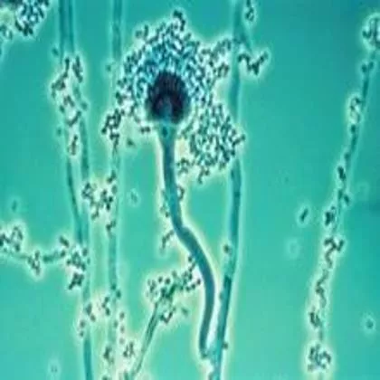
Culture examination:
Media: Sabouraud dextrose CC (chloramphenicol/cyclohexamide)
Inhibiting the growth of bacteria/ saprophytic fungi
Macroscopic colony morphology
Identification can be further confirmed by visualizing the
Microscopic morphology using either a slide culture /a sticky tape
prep stained with
lactophenol cotton blue
Diagnosis
Tinae growth on a Sabouraud
dextrose CC agar plate.
Lactophenol Cotton blue staining
Various antifungal treatments can be effective against tineas
Depending upon the extent of lesions: local/ systemic treatments
•
Topical application:
First removal of the crust and application of weak solution of
Iodine, Whitfield's ointment, 10% ammoniated mercury ointment,
Ointments containing propionic and undecylenic acids,
Hexetidine etc.
Natamycin and nanomycin weak solution of iodine
Allylamine (Contais terbinafine)
Miconazole
Clotrimazole
•
Orally:
Griseofulvin
•
Systemic application
: Inj. of Sodium Iodide
Treatment
•
Maintain
General hygienic measures
•
Isolation and treatment of infected
animals
•
Disinfection of contaminated
equipment and premises
•
Oral administration of griseofulvin
also has protective effect
•
Proper diet with vitamin A
supplementation is considered
desirable
•
A vaccine against ringworm,
containing the inactivated
Trichophyton verrucosum
strain
Prevention and control
Questions?
1. ________ is a superficial fungal infection found on the head.
a)
Tinea cruris
b)
Tinea capitis
c)
Tinea pedis
d)
Tinea corporis
2. Sabouraud dextrose agar CC is selective for ________.
a)
All fungi
b)
Non-saprophytic fungi
c)
Bacteria
d)
Viruses
3. ___________Dermatophytes effect the skin, hair and nails
a)
Epidermaphyton
b)
Microsporum
c)
Trichophyton
d)
All
Zoonotic diseases, caused by fungi, have the potential to affect both animals and humans and are often transmitted through various environmental factors. These diseases can lead to a range of conditions affecting different organs and structures of the body. Mycoses, such as superficial and systemic mycoses, are a significant aspect of these diseases with different types like cutaneous, subcutaneous, and systemic mycoses each affecting the body differently. Cutaneous mycoses, caused by dermatophytes, are commonly transmitted through contact with the environment and can lead to infections of the skin, hair, and nails.

Download Presentation

Please find below an Image/Link to download the presentation.
The content on the website is provided AS IS for your information and personal use only. It may not be sold, licensed, or shared on other websites without obtaining consent from the author. Download presentation by click this link. If you encounter any issues during the download, it is possible that the publisher has removed the file from their server.
E N D
Presentation Transcript
UNIT-3 Zoonotic disease (Credit Hours-3+1)
Superficial and Systemic mycoses
Features of Fungi Eukaryotic cell/ Prokaryotic cells? Autophagy/hetrophagy ? Unicelluler/Multicelluler? Are they motile/non motile? They multiply by mitotic/meiotic division? Classification- Depending on cell morphology 1. Yeasts eg. Yeast, Cryptococcus neoformans 2. Yeast like fungi eg. Candida albicans 3. Molds eg. Dermatophytes 4. Dimorphic fungi eg. Histoplasma capsulatum Blastomyces dermatidis
Introduction: Fungus are: Normal skin microbiota Opportunistic infection: Entry through a wound Immunocompromised patients Causes a variety of disease conditions:Animals & man Infections, also K/ as mycoses Most of mycoses: zoonotic in nature Effect all organs & structures of the body Moist environment: Promote growth & transmission (sweaty shoes, communal showers, and locker rooms)
Types of Mycoses Mycoses: 3 types (Based on their invasiveness) 1. Cutaneous mycoses Infections of the epidermis, hair, & nail Ex. Tinea 2. Subcutaneous mycoses Penetrates epidermis/dermis to infect deeper tissues Ex. Sporotrichosis 3. Systemic mycoses Spreads throughout body Ex. Aspergillosis
Cutaneous Mycoses *A group of cutaneous mycoses called tineas* o Caused by Dermatophytes (fungal molds) Keratinophilic - "keratin loving (keratin of horns, hooves, nails, hair and skin Commonly found : Environment & soils Frequently transferred to the skin via contact o There are three genera of dermatophytes: Epidermophyton: Infects skin, nails but never hair Trichophyton: Infects hair, skin and nails Microsporum: Infects hair and skin, rare cases of nail o Several species of Microsporum & Trichophyton causes zoonotic ring worm infection
Cutaneous Mycoses These genera have been categorized into three groups (On the basis of their host preferences) (i) Zoophilic dermatophytes: Primarily parasites of animals but mostly are also pathogenic for man (ii) Geophilic dermatophytes: Inhabitants of soil and rarely infect man & animals (iii) Anthropophilic dermatophytes: Primarily parasites of man but cause disease in animals
Cutaneous Mycoses Based on hosts affected by different dermatophytes Fungus (A)Zoophilic dermatophyte Trichophyton equinum T. mentagrophytes Horse, donkey, cattle, pig, sheep, dog, rabbit, mouse, rat, & man Cattle, buffalo, horse, donkey, pig, sheep, goat, & man Bird, dog, cat, & man Horse and man Cat, dog, sheep, horse, cattle, monkey & man Cat, dog, monkey and man (B) Geophilic dermatophytes M. nanum M. gypseum Dog, horse and rarely man (C) Anthropophilic dermatophytes T. rubrum M. audouinii Man, rarely animals Hosts Horse, mule, monkey, and man T. verrucosum T. gallinae Microsporum equinum M. canis M. distortum Pig and rarely man Man, rarely animals
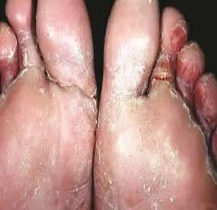
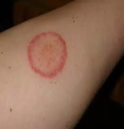
Cutaneous Mycoses Tinea s on most areas of the body are generally called ringworm Some Common Tineas and Location on the Body Tinea corporis (ringworm) Body Tinea capitis (ringworm) Scalp Tinea pedis (athlete s foot) Feet Tinea barbae (barber s itch) Beard Tinea cruris (jock itch) Groin Toenails, fingernails Tinea unguium (onychomycosis)
Epidemiology Direct contact; infected host Contact with Inanimate objects; carrying fungal spores More prevalent in animals, kept confined i.e. in winter season High humidity in the environment; favorable for multiplication Young animals; most susceptible
Disease in animals o Common in: Cattle, horse, pig & many other species o Rare in: Sheep & very rare in goats In cattle: Calves are more susceptible Effected parts: Neck, head & perineum sometimes other parts Lesions: Grey-white crust followed by alopecia In horses Lesions: Axillary girth area & later spread over the trunk & rump, and to the neck, head and limbs Lesions with scabs followed by alopecia appear which vary in size with the species of fungus involved In Pigs Similar lesions on the back and sides In sheep lesions consist of round, bald patches covered with greyish crust, in head In goats Similar lesions are observed generally all over the body
Disease in human Human being can contract ringworm infection from animals In rural areas: 80% of human cases are of animal origin In urban areas:10% of human cases are of animal origin Horses and cattle: Trichophyton spp. infection Dogs: M. canis infection Infected pet animals (Guinea pigs/hamsters/cats/rabbits) to farmers, zoo- keepers, veterinarians & children Children are particularly susceptible M. canis(microsporia): Develop single/ multiple, round, oval or polycyclic reddened scaly lesions, primarily on face, neck, & arms M. distortum: lesions on scalp, neck, face, trunk & other parts of the body T. rubrum: Superficial mycosis of feet, toe-webs, & hands T. mentagrophytes/T. verrucosum/M. gypseum:Hairy skin on the scalp, neck, face and rarely on extremities
Diagnosis By characteristic lesion: Ring like lesion Wood s lamp (also called a black lamp) examination UV Wavelength - 365 nm Used to examine dermatophyte infections of M. canis, M. distortum and M. equinum Fungal elements (spores and hyphae) show greenish fluorescence Wood s lamp
Diagnosis Direct microscopic evaluation: Specimens: Skin scrapings, hair, or nails Preparation of specimens: Wet mount slide Skin/Hair 10% 20% aqueous KOH Dissolves keratin Dissolves keratin visualization of the hyphae & fungal spores
Diagnosis Culture examination: Media: Sabouraud dextrose CC (chloramphenicol/cyclohexamide) Inhibiting the growth of bacteria/ saprophytic fungi Macroscopic colony morphology Identification can be further confirmed by visualizing the Microscopic morphology using either a slide culture /a sticky tape prep stained with lactophenol cotton blue Tinae growth on a Sabouraud dextrose CC agar plate. Lactophenol Cotton blue staining
Treatment Various antifungal treatments can be effective against tineas Depending upon the extent of lesions: local/ systemic treatments Topical application: First removal of the crust and application of weak solution of Iodine, Whitfield's ointment, 10% ammoniated mercury ointment, Ointments containing propionic and undecylenic acids, Hexetidine etc. Natamycin and nanomycin weak solution of iodine Allylamine (Contais terbinafine) Miconazole Clotrimazole Orally: Griseofulvin Systemic application: Inj. of Sodium Iodide
Prevention and control MaintainGeneral hygienic measures Isolation and treatment of infected animals Disinfection of equipment and premises Oral administration of griseofulvin also has protective effect Proper diet with supplementation desirable A vaccine against containing the Trichophyton verrucosum strain contaminated vitamin considered A is ringworm, inactivated
Questions? 1. ________ is a superficial fungal infection found on the head. a) Tinea cruris b) Tinea capitis c) Tinea pedis d) Tinea corporis 2. Sabouraud dextrose agar CC is selective for ________. a) All fungi b) Non-saprophytic fungi c) Bacteria d) Viruses 3. ___________Dermatophytes effect the skin, hair and nails a) Epidermaphyton b) Microsporum c) Trichophyton d) All