Zoonotic disease

Zoonotic disease
Lecturer muna tawfeeq

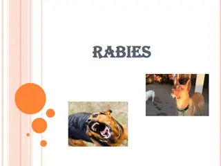
Newcastle disease
Etiology:
Avian paramyxoviruses (APMV ) belong
to the genus
Avulavirus
in the family
Paramyxoviridae
.
Epidemiology
endemic among poultry in much of Asia, Africa
and the Middle East, and some countries in
Central and South America.
transmitted by inhalation or ingestion, and birds
shed these viruses in both feces and respiratory
secretions
Disease in poultry
1.
Respiratory form
, with signs such as coughing, gasping, sneezing
and rales.
2.
neurological form
with signs such as tremors, clonic spasms,
paresis or paralysis of the wings and/or legs, torticollis, circling
and
3.
alimentary form
which include lethargy, inappetence, ruffled
feathers, and conjunctival reddening and edema. Some birds
develop watery, greenish or white diarrhea
4.
Egg laying often declines , Sudden death.
Disease in human
Newcastle disease viruses can infect humans, although
this seems to occur only after exposure to particularly
high concentrations of virus.
can cause conjunctivitis in humans The conjunctivitis
usually resolves rapidly without treatment,.
Mild, self-limiting influenza-like disease.
Influenza
Influenza viruses are RNA orthomyxoviruses
that are categorized into three types (A, B,
and C) based upon antigenic variation, host
specificity, and pathogenicity. Type A
influenza naturally infects humans as well as
a wide range of avian and mammalian
species including swine, horses, ferrets, dogs,
large felids, domestic cats, mink, and seals,
and is further subclassified into subtypes
based upon two viral surface glycoproteins:
hemagglutinin and neuraminidase. Type B
and C influenza viruses infect only humans
Swine influenza
synonym : (hog flu, pig flu)
Etiology
: Orthomyxoviruses
The most common subtypes of influenza virus in swine are H1N1,
H1N2, and H3N2. One relatively stable subtype, H1N1
Swine influenza viruses are usually introduced into a herd by an
infected pig. Morbidity reach up to 100%, but most animals recover
within 3–7 days .
In uncomplicated cases, the case fatality rate ranges from less than
1% to 4%.
in the emergence of new human strains of influenza virus and may
serve as disease reservoirs. Animals can be infected with influenza
strains that then undergo mutational and reassortment events
resulting in the emergence of a novel, pathogenic virus transmissible
to humans. Pigs are well-known examples and are believed to serve
as ‘mixing vessels’ during the adaptation of avian influenza viruses to
human hosts, although their full role in this capacity is still unknown
Transmission
The primary route of virus transmission is through pig to pig
contact via the nasopharyngeal route, the virus is shed in nasal
secretions and disseminated through droplets or aerosols.
•
Pigs are the principal hosts of classic swine influenza virus.
•
(Human infections have been reported, but porcine strains of influenza
A do not appear to easily spread in the human population.
•
However, deaths have occurred in immunocompromised people.)
•
In 2009, a pandemic strain of H1N1 influenza A virus spread globally; it
infected people, swine, and poultry, as well as a small number of dogs,
cats, and other animals.
•
The disease in swine occurs commonly in the midwestern USA (and
occasionally in other states), Mexico, Canada, South America, Europe
(including the UK, Sweden, and Italy), Kenya, China, Japan, Taiwan, and
other parts of eastern Asia.
Disease in animal
fever, lethargy, anorexia, weight loss, nasal
discharge, and labored breathing, Coughing,
sneezing,
Conjunctivitis is a less common clinical sign.
Abortions.
Some strains can circulate in pigs with few or no
clinical signs.
Complications may include secondary bacterial
or viral infections.
Severe, fatal bronchopneumonia.
control
•
strict import controls are the only specific preventive
measures.
•
Good management practices and freedom from
stress, particularly due to crowding and dust, help
reduce losses.
•
Vaccination Commercially available killed vaccines
that contain both H1N2 and H3N2 subtypes appear to
induce a strong protective immune response
.
Equine influenza
Etiology
the common type of influenza A virus that currently
circulates in horse populations is the subtype A2
(H3N8). An earlier subtype,A1 (H7N7) members of the
influenza virus A genus of the family Orthomyxoviridae.
T
r
a
n
s
m
i
s
s
i
o
n
:
•
Highly contagious, Transmission of equine influenza virus
occurs by direct contact, inhalation of aerosols of infected
material, and on fomites. Survival of the virus on clothing
and surfaces, including vehicles used to transport horses
shedding the virus,
•
Once introduced into an area with a susceptible
population, the disease, with an incubation period of only
one to three days, spreads quickly and is capable of
causing explosive outbreaks. Crowding and transportation
are factors that favor the spread of influenza.
Disease in animals
fever (38.5-41°C) after an incubation period of 24-72 hours. Horses
may be depressed, refuse feed, and reluctant to move.
Coughing, serous (clear, runny) nasal discharge,
mild swelling of the submandibular lymph nodes.
Abnormal lung sounds
, severe bronchitis, pneumonia and edema of the legs may develop.
Complications
are usually associated with secondary bacterial
infection, usually
Strep. zooepidemicus,
that results in a
mucopurulent nasal discharge, persistent fever, and markedly
abnormal lung sounds.
Public health risk
There is little risk to public health. In experimental
settings the virus has shown the ability to infect humans,
and a few people in contact with infected horses
developed antibodies to equine influenza viruses, but no
humans exposed to the virus have become ill.
Prevention and Control
•
Vaccination
•
When the disease appears, efforts are placed on movement
control and isolation of infected horses.
•
The virus is easily killed by common disinfectants, so thorough
cleaning and disinfection is part of biosecurity measures in
responding to the disease.
Q fever
Synonyms:
Pneumorickettsiosis, Balkan influenza, coxiellosis,
abattoir fever,Australian Q fever, hiberno-vernal
bronchopneumonia, nine-mile fever, quadrilateral fever
Etiology:
fever is caused by the bacteria
Coxiella burnetii
(
rickettsiae non-
motile, pleomorphic Gram-negative bacteria which replicate
only in host cells.).
What are the risk factors for Q Fever infection?
domestic species - cattle, sheep, goats, dogs, cats, rodents,
rabbits, birds in additions to wild animals.
Transmission:
•
It is transferred by ticks between animals. Ticks are a
temporary host
•
Dogs and cats can be infected by eating infected material -
e.g. placentas and newborn of wildlife, rabbits, rodents.
•
Pregnant animals can be a major source of
Coxiella
- in urine,
birth fluids, placenta,
•
Aerosol transfer to people occurs- via droplets or desiccated
motes - in dust fetuses,
•
milk, and manure Milk transfer to people by drinking
unpasteurized milk.
•
Smoking in an area where infected animals have been
•
Person-to-person transmission are possible but rarely
reported.
Clinical signs in people:
Q fever usually presents in people as a flu-like illness
but can progress to a potentially fatal atypical
pneumonia, hepatitis and endocarditis. Approximately
half of all human infections are asymptomatic. In
pregnant women, infections can cause premature
delivery, abortion and infection of the placenta .Most
cases of acute disease heal spontaneously..
Disease in animals:
The most common sign of infection in animals is abortion during
late pregnancy. However, most animals do not show any signs of
illness with Q fever. In ruminants, after
C. burnetii
has invaded the
bloodstream, it becomes localized in the mammary glands, the
supramammary lymph nodes, and the placenta.
Control and prevention
Keep pregnant livestock separate from other animals.
Burn or bury the remaining reproductive tissues after
abortions or delivery of newborn animals to reduce the
spread of the disease between animals. Take great care
when handling these tissues to avoid your exposure to Q
fever
Psittacosis
Synonym:
Avian chlamydiosis & ornithosis or
parrot fever
in humans
.
Etiology:
Chlamydophila psittaci
Ornithosis is a worldwide zoonosis that is carried
in a latent state in wild and domesticated birds but
becomes active under stressful conditions such as
overcrowding
Disease in birds
There are no specific signs of disease that are characteristic of
psittacosis. Some birds may show general ‘sick’ symptoms – lack of
appetite, weight loss, depression and lethargy, watery green
droppings, discharge from eyes or nose, or even sudden death
Disease in man
fever, chills, frontal headache, and muscle aches, and
later ones are coughing and lung consolidation.
Unchecked, infection can lead to systemic
complications involving the meninges, brain, heart,
or liver. Although most patients respond well to
tetracycline or erythromycin therapy, recovery is
often slow and fraught with relapses.
Control
:
quarantining imported birds
taking precautions in handling birds, feathers, and
droppings Prevent overcrowding and maintain good
sanitation and ventilation where birds are kept.
Do not purchase ill birds or those which were kept in
dirty or crowded conditions.
Before adding a new bird into a group, have it
examined by a veterinarian.
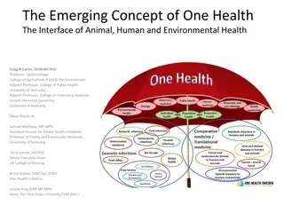
Zoonotic diseases like Newcastle Disease and Influenza pose risks to both animals and humans. Learn about their etiology, symptoms in poultry and humans, and how they can affect different species. Stay informed to prevent transmission and protect health.

Download Presentation

Please find below an Image/Link to download the presentation.
The content on the website is provided AS IS for your information and personal use only. It may not be sold, licensed, or shared on other websites without obtaining consent from the author. Download presentation by click this link. If you encounter any issues during the download, it is possible that the publisher has removed the file from their server.
E N D
Presentation Transcript
Zoonotic disease Lecturer muna tawfeeq
Newcastle disease Etiology: Avian paramyxoviruses (APMV ) belong to the genus Avulavirus in the family Paramyxoviridae. Epidemiology endemic among poultry in much of Asia, Africa and the Middle East, and some countries in Central and South America. transmitted by inhalation or ingestion, and birds shed these viruses in both feces and respiratory secretions
Disease in poultry 1. Respiratory form, with signs such as coughing, gasping, sneezing and rales. 2. neurological form with signs such as tremors, clonic spasms, paresis or paralysis of the wings and/or legs, torticollis, circling and 3. alimentary form which include lethargy, inappetence, ruffled feathers, and conjunctival reddening and edema. Some birds develop watery, greenish or white diarrhea 4. Egg laying often declines , Sudden death.
Disease in human Newcastle disease viruses can infect humans, although this seems to occur only after exposure to particularly high concentrations of virus. can cause conjunctivitis in humans The conjunctivitis usually resolves rapidly without treatment,. Mild, self-limiting influenza-like disease.
Influenza Influenza viruses are RNA orthomyxoviruses that are categorized into three types (A, B, and C) based upon antigenic variation, host specificity, and pathogenicity. Type A influenza naturally infects humans as well as a wide range of avian and mammalian species including swine, horses, ferrets, dogs, large felids, domestic cats, mink, and seals, and is further subclassified into subtypes based upon two viral surface glycoproteins: hemagglutinin and neuraminidase. Type B and C influenza viruses infect only humans
Swine influenza synonym : (hog flu, pig flu) Etiology: Orthomyxoviruses The most common subtypes of influenza virus in swine are H1N1, H1N2, and H3N2. One relatively stable subtype, H1N1 Swine influenza viruses are usually introduced into a herd by an infected pig. Morbidity reach up to 100%, but most animals recover within 3 7 days . In uncomplicated cases, the case fatality rate ranges from less than 1% to 4%. in the emergence of new human strains of influenza virus and may serve as disease reservoirs. Animals can be infected with influenza strains that then undergo mutational and reassortment events resulting in the emergence of a novel, pathogenic virus transmissible to humans. Pigs are well-known examples and are believed to serve as mixing vessels during the adaptation of avian influenza viruses to human hosts, although their full role in this capacity is still unknown
Transmission The primary route of virus transmission is through pig to pig contact via the nasopharyngeal route, the virus is shed in nasal secretions and disseminated through droplets or aerosols.
Pigs are the principal hosts of classic swine influenza virus. (Human infections have been reported, but porcine strains of influenza A do not appear to easily spread in the human population. However, deaths have occurred in immunocompromised people.) In 2009, a pandemic strain of H1N1 influenza A virus spread globally; it infected people, swine, and poultry, as well as a small number of dogs, cats, and other animals. The disease in swine occurs commonly in the midwestern USA (and occasionally in other states), Mexico, Canada, South America, Europe (including the UK, Sweden, and Italy), Kenya, China, Japan, Taiwan, and other parts of eastern Asia.
Disease in animal fever, lethargy, anorexia, weight loss, nasal discharge, and labored breathing, Coughing, sneezing, Conjunctivitis is a less common clinical sign. Abortions. Some strains can circulate in pigs with few or no clinical signs. Complications may include secondary bacterial or viral infections. Severe, fatal bronchopneumonia.
control strict import controls are the only specific preventive measures. Good management practices and freedom from stress, particularly due to crowding and dust, help reduce losses. Vaccination Commercially available killed vaccines that contain both H1N2 and H3N2 subtypes appear to induce a strong protective immune response.
Equine influenza Etiology the common type of influenza A virus that currently circulates in horse populations is the subtype A2 (H3N8). An earlier subtype,A1 (H7N7) members of the influenza virus A genus of the family Orthomyxoviridae.
Transmission: Highly contagious, Transmission of equine influenza virus occurs by direct contact, inhalation of aerosols of infected material, and on fomites. Survival of the virus on clothing and surfaces, including vehicles used to transport horses shedding the virus, Once introduced into an area with a susceptible population, the disease, with an incubation period of only one to three days, spreads quickly and is capable of causing explosive outbreaks. Crowding and transportation are factors that favor the spread of influenza.
Disease in animals fever (38.5-41 C) after an incubation period of 24-72 hours. Horses may be depressed, refuse feed, and reluctant to move. Coughing, serous (clear, runny) nasal discharge, mild swelling of the submandibular lymph nodes. Abnormal lung sounds , severe bronchitis, pneumonia and edema of the legs may develop. Complications are usually associated with secondary bacterial infection, usually Strep. zooepidemicus, that results in a mucopurulent nasal discharge, persistent fever, and markedly abnormal lung sounds.
Public health risk There is little risk to public health. In experimental settings the virus has shown the ability to infect humans, and a few people in contact with infected horses developed antibodies to equine influenza viruses, but no humans exposed to the virus have become ill.
Prevention and Control Vaccination When the disease appears, efforts are placed on movement control and isolation of infected horses. The virus is easily killed by common disinfectants, so thorough cleaning and disinfection is part of biosecurity measures in responding to the disease.
Q fever Synonyms: Pneumorickettsiosis, Balkan influenza, coxiellosis, abattoir fever,Australian Q fever, hiberno-vernal bronchopneumonia, nine-mile fever, quadrilateral fever Etiology: fever is caused by the bacteria Coxiella burnetii ( rickettsiae non- motile, pleomorphic Gram-negative bacteria which replicate only in host cells.). What are the risk factors for Q Fever infection? domestic species - cattle, sheep, goats, dogs, cats, rodents, rabbits, birds in additions to wild animals.
Transmission: It is transferred by ticks between animals. Ticks are a temporary host Dogs and cats can be infected by eating infected material - e.g. placentas and newborn of wildlife, rabbits, rodents. Pregnant animals can be a major source of Coxiella - in urine, birth fluids, placenta, Aerosol transfer to people occurs- via droplets or desiccated motes - in dust fetuses, milk, and manure Milk transfer to people by drinking unpasteurized milk. Smoking in an area where infected animals have been Person-to-person transmission are possible but rarely reported.
Clinical signs in people: Q fever usually presents in people as a flu-like illness but can progress to a potentially fatal atypical pneumonia, hepatitis and endocarditis. Approximately half of all human infections are asymptomatic. In pregnant women, infections can cause premature delivery, abortion and infection of the placenta .Most cases of acute disease heal spontaneously..
Disease in animals: The most common sign of infection in animals is abortion during late pregnancy. However, most animals do not show any signs of illness with Q fever. In ruminants, after C. burnetii has invaded the bloodstream, it becomes localized in the mammary glands, the supramammary lymph nodes, and the placenta.
Control and prevention Keep pregnant livestock separate from other animals. Burn or bury the remaining reproductive tissues after abortions or delivery of newborn animals to reduce the spread of the disease between animals. Take great care when handling these tissues to avoid your exposure to Q fever
Psittacosis Synonym: Avian chlamydiosis & ornithosis or parrot fever in humans. Etiology: Chlamydophila psittaci Ornithosis is a worldwide zoonosis that is carried in a latent state in wild and domesticated birds but becomes active under stressful conditions such as overcrowding
Disease in birds There are no specific signs of disease that are characteristic of psittacosis. Some birds may show general sick symptoms lack of appetite, weight loss, depression and lethargy, watery green droppings, discharge from eyes or nose, or even sudden death
Disease in man fever, chills, frontal headache, and muscle aches, and later ones are coughing and lung consolidation. Unchecked, infection can lead to systemic complications involving the meninges, brain, heart, or liver. Although most patients respond well to tetracycline or erythromycin therapy, recovery is often slow and fraught with relapses.
Control: quarantining imported birds taking precautions in handling birds, feathers, and droppings Prevent overcrowding and maintain good sanitation and ventilation where birds are kept. Do not purchase ill birds or those which were kept in dirty or crowded conditions. Before adding a new bird into a group, have it examined by a veterinarian.