Rabies: A Comprehensive Overview of the Disease
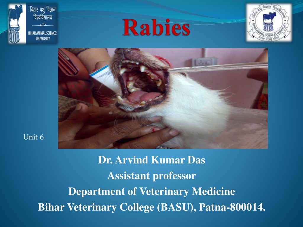


Dr. Arvind Kumar Das
Assistant professor
Department of Veterinary Medicine
Bihar Veterinary College (BASU), Patna-800014.
Unit 6


Introduction
Synonym:
Hydrophobia, Lyssa, Mad dog disease,
Rage, Tollwut
The disease occurs as paralytic rabies, known as Derriengue
Acute viral disease causes acute encephalitis.
Rabies occurs in all warm blooded animals including human
Rabies is one of the most deadly zoonoses. Each year, it kills nearly 60,000
people worldwide, mostly children in developing countries
1 person die from rabies every 10 min in world.
Rabies 100% preventable.
>95% of human deaths caused by dog-mediated rabies


ETIOLOGY
Classification
Family
Rhabdoviridae
Genus
Lyssavirus
(including
Rabies virus)
Many viruses with
broad
host ranges
Vertebrates
Invertebrates
Plants
Features
Bullet-shaped with one flat
end
(75 x 180 nm)
,
Enveloped
having 10 nm
spike like glycoprotein
peplomers covering the
surface.
Image, Curtsey with google
ETIOLOGY
There is a leader sequence (LDR) of approximately 50 nucleotides, followed by
N, P, M, G, and L genes that encode 5 proteins. i.e. N-Nucleoprotein, P-
Phosphoprotein; M-Matrix protein; G-Glycoprotein; L-large polymerase or
transcriptase protein.
In the nature, 7 distinct genotypes of rabies virus circulating
Genotype 1 - classical rabies virus (CRV)
strains, including field and
laboratory – fixed strains
Rabies related viruses (RRVs) –
Genotype 2 - Lagos bat virus
Genotype 3 - Mokola virus
Genotype 4 - Duvenhage virus
Genotypes 5 and 6 - European bat Lyssavirus 1 and 2
Genotype 7 - Australian bat Lyssavirus (ABLV)
Image, Curtsey with google
Features of Virus
Rhabdovirus – lipid containing, single stranded
non segmented nucleotide RNA of
approximately 12 kb.
Sensitive to lipid solvents (Soap, ether, chloroform, acetone)
45-75% ethanol, iodine preparation and quaternary ammonium compounds
Relatively stable at pH between 5 & 10
Sensitive to pasteurization and UV light
The nucleic acid readily inactivated by
β
-propiolactone
Virus may persist up to 18 days at inoculation site
Virus travels along the nerves centripetally at a rate of roughly 3mm/hr
Once disease is established it is nearly always fatal
The virus can not live outside its host for more than a couple of seconds but has been found in
animals as long as 48 hrs after death
Epidemiology
Widely distributed throughout the world .
In USA, Canada and Western Europe (rabies in dogs is controlled by
vaccination), rabies is endemic in wild life – Skunks, Foxes, Racoons,
bats
Cats are the most affected animal in USA
In Asia, Latin America and Africa, rabies is endemic in dogs and wild life
Vampire bats are important in the spread of rabies in cattle in South
America
With the exception of Antarctica, rabies is endemic on all continents.
Almost 95% of cases are reported in Asia and Africa.
Rabies Transmission
Hosts:
All
warm-blooded animals
can be infected with varying susceptibility
High - wolves, coyotes, foxes, dogs
Intermediate - skunks, raccoons, bats
Low - opossums
Reptiles due to being cold blooded and birds don’t get rabies
Reptiles due to being cold blooded and birds don’t get rabies
Vampire bats can transmit virus for months
Insectivorous and frugivorous bats may also harbour and transmit
virus
Young animals are more susceptible than adults
Domestic animals most likely to be diagnosed with rabies :
Dog > Cow > Horse/mule > sheep/goat
Rodents such as mice , rats, squirrels and chipmunks may be
exposed to rabies virus but are rare source to transmit.
Virus occurs in
saliva
, nervous system, urine, lymph, milk
Image, Curtsey with google
Transmission
The virus is typically transmitted
by bites
by bites
wound
wound
or contact of infected saliva with
or contact of infected saliva with
mucous membranes
mucous membranes
(eyes, mouth, etc)
The virus cannot infiltrate intact skin
Saliva becomes non-infectious when it dries
People have been infected by aerosol in bat caves
Requires several weeks for infection to become apparent
Replication in
muscle
in myocytes
and connective tissues
at site of
inoculation
and shed into extracellular spaces.
Enters
peripheral nervous system
at
neuromuscular junctions
Spreads up the peripheral nerves to the
central nervous system
Entering peripheral or cranial nerves, progress to axons then enter spinal or
cranial ganglia
Virus replicates rapidly in ganglion and spread throughout the CNS
Virus spread centrifugally by nerves throughout the body including the
salivary glands
Encephalitis
Virus grows to high titers in the
salivary glands
Negri bodies
appear in neuron cell bodies
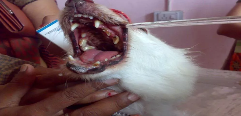
Symptoms
Dumb or the furious form
In dumb form the animals falls into a stupor and has a peculiar staring
expression
Paralysis of the mastication muscles
In furious form animal goes into rages, biting and slashing at any moving
objects or even inanimate objects
Champing of jaws, excessive salivation
Paralysis may follow either the furious or dumb form
Death occurs within 10 days of the 1st symptom
Symptoms
Animals (domestic)
Fearfulness
Aggression
Excessive drooling
Difficulty swallowing
Staggering
Seizures
Depression
Self-mutilation
Light sensitivity
Animals (wildlife)
Any of above
Unusual behavior
Nocturnal animal active
during day
Lose fear of humans
CLINICAL SIGNS
Dog:
The incubation period in natural outbreak of
dog rabies averages from
3-8 weeks
. But it may be
as short as 10 days
to as long as one year.
The clinical features divided into three phases.
1)
Prodromal phase
: No externally visible signs are
seen
2)
Furious form
:
Mad dog syndrome,
Animal bites,
attack and show signs of aggression and madness,
irritable, restless, nervous, deprived appetite, and
often dangerous as it loses all fear of humans and
bites at anything. By about 3rd day after the onset of
illness the dog enters the furious stage which lasts
for 3-7 days.
3)
Paralytic form
:
Early paralysis of throat muscles.
Animals shows head drooling, dropped lower jaw
and salivary discharge,
lasts in1-3 days.
Animals show only vague CNS signs, which
intensify rapidly.
Image, Curtsey with google
CLINICAL SIGNS
C
attle
P
aralytic form
:
K
nuckling of the hind fetlocks, sagging and swaying of the hindquarters while
walking, often deviation or flaccidity of the tail to one side, are common signs.
Decreased sensation over the hindquarters
D
rooling of saliva, tenesmus, pumping of anus and followed by recumbency
in later stages.
F
urious
form:
A
nimal alert, hypersensitive, violently attack, loud and coarse bellowing,
sexual excitement and collapses suddenly.
Cattle are very restless, excited and aggressive with salivation, abdominal
pain, diarrhoea and rectal straining. Paralysis of hind quarters occurs followed
by death in 3-6 days after the first signs of illness.
Sheep
and Goats
Clinically similar to cattle. Sexual excitement, violent attack, vigorous
wool pulling, sudden falling and salivation are characteristic.
Goats are commonly aggressive, and continuous bleating is common.
Horse
Muzzle tremors and pharyngeal paresis are common.
In addition to these abnormal postures, kicking, biting, colic, sudden
onset of lameness in one limb followed by recumbency, high stepping
gait, blindness, paddling, convulsions and terminally paralysis.
Pigs
Tendency to attack, twitching of the nose, rapid chewing movements,
excessive salivation, walk backward and terminally paralysis.
Gross pathology
In majority of cases , CNS lesions are mild .
Leptomeningeal congestion and mild oedema.
The meninges may be cloudy .
In some case uneatable objects like straw, wood, leather pieces, rubber, etc may
be found in the stomach of carnivores.
Histologic features
Typical changes are seen only when rabid animal is allowed to die on its own
The CNS lesions includes:
•
Perivascular cuffing with lymphocytes.
•
Vascular congestion and even perivascular haemorrhages
LESIONS
(A)GROSS:
no lesion.
(B)MICROSCOPIC :
1.
Necrosis of neuron with specific cytoplasmic inclusion body in
affected nerve cell.
2.
Encephalitis:It is characterised by 1)perivascular cuffing
2)neuronophagic nodules 3)Distraction of neurons.
3.
Change in 1)Brain stem 2)hippocampus 3)proliferation of glial
cell known as babes nodules
.
Sample to be collected
Live animals :
Saliva, corneal/ Conjunctival smear.
Dead animals
The whole carcass or the severed head of the animal suspected to
have died of rabies.
Alternatively, the brain may be removed carefully and two
portions, one in 50% glycerol saline and the other in Zenker’s
fixative, sent for biological test and microscopy, respectively.
The brain tissue selected should include portions of hippocampus,
cerebellum, cerebral cortex, and placed in 50% glycerol saline to
preserve the virus.
No refrigeration is required.
DIAGNOSIS
It based on clinical sign , history ,symptoms,
1.
FAT-fluorescent antibody test.
(
This test is recommended by both WHO and OIE
)
2
.
Seller’s stain
– negri body seen in hippocampus.
3. Histological
search of negri body.
4. Serological test
of –
Immunoperoxidase test
ELISA
Rapid fluorescence focus inhibition test (RFFIT)
Virus neutralization test
Rapid rabies enzyme immuno diagnosis test (RREID) based on the use of
antinucleocapsid lgG are highly useful
RT-PCR amplification technique is 1000 times more sensitive than other
tests.
The virus can be detected in saliva and skin biopsy samples by RT-PCR..
TREATMENT
1.
N
o specific treatment for rabies.
2.
Dogs usually die after showing clinical signs.
3.
The site of bite should be washed with water or soa
p.
Sodium
bicarbonate or caustic soda may be used.
4.
W
ound is treated with 2% quaternary ammonium compound or
tincture iodine or 40-70% alcohol.
5.
Wound may also be cauterized with carbolic acid or nitric acid.
6.
Antirabies serum may be applied topically or infiltrated around the
wound.
7.
Treatment with antirabies serum is also effective.
D
ose
recommended is 40 I.U. per kg body weight.
8.
It should be given as early as possible after exposure and in any case
within five days, after which it may not be beneficial.
Vaccine types:
live or inactivated, according to the strain
of rabies virus used
More recently a
third generation of live veterinary
rabies vaccine
has been developed using recombinant
technology. These vaccines are used
either parentrally or
orally
.
Vaccination of Animals
Vaccination of Animals
Vaccine types:
live or inactivated, according to the strain of rabies virus
used
More recently a
third generation of live veterinary rabies vaccine
has been developed using recombinant technology. These vaccines are
used
either parentrally or orally
.
Available for Humans,
dogs, cats, ferrets
dogs, cats, ferrets
, cattle and horses.
Oral rabies vaccine is offered to wildlife
Pre exposure vaccination
- Pre exposure vaccination schedule for
dogs starts at 12th week age – first dose,
28 days later – second dose,
one year later- third dose,
three years later- fourth dose.
Post exposure immunization
: 0, 3, 7, 14, 21, 28 days after exposure.
1.
Raksha rab –Indian immunological.
2.
Rabigen :serum institute.
Rabigen
Composition
Virus titre per dose(1ml)
Inactivated VP12 Rabies virus strain > 1.0 I.U
Aluminum hydroxide gel (as adjuvant) 10 % v/v
Thiomersal IP (as preservative) 0.01 % w/v
Indication-
Active immunization of Dogs, Cats,
Cattle and Horses, and in principle
all mammal against Rabies.
As a Post Exposure Prophylaxis
Image, Curtsey with google
Control
(as per the WHO recommendations)
Notification of suspected cases, and destruction of dogs
with clinical signs and dogs bitten by a suspected rabid
animal.
Compulsory immunization of dogs
Sterilization and vaccination of stray dogs
Epidemiological Surveillance
Education of Public
Development of cost effective vaccine
World Rabies Day is a worldwide campaign against rabies. One person in
the world dies from rabies every 10 MINUTES, equaling 60,000 each
year!
The disease is 100% fatal, but 100% preventable.
Image, Curtsey with google
Thank you
D
u
n
n
e
-
C
I
D
P
J
a
n
u
a
r
y
1
8
,
2
0
0
5
Rabies, also known as hydrophobia or mad dog disease, is a deadly viral infection that affects warm-blooded animals, including humans. This article delves into the etiology, features of the virus, and epidemiology of rabies, emphasizing its prevention and control measures to combat this fatal zoonotic disease.

Download Presentation

Please find below an Image/Link to download the presentation.
The content on the website is provided AS IS for your information and personal use only. It may not be sold, licensed, or shared on other websites without obtaining consent from the author.If you encounter any issues during the download, it is possible that the publisher has removed the file from their server.
You are allowed to download the files provided on this website for personal or commercial use, subject to the condition that they are used lawfully. All files are the property of their respective owners.
The content on the website is provided AS IS for your information and personal use only. It may not be sold, licensed, or shared on other websites without obtaining consent from the author.
E N D
Presentation Transcript
Unit 6 Dr. Arvind Kumar Das Assistant professor Department of Veterinary Medicine Bihar Veterinary College (BASU), Patna-800014.
Introduction Synonym: Hydrophobia, Lyssa, Mad dog disease, Rage, Tollwut The disease occurs as paralytic rabies, known as Derriengue Acute viral disease causes acute encephalitis. Rabies occurs in all warm blooded animals including human Rabies is one of the most deadly zoonoses. Each year, it kills nearly 60,000 people worldwide, mostly children in developing countries 1 person die from rabies every 10 min in world. Rabies 100% preventable. >95% of human deaths caused by dog-mediated rabies
ETIOLOGY Classification Family Rhabdoviridae Genus Lyssavirus (including Rabies virus) Many viruses with broad host ranges Vertebrates Invertebrates Plants Features Bullet-shaped with one flat end (75 x 180 nm), Enveloped having 10 nm spike like glycoprotein peplomers covering the surface. Image, Curtsey with google
ETIOLOGY There is a leader sequence (LDR) of approximately 50 nucleotides, followed by N, P, M, G, and L genes that encode 5 proteins. i.e. N-Nucleoprotein, P- Phosphoprotein; M-Matrix protein; G-Glycoprotein; L-large polymerase or transcriptase protein. In the nature, 7 distinct genotypes of rabies virus circulating Genotype 1 - classical rabies virus (CRV) strains, including field and laboratory fixed strains Rabies related viruses (RRVs) Genotype 2 - Lagos bat virus Genotype 3 - Mokola virus Genotype 4 - Duvenhage virus Genotypes 5 and 6 - European bat Lyssavirus 1 and 2 Genotype 7 - Australian bat Lyssavirus (ABLV) Image, Curtsey with google
Features of Virus Rhabdovirus lipid containing, single strandednon segmented nucleotide RNA of approximately 12 kb. Sensitive to lipid solvents (Soap, ether, chloroform, acetone) 45-75% ethanol, iodine preparation and quaternary ammonium compounds Relatively stable at pH between 5 & 10 Sensitive to pasteurization and UV light The nucleic acid readily inactivated by -propiolactone Virus may persist up to 18 days at inoculation site Virus travels along the nerves centripetally at a rate of roughly 3mm/hr Once disease is established it is nearly always fatal The virus can not live outside its host for more than a couple of seconds but has been found in animals as long as 48 hrs after death
Epidemiology Widely distributed throughout the world . In USA, Canada and Western Europe (rabies in dogs is controlled by vaccination), rabies is endemic in wild life Skunks, Foxes, Racoons, bats Cats are the most affected animal in USA In Asia, Latin America and Africa, rabies is endemic in dogs and wild life Vampire bats are important in the spread of rabies in cattle in South America With the exception of Antarctica, rabies is endemic on all continents. Almost 95% of cases are reported in Asia and Africa.
Rabies Transmission Hosts: All warm-blooded animals can be infected with varying susceptibility High - wolves, coyotes, foxes, dogs Intermediate - skunks, raccoons, bats Low - opossums Reptiles due to being cold blooded and birds don t get rabies Vampire bats can transmit virus for months Insectivorous and frugivorous bats may also harbour and transmit virus Young animals are more susceptible than adults Domestic animals most likely to be diagnosed with rabies : Dog > Cow > Horse/mule > sheep/goat Rodents such as mice , rats, squirrels and chipmunks may be exposed to rabies virus but are rare source to transmit. Virus occurs in saliva, nervous system, urine, lymph, milk Image, Curtsey with google
Transmission The virus is typically transmitted by bites wound or contact of infected saliva with mucous membranes (eyes, mouth, etc) The virus cannot infiltrate intact skin Saliva becomes non-infectious when it dries People have been infected by aerosol in bat caves Requires several weeks for infection to become apparent Replication in muscle in myocytes and connective tissues at site of inoculation and shed into extracellular spaces. Enters peripheral nervous system at neuromuscular junctions Spreads up the peripheral nerves to the central nervous system Entering peripheral or cranial nerves, progress to axons then enter spinal or cranial ganglia Virus replicates rapidly in ganglion and spread throughout the CNS Virus spread centrifugally by nerves throughout the body including the salivary glands Encephalitis Virus grows to high titers in the salivary glands Negri bodies appear in neuron cell bodies
Symptoms Dumb or the furious form In dumb form the animals falls into a stupor and has a peculiar staring expression Paralysis of the mastication muscles In furious form animal goes into rages, biting and slashing at any moving objects or even inanimate objects Champing of jaws, excessive salivation Paralysis may follow either the furious or dumb form Death occurs within 10 days of the 1st symptom
Symptoms Animals (domestic) Fearfulness Aggression Excessive drooling Difficulty swallowing Staggering Seizures Depression Self-mutilation Light sensitivity Animals (wildlife) Any of above Unusual behavior Nocturnal animal active during day Lose fear of humans
CLINICAL SIGNS Dog: The incubation period in natural outbreak of dog rabies averages from 3-8 weeks. But it may be as short as 10 days to as long as one year. The clinical features divided into three phases. 1) Prodromal phase: No externally visible signs are seen 2) Furious form: Mad dog syndrome, Animal bites, attack and show signs of aggression and madness, irritable, restless, nervous, deprived appetite, and often dangerous as it loses all fear of humans and bites at anything. By about 3rd day after the onset of illness the dog enters the furious stage which lasts for 3-7 days. 3) Paralytic form: Early paralysis of throat muscles. Animals shows head drooling, dropped lower jaw and salivary discharge, lasts in1-3 days. Animals show only vague CNS signs, which intensify rapidly. Image, Curtsey with google
CLINICAL SIGNS Cattle Paralytic form: Knuckling of the hind fetlocks, sagging and swaying of the hindquarters while walking, often deviation or flaccidity of the tail to one side, are common signs. Decreased sensation over the hindquarters Drooling of saliva, tenesmus, pumping of anus and followed by recumbency in later stages. Furious form: Animal alert, hypersensitive, violently attack, loud and coarse bellowing, sexual excitement and collapses suddenly. Cattle are very restless, excited and aggressive with salivation, abdominal pain, diarrhoea and rectal straining. Paralysis of hind quarters occurs followed by death in 3-6 days after the first signs of illness.
Sheep and Goats Clinically similar to cattle. Sexual excitement, violent attack, vigorous wool pulling, sudden falling and salivation are characteristic. Goats are commonly aggressive, and continuous bleating is common. Horse Muzzle tremors and pharyngeal paresis are common. In addition to these abnormal postures, kicking, biting, colic, sudden onset of lameness in one limb followed by recumbency, high stepping gait, blindness, paddling, convulsions and terminally paralysis. Pigs Tendency to attack, twitching of the nose, rapid chewing movements, excessive salivation, walk backward and terminally paralysis.
Gross pathology In majority of cases , CNS lesions are mild . Leptomeningeal congestion and mild oedema. The meninges may be cloudy . In some case uneatable objects like straw, wood, leather pieces, rubber, etc may be found in the stomach of carnivores. Histologic features Typical changes are seen only when rabid animal is allowed to die on its own The CNS lesions includes: Perivascular cuffing with lymphocytes. Vascular congestion and even perivascular haemorrhages
LESIONS (A)GROSS: no lesion. (B)MICROSCOPIC : 1.Necrosis of neuron with specific cytoplasmic inclusion body in affected nerve cell. 2.Encephalitis:It is characterised by 1)perivascular cuffing 2)neuronophagic nodules 3)Distraction of neurons. 3.Change in 1)Brain stem 2)hippocampus 3)proliferation of glial cell known as babes nodules.
Sample to be collected Live animals : Saliva, corneal/ Conjunctival smear. Dead animals The whole carcass or the severed head of the animal suspected to have died of rabies. Alternatively, the brain may be removed carefully and two portions, one in 50% glycerol saline and the other in Zenker s fixative, sent for biological test and microscopy, respectively. The brain tissue selected should include portions of hippocampus, cerebellum, cerebral cortex, and placed in 50% glycerol saline to preserve the virus. No refrigeration is required.
DIAGNOSIS It based on clinical sign , history ,symptoms, 1. FAT-fluorescent antibody test. (This test is recommended by both WHO and OIE) 2. Seller s stain negri body seen in hippocampus. 3. Histological search of negri body. 4. Serological test of Immunoperoxidase test ELISA Rapid fluorescence focus inhibition test (RFFIT) Virus neutralization test Rapid rabies enzyme immuno diagnosis test (RREID) based on the use of antinucleocapsid lgG are highly useful RT-PCR amplification technique is 1000 times more sensitive than other tests. The virus can be detected in saliva and skin biopsy samples by RT-PCR..
TREATMENT 1.No specific treatment for rabies. 2.Dogs usually die after showing clinical signs. 3.The site of bite should be washed with water or soap. Sodium bicarbonate or caustic soda may be used. 4.Wound is treated with 2% quaternary ammonium compound or tincture iodine or 40-70% alcohol. 5.Wound may also be cauterized with carbolic acid or nitric acid. 6.Antirabies serum may be applied topically or infiltrated around the wound. 7.Treatment with antirabies serum is also effective. Dose recommended is 40 I.U. per kg body weight. 8.It should be given as early as possible after exposure and in any case within five days, after which it may not be beneficial.
Vaccination of Animals Vaccine types: live or inactivated, according to the strain of rabies virus used More recently a third generation of live veterinary rabies vaccine has been developed using recombinant technology. These vaccines are used either parentrally or orally.
Vaccination of Animals Vaccine types: live or inactivated, according to the strain of rabies virus used More recently a third generation of live veterinary rabies vaccine has been developed using recombinant technology. These vaccines are used either parentrally or orally. Available for Humans, dogs, cats, ferrets, cattle and horses. Oral rabies vaccine is offered to wildlife Pre exposure vaccination- Pre exposure vaccination schedule for dogs starts at 12th week age first dose, 28 days later second dose, one year later- third dose, three years later- fourth dose. Post exposure immunization : 0, 3, 7, 14, 21, 28 days after exposure. 1. Raksha rab Indian immunological. 2. Rabigen :serum institute.
Rabigen Composition Virus titre per dose(1ml) Inactivated VP12 Rabies virus strain > 1.0 I.U Aluminum hydroxide gel (as adjuvant) 10 % v/v Thiomersal IP (as preservative) 0.01 % w/v Indication- Active immunization of Dogs, Cats, Cattle and Horses, and in principle all mammal against Rabies. As a Post Exposure Prophylaxis Image, Curtsey with google
Control (as per the WHO recommendations) Notification of suspected cases, and destruction of dogs with clinical signs and dogs bitten by a suspected rabid animal. Compulsory immunization of dogs Sterilization and vaccination of stray dogs Epidemiological Surveillance Education of Public Development of cost effective vaccine
World Rabies Day is a worldwide campaign against rabies. One person in the world dies from rabies every 10 MINUTES, equaling 60,000 each year! The disease is 100% fatal, but 100% preventable. Image, Curtsey with google