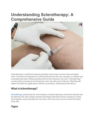
Varicose Veins: Anatomy, Epidemiology, and Management
Varicose veins, a common venous disorder, affect a significant percentage of the population, particularly women. Learn about the anatomy, epidemiology, and pathophysiology of varicose veins. Explore how to assess and investigate patients with varicose veins, along with the management and treatment outcomes. Discover the types of varicose veins and other venous disorders, along with the underlying pathophysiology and clinical presentation. Educate yourself on this condition to better understand its impact on patients and available treatment options.

Download Presentation

Please find below an Image/Link to download the presentation.
The content on the website is provided AS IS for your information and personal use only. It may not be sold, licensed, or shared on other websites without obtaining consent from the author.If you encounter any issues during the download, it is possible that the publisher has removed the file from their server.
You are allowed to download the files provided on this website for personal or commercial use, subject to the condition that they are used lawfully. All files are the property of their respective owners.
The content on the website is provided AS IS for your information and personal use only. It may not be sold, licensed, or shared on other websites without obtaining consent from the author.
E N D
Presentation Transcript
Miss Julie Reid Consultant Vascular Surgeon
Objectives Know about the anatomy, epidemiology and pathophysiology of varicose veins Know how to assess a patients with varicose veins Know how to investigate a patient with varicose veins Know about the management and outcomes of treatment for varicose veins
Epidemiology Venous disease 80% 35% trunk varices Women > men, 2:1 Prevalence increases with age Positive family history Obesity Occupation
Types of varicose vein Primary trunk varicose veins Secondary trunk varicose veins Reticular veins Spider veins/venous flares Venous malformations
Types of varicose vein Primary trunk varicose veins Secondary trunk varicose veins Reticular veins Spider veins/venous flares Venous malformations
Other venous disorders Primary trunk varicose veins Secondary trunk varicose veins Reticular veins Spider veins/venous flares Venous malformations
Other venous disorders Primary trunk varicose veins Secondary trunk varicose veins Reticular veins Spider veins/venous flares Venous malformations
Other venous disorders Primary trunk varicose veins Secondary trunk varicose veins Reticular veins Spider veins/venous flares Venous malformations
Pathophysiology Primary valve failure Primary degenerative changes in the valve leaflets Secondary valve failure Developmental weakness in vein wall and secondary vein widening and valve incompetence
Presentation Uncomplicated veins Cosmesis Symptoms For reassurance
Presentation Complications of varicose veins Superficial thrombophlebitis Lipodermatosclerosis and pigmentation Varicose eczema Ulceration Haemorrhage
Presentation Complications of varicose veins Superficial thrombophlebitis Lipodermatosclerosis and pigmentation Varicose eczema Ulceration Haemorrhage
Presentation Complications of varicose veins Superficial thrombophlebitis Lipodermatosclerosis and pigmentation Varicose eczema Ulceration Haemorrhage
Presentation Complications of varicose veins Superficial thrombophlebitis Lipodermatosclerosis and pigmentation Varicose eczema Ulceration Haemorrhage
Presentation Complications of varicose veins Superficial thrombophlebitis Lipodermatosclerosis and pigmentation Varicose eczema Ulceration Haemorrhage
Clinical Assessment History Are the symptoms due to venous disease Is the presentation complicated or uncomplicated Is there any other significant pathology such as previous DVT or arterial disease Any other health problems Previous varicose vein surgery?
Clinical Assessment General examination Examination standing and adequately exposed (leg and lower abdomen) Inspection Distribution of varicose veins (GSV/SSV) Signs of chronic venous insufficiency Scars
Clinical Assessment Special tests Trendelenberg s test Perthes Test
Clinical Assessment Special tests Trendelenberg s test Perthes Test
Clinical Assessment Hand Held Doppler
Clinical Assessment DuplexUltrasonography Vascular surgeons should have the necessary skills to perform Duplex US in the out-patient clinic Should ideally be performed in all patients Determines site and extent of reflux Determines suitability of vein for endovenoustreatment Essential In cases of chronic venous insufficiency Previous DVT or suspected deep venous insufficiency Recurrent varicose veins Suspect sapheno-popliteal incompetence
Treatment Reassurance Compression hosiery Foam sclerotherapy Endovenous ablation Conventional surgery
Treatment Compression Hosiery Graduated compression stockings Class I - Class II - Class III - 14 17mmHg 18 24mmHg 25 35mmHg May relieve symptoms Useful where there is uncertainty about symptoms or patient unfit for other treatment Prevents skin deterioration and recurrent ulceration in CVI
Treatment Foam Sclerotherapy Foam (sodium tetradecyl sulphate mixed with air in ratio 1:4 Causes phlebitis and vein occlusion Injection of foam into truncal vein or varicose vein GSV/SSV cannulated under US guidance Compression hosiery The late results of foam are unknown but occlusion of a truncal vein occurs in about 80% after 1 year
Treatment Endovenous ablation Radiofrequency Ablation delivery of thermal energy from an electric current to the vein wall Generates temperatures of 85 120 C Endovenous Laser Ablation Delivery of light energy at 810 1470 nm Thermal damage to vein wall Both damage the vein wall causing subsequent thrombosis
Treatment Endovenous ablation Outpatient setting Cannulate vein GSV/SSV US guidance Pass guidewire/catheter Position catheter Tumescent anaesthesia Catheter withdrawn
Treatment Endovenous ablation Outpatient setting Cannulate vein GSV/SSV US guidance Pass guidewire/catheter Position catheter Tumescent anaesthesia Catheter withdrawn
Treatment Endovenous ablation Outpatient setting Cannulate vein GSV/SSV US guidance Pass guidewire/catheter Position catheter Tumescent anaesthesia Catheter withdrawn
Treatment Endovenous ablation Outpatient setting Cannulate vein GSV/SSV US guidance Pass guidewire/catheter Position catheter Tumescent anaesthesia Catheter withdrawn
Treatment Endovenous ablation Outpatient setting Cannulate vein GSV/SSV US guidance Pass guidewire/catheter Position catheter Tumescent anaesthesia Catheter withdrawn
Treatment Endovenous ablation Compression hoisery for 6 weeks Analgesia NSAID Review 6 weeks 20% residual varicosities Foam sclerotherapy
Treatment Conventional Surgery Gold standard Saphenofemoralligation and stripping GSV Saphenopopliteal ligation Phlebectomies Complications Bruising, bleeding Wound infection Nerve injury DVT