Large Submucous Pedunculated Myomatous Polyp with Chronic Partial Uterine Inversion

undefined
 
A
 
c
a
s
e
 
o
f
 
l
a
r
g
e
 
s
u
b
m
u
c
o
u
s
p
e
d
u
n
c
u
l
a
t
e
d
 
m
y
o
m
a
t
o
u
s
 
p
o
l
y
p
 
w
i
t
h
c
h
r
o
n
i
c
 
p
a
r
t
i
a
l
 
u
t
e
r
i
n
e
 
i
n
v
e
r
s
i
o
n
 
 
B
y
 
D
r
 
A
l
i
s
h
a
 
D
a
s
 
U
n
d
e
r
 
g
u
i
d
a
n
c
e
 
o
f
 
D
r
 
H
e
m
a
n
t
D
e
s
h
p
a
n
d
e
,
 
D
e
p
t
.
 
o
f
 
O
b
s
t
e
t
r
i
c
s
 
&
 
G
y
n
a
e
c
o
l
o
g
y
,
D
r
 
D
.
 
Y
.
 
P
a
t
i
l
 
M
e
d
i
c
a
l
 
C
o
l
l
e
g
e
 
a
n
d
R
e
s
e
a
r
c
h
 
C
e
n
t
r
e
,
 
P
i
m
p
r
i
,
 
P
u
n
e
.
 
HISTORY OF PRESENTING COMPLAINTS
 
A 41 year old female Para 1 living 1 with previous LSCS came on 16
th
 August
2019 with complaint of
Bleeding P/V since 1 day
Foul smelling discharge P/V since 1 month
Pain in abdomen and genital region since 3 months
Menorrhagia associated with dysmenorrhea since 1 year
No H/O –
white discharge
Bladder bowel complaints , urinary frequency or urgency
Fever , nausea , vomitting
Mass coming out PV
 
Menstrual history
Menarche attained at 14 years of age
Last menstrual period – 8
th
 August (1 week prior)
Bled for 5 days.  3 pads soaked per day associated with passage of clots and
dysmenorrhea
Past menstrual history since 1 year: 5-7 days / 28-32 days / increased menstrual
flow . Associated with clot passage and dysmenorrhoea


Past menstrual history prior to 1 year: 4 to 5 days / 28-32 days / normal flow (2 to
3 pads per day).
 
O
b
s
t
e
t
r
i
c
 
h
i
s
t
o
r
y
Married life 16 years
P1L1 – 14 year old female child . LSCS done in view of non progress of labour
P
u
e
r
p
e
r
a
l
 
p
e
r
i
o
d
 
u
n
e
v
e
n
t
f
u
l
 
P
A
S
T
 
M
E
D
I
C
A
L
 
A
N
D
 
S
U
R
G
I
C
A
L
 
H
I
S
T
O
R
Y
:
No history of Diabetes mellitus , hypertension, tuberculosis , thyroid disorder.
No history of intake of OC pills routinely
No major surgery in the past and no prior blood transfusion.
 
Personal history
Normal sleep and bowel bladder habits
No history of smoking or alcohol consumption


GENERAL PHYSICAL EXAMINATION
 
Moderately built and nourished
Comfortable and co-operative at the time of examination
Height-165 cm
Weight- 51kg
BMI – 18.7 kg/m
²
Skin – normal in texture and colour
Pulse – 76 bpm regular in rate, rhythm and normal volume measured in left
radial artery. No apex pulse deficit and radio radial delay
BP – 110/70 mmHg taken in left brachial artery in supine position
Breasts  - clinically normal
Thyroid - clinically normal
Spine  - clinically normal
 
No pallor, icterus, edema, cyanosis, lymphadenopathy, clubbing
 
CVS – S1 S2 normal . No murmurs
RS air entry bilaterally equal . No crepts , rales , rhonchi or abnormal breath
sounds
 
No cervical lymphadenopathy
 
 
A
B
D
O
M
I
N
A
L
 
E
X
A
M
I
N
A
T
I
O
N
Inspection:
Umbilicus central and inverted
All quadrants move equally with respiration
Scar of previous LSCS noted on lower abdomen. Well healed
No striae , dilated veins, sinuses
 
Palpation 
:
Soft , scaphoid
No tenderness guarding and rigidity
No organomegaly (hepatomegaly and splenomegaly)
Uterus just palpable in suprapubic region (12 to 14 weeks size)
Consistency firm , no tenderness , mobility present from side to side(no fixity
to skin and underlying structures) and lower pole could not be reached
 
GYNAECOLOGICAL EXAMINATION
 
Per speculum examination : large bosselated heterogenous mass
noted of size about 8x8 cm stretching the vaginal walls . Foul smelling
necrotic areas with hemorrhage and ulceration suggestive of infection
noted on the mass. No active bleeding noted
Cervix not visualized as vision was obscured by the lesion
 
Per vaginal examination :
Large mass noted protruding through cervix and extending into the
vagina
Fornices obliterated due to stretching effect of the mass
Cervix was not palpable as the mass was impacting the vaginal walls
Uterus size not assessed as bimanual examination was not possible
due to mass
 
PROVISIONAL DIFFERENTIAL CLINICAL DIAGNOSIS
? Submucous pedunculated myomatous polyp
? Hypervascular endometrial polyp
? Cervical polypoidal growth
 
I
N
V
E
S
T
I
G
A
T
I
O
N
S
 
 
H
e
m
o
g
r
a
m
,
 
b
l
o
o
d
 
s
u
g
a
r
 
l
e
v
e
l
s
 
a
n
d
 
l
i
v
e
r
 
a
n
d
 
r
e
n
a
l
f
u
n
c
t
i
o
n
 
t
e
s
t
 
w
i
t
h
 
b
l
o
o
d
 
e
l
e
c
t
r
o
l
y
t
e
s
 
:
 
W
N
L
H
I
V
 
,
 
H
b
s
A
g
 
 
n
e
g
a
t
i
v
e
U
S
G
 
:
 
B
u
l
k
y
 
u
t
e
r
u
s
 
1
0
x
6
x
6
 
c
m
 
w
i
t
h
 
t
h
i
c
k
e
n
e
d
 
E
n
d
o
m
e
t
r
i
a
l
e
c
h
o
 
c
o
m
p
l
e
x
 
o
f
 
2
.
7
 
c
m
 
.
 
L
a
r
g
e
 
m
a
s
s
 
o
f
 
7
.
3
x
6
.
9
x
6
.
9
 
c
m
n
o
t
e
d
 
e
x
t
e
n
d
i
n
g
 
u
p
p
e
r
 
v
a
g
i
n
a
 
s
h
o
w
s
 
v
a
s
c
u
l
a
r
 
p
e
d
i
c
l
e
c
o
n
t
i
n
u
o
u
s
 
w
i
t
h
 
t
h
i
c
k
e
n
e
d
 
e
n
d
o
m
e
t
r
i
u
m
 
.
 
S
u
g
g
e
s
t
i
v
e
 
o
f
f
i
b
r
o
v
a
s
c
u
l
a
r
 
e
n
d
o
m
e
t
r
i
a
l
 
p
o
l
y
p
 
o
r
 
s
u
b
m
u
c
o
u
s
 
f
i
b
r
o
i
d
p
r
o
l
a
p
s
i
n
g
 
t
h
r
o
u
g
h
 
c
e
r
v
i
c
a
l
 
c
a
n
a
l
 
i
n
t
o
 
u
p
p
e
r
 
v
a
g
i
n
a
.


M
R
I
 
f
i
n
d
i
n
g
s
 
s
u
g
g
e
s
t
i
v
e
 
o
f
B
u
l
k
y
 
u
t
e
r
u
s
 
1
0
x
7
x
8
 
c
m
w
i
t
h
 
l
a
r
g
e
 
w
e
l
l
 
d
e
f
i
n
e
d
l
o
b
u
l
a
t
e
d
 
s
o
l
i
d
 
e
n
h
a
n
c
i
n
g
p
e
d
u
n
c
u
l
a
t
e
d
 
h
y
p
e
r
v
a
s
c
u
l
a
r
m
a
s
s
 
p
r
o
j
e
c
t
i
n
g
 
i
n
f
e
r
i
o
r
l
y
f
r
o
m
 
e
n
d
o
m
e
t
r
i
a
l
 
c
a
v
i
t
y
,
t
h
r
o
u
g
h
 
c
e
r
v
i
c
a
l
 
c
a
n
a
l
 
o
f
1
0
x
8
.
7
x
8
.
5
 
c
m
 
w
i
t
h
e
x
t
e
n
s
i
v
e
 
a
r
e
a
s
 
o
f
h
e
m
o
r
r
h
a
g
e
 
a
n
d
 
n
e
c
r
o
s
i
s
.
N
o
 
m
y
o
m
e
t
r
i
a
l
 
i
n
v
o
l
v
e
m
e
n
t
.
 
P
a
r
a
m
e
t
r
i
a
l
 
p
l
a
n
e
s
 
a
r
e
i
n
t
a
c
t
 
.
 
V
a
g
i
n
a
 
i
s
 
s
t
r
e
t
c
h
e
d
a
n
d
 
p
a
t
e
n
t
.
 
MANAGEMENT
 
In view of the bleeding, malignancy was considered and
a core biopsy of the vaginal mass was done.
Histopathological study revealed fragments composed of
spindle cells with areas of haemorrhage and edema
.There was no evidence of malignancy. The pathological
diagnosis was “fibroid polyp- leiomyoma with secondary
changes.”
 
The patient was taken up for definitive surgery.
 
Patient was taken for total
abdominal hysterectomy
with preservation of
bilateral ovaries.
Fundal dimpling due to the
mass with chronic
inversion of uterus was
noted intra operatively
(Flower vase appearance
of uterus and adnexa)
 
Specimen was resected
en bloc
Vascular stalk extending
from endometrium was
noted
Specimen was sent for
histopathological
examination
 
The patient had an
uneventful post-
operative period and
was discharged after
15 days with full
recovery.
Histopathology report
was suggestive of
leiomyoma
 
DISCUSSION
 
A
 
l
e
i
o
m
y
o
m
a
,
 
a
l
s
o
 
k
n
o
w
n
 
a
s
 
f
i
b
r
o
i
d
s
,
 
i
s
 
a
 
b
e
n
i
g
n
 
s
m
o
o
t
h
 
m
u
s
c
l
e
t
u
m
o
u
r
.
Uterine fibroids are leiomyomata of the uterine smooth muscle. As other
leiomyomata, they are benign, but may lead to excessive menstrual
bleeding (menorrhagia), often cause anaemia and may lead to infertility.
 
They are usually slow growing tumors and take a few years to become
clinically palpable.
 
Each leiomyoma is derived from smooth muscle rests from either vessel
walls or uterine musculature.
Evidence suggests that the steroidal sex hormones (estrogen and
progesterone) play a vital role in growth of fibroids.
They are usually seen in women of reproductive age group and cease to
grow after menopause.
They are associated with hyperestrogenism and may be diagnosed along
with endometrial hyperplasia, abnormal uterine bleeding and carcinoma.


A typical leiomyoma is a well circumscribed tumour with a
pseudocapsule. Cut surface has a pinkish white appearance with
whorled pattern. Consistency is generally firm and blood vessels are
found in the periphery and supply blood to the fibroid radially.
Microscopically they demonstrate spindle shaped muscle cells
seperated by fibrous strands.
 
Degenerative changes like necrosis begin centrally while calcification
begins peripherally and spreads inwards.
 
Leiomyomas are classified based on their anatomic location
I
n
t
r
a
m
u
r
a
l
 
7
5
%
 
 
T
h
e
y
 
g
r
o
w
 
s
y
m
m
e
t
r
i
c
a
l
l
y
 
l
y
i
n
g
 
w
i
t
h
i
n
 
t
h
e
 
m
y
o
m
e
t
r
i
a
l
 
w
a
l
l
S
u
b
s
e
r
o
u
s
 
1
0
%
 
-
 
T
h
e
y
 
g
r
o
w
 
o
u
t
w
a
r
d
 
a
n
d
 
m
a
y
 
p
r
e
s
e
n
t
 
a
s
 
a
 
b
o
s
s
e
l
a
t
e
d
g
r
o
w
t
h
 
f
r
o
m
 
u
t
e
r
u
s
 
t
o
w
a
r
d
s
 
p
e
r
i
t
o
n
e
u
m
(they may also be pedunculated and may form parasitic fibroids)
S
u
b
m
u
c
o
u
s
 
1
5
%
 
-
 
T
h
e
y
 
g
r
o
w
 
t
o
w
a
r
d
s
 
t
h
e
 
u
t
e
r
i
n
e
 
c
a
v
i
t
y
 
a
n
d
 
m
a
y
 
b
e
 
c
o
v
e
r
e
d
b
y
 
o
n
l
y
 
a
 
t
h
i
n
 
l
a
y
e
r
 
o
f
 
e
n
d
o
m
e
t
r
i
u
m
 
(
t
h
e
y
 
m
a
y
 
g
r
o
w
 
t
o
w
a
r
d
s
 
t
h
e
 
v
a
g
i
n
a
 
a
l
s
o
a
n
d
 
b
e
 
s
u
s
p
e
n
d
e
d
 
b
y
 
a
 
p
e
d
i
c
l
e
 
a
s
 
w
a
s
 
s
e
e
n
 
i
n
 
t
h
e
 
p
r
e
s
e
n
t
 
c
a
s
e
 
w
h
e
r
e
i
n
 
t
h
e
y
a
r
e
 
c
a
l
l
e
d
 
s
u
b
m
u
c
o
u
s
 
m
y
o
m
a
t
o
u
s
 
p
o
l
y
p
)


The occurrence of chronic inversion of uterus in a
non pregnant woman is rare.
 Non gravid uterine inversion is usually associated
with uterine pathology.
Prolapse and extrusion of fibroids especially a sub
mucous myoma of the fundus tends to be the most
common factor inciting the inversion.
Other less common causes are endometrial
polyps and inversion associated with uterine
neoplasm.
Leiomyoma, leiomyosarcoma,
rhabdomyosarcoma, endometrial carcinoma, all
have been known to be the preceding factors .
 
 
The underlying cause of uterine inversion
in 80%-85% of cases is uterine leiomyoma
making it the most common cause.
The proposed factors thought to contribute
to uterine inversion are
(a) a uterus previously distended by a
tumor undergoing sudden emptying;
(b) Intra uterine tumor causing thinning of
uterus;
 (c) dilatation of the cervix.
 
 
The present case report is that of a
woman presenting with nonpuerperal
partial chronic uterine inversion secondary
to a prolapsed necrotic sub mucous
fibroid. In our patient hysterectomy was
carried out as there was extensive
necrosis with secondary degenerative
changes.
 
CONCLUSION
 
The occurrence of chronic inversion of uterus in the non
puerperal state is a rarely encountered entity.
 
On encountering a large prolapsed fibroid one should
suspect the presence of chronic nonpuerperal uterine
inversion.
 
It is advisable to perform biopsy of the mass in view of its
association with uterine malignancy.
 
In chronic inversion secondary to a fibroid, the clinician
should suspect infection of the fibroid and uterus.
Vaginal restoration and removal is difficult.
 
Slide Note
Embed
Share

A 41-year-old female presented with complaints of bleeding, foul-smelling discharge, abdominal pain, and a mass protruding from the vagina. History revealed menorrhagia, dysmenorrhea, and recent onset symptoms. Physical examination showed no significant abnormalities except for a firm palpable uterus. Further evaluation and management are necessary due to the suspected diagnosis of a large submucous pedunculated myomatous polyp with chronic partial uterine inversion.

  • Uterine Inversion
  • Myomatous Polyp
  • Bleeding
  • Abdominal Pain

Uploaded on Aug 15, 2024 | 2 Views


Download Presentation

Please find below an Image/Link to download the presentation.

The content on the website is provided AS IS for your information and personal use only. It may not be sold, licensed, or shared on other websites without obtaining consent from the author. Download presentation by click this link. If you encounter any issues during the download, it is possible that the publisher has removed the file from their server.

E N D

Presentation Transcript


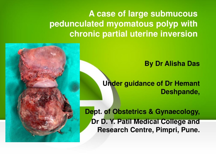
  1. A case of large submucous pedunculated myomatous polyp with chronic partial uterine inversion By Dr Alisha Das Under guidance of Dr Hemant Deshpande, Dept. of Obstetrics & Gynaecology, Dr D. Y. Patil Medical College and Research Centre, Pimpri, Pune.

  2. HISTORY OF PRESENTING COMPLAINTS A 41 year old female Para 1 living 1 with previous LSCS came on 16th August 2019 with complaint of Bleeding P/V since 1 day Foul smelling discharge P/V since 1 month Pain in abdomen and genital region since 3 months Menorrhagia associated with dysmenorrhea since 1 year No H/O white discharge Bladder bowel complaints , urinary frequency or urgency Fever , nausea , vomitting Mass coming out PV Menstrual history Menarche attained at 14 years of age Last menstrual period 8th August (1 week prior) Bled for 5 days. 3 pads soaked per day associated with passage of clots and dysmenorrhea Past menstrual history since 1 year: 5-7 days / 28-32 days / increased menstrual flow . Associated with clot passage and dysmenorrhoea

  3. Past menstrual history prior to 1 year: 4 to 5 days / 28-32 days / normal flow (2 to 3 pads per day). Obstetric history Married life 16 years P1L1 14 year old female child . LSCS done in view of non progress of labour Puerperal period uneventful PAST MEDICAL AND SURGICAL HISTORY: No history of Diabetes mellitus , hypertension, tuberculosis , thyroid disorder. No history of intake of OC pills routinely No major surgery in the past and no prior blood transfusion. Personal history Normal sleep and bowel bladder habits No history of smoking or alcohol consumption

  4. GENERAL PHYSICAL EXAMINATION Moderately built and nourished Comfortable and co-operative at the time of examination Height-165 cm Weight- 51kg BMI 18.7 kg/m Skin normal in texture and colour Pulse 76 bpm regular in rate, rhythm and normal volume measured in left radial artery. No apex pulse deficit and radio radial delay BP 110/70 mmHg taken in left brachial artery in supine position Breasts - clinically normal Thyroid - clinically normal Spine - clinically normal No pallor, icterus, edema, cyanosis, lymphadenopathy, clubbing CVS S1 S2 normal . No murmurs RS air entry bilaterally equal . No crepts , rales , rhonchi or abnormal breath sounds No cervical lymphadenopathy

  5. ABDOMINAL EXAMINATION Inspection: Umbilicus central and inverted All quadrants move equally with respiration Scar of previous LSCS noted on lower abdomen. Well healed No striae , dilated veins, sinuses Palpation : Soft , scaphoid No tenderness guarding and rigidity No organomegaly (hepatomegaly and splenomegaly) Uterus just palpable in suprapubic region (12 to 14 weeks size) Consistency firm , no tenderness , mobility present from side to side(no fixity to skin and underlying structures) and lower pole could not be reached

  6. GYNAECOLOGICAL EXAMINATION Per speculum examination : large bosselated heterogenous mass noted of size about 8x8 cm stretching the vaginal walls . Foul smelling necrotic areas with hemorrhage and ulceration suggestive of infection noted on the mass. No active bleeding noted Cervix not visualized as vision was obscured by the lesion Per vaginal examination : Large mass noted protruding through cervix and extending into the vagina Fornices obliterated due to stretching effect of the mass Cervix was not palpable as the mass was impacting the vaginal walls Uterus size not assessed as bimanual examination was not possible due to mass PROVISIONAL DIFFERENTIAL CLINICAL DIAGNOSIS ? Submucous pedunculated myomatous polyp ? Hypervascular endometrial polyp ? Cervical polypoidal growth

  7. INVESTIGATIONS Hemogram, blood sugar levels and liver and renal function test with blood electrolytes : WNL HIV , HbsAg negative USG : Bulky uterus 10x6x6 cm with thickened Endometrial echo complex of 2.7 cm . Large mass of 7.3x6.9x6.9 cm noted extending upper vagina shows vascular pedicle continuous with thickened endometrium . Suggestive of fibrovascular endometrial polyp or submucous fibroid prolapsing through cervical canal into upper vagina.

  8. MRI findings suggestive of Bulky uterus 10x7x8 cm with large well defined lobulated solid enhancing pedunculated hypervascular mass projecting inferiorly from endometrial cavity, through cervical canal of 10x8.7x8.5 cm with extensive areas of hemorrhage and necrosis. No myometrial involvement . Parametrial planes are intact . Vagina is stretched and patent.

  9. MANAGEMENT In view of the bleeding, malignancy was considered and a core biopsy of the vaginal mass was done. Histopathological study revealed fragments composed of spindle cells with areas of haemorrhage and edema .There was no evidence of malignancy. The pathological diagnosis was fibroid polyp- leiomyoma with secondary changes. The patient was taken up for definitive surgery.

  10. Patient was taken for total abdominal hysterectomy with preservation of bilateral ovaries. Fundal dimpling due to the mass with chronic inversion of uterus was noted intra operatively (Flower vase appearance of uterus and adnexa)

  11. Specimen was resected en bloc Vascular stalk extending from endometrium was noted Specimen was sent for histopathological examination

  12. The patient had an uneventful post- operative period and was discharged after 15 days with full recovery. Histopathology report was suggestive of leiomyoma

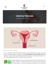
  13. DISCUSSION A leiomyoma, also known as fibroids, is a benign smooth muscle tumour. Uterine fibroids are leiomyomata of the uterine smooth muscle. As other leiomyomata, they are benign, but may lead to excessive menstrual bleeding (menorrhagia), often cause anaemia and may lead to infertility. They are usually slow growing tumors and take a few years to become clinically palpable. Each leiomyoma is derived from smooth muscle rests from either vessel walls or uterine musculature. Evidence suggests that the steroidal sex hormones (estrogen and progesterone) play a vital role in growth of fibroids. They are usually seen in women of reproductive age group and cease to grow after menopause. They are associated with hyperestrogenism and may be diagnosed along with endometrial hyperplasia, abnormal uterine bleeding and carcinoma.

  14. A typical leiomyoma is a well circumscribed tumour with a pseudocapsule. Cut surface has a pinkish white appearance with whorled pattern. Consistency is generally firm and blood vessels are found in the periphery and supply blood to the fibroid radially. Microscopically they demonstrate spindle shaped muscle cells seperated by fibrous strands. Degenerative changes like necrosis begin centrally while calcification begins peripherally and spreads inwards. Leiomyomas are classified based on their anatomic location Intramural 75% They grow symmetrically lying within the myometrial wall Subserous 10% - They grow outward and may present as a bosselated growth from uterus towards peritoneum (they may also be pedunculated and may form parasitic fibroids) Submucous 15% - They grow towards the uterine cavity and may be covered by only a thin layer of endometrium (they may grow towards the vagina also and be suspended by a pedicle as was seen in the present case wherein they are called submucous myomatous polyp)

  15. The occurrence of chronic inversion of uterus in a non pregnant woman is rare. Non gravid uterine inversion is usually associated with uterine pathology. Prolapse and extrusion of fibroids especially a sub mucous myoma of the fundus tends to be the most common factor inciting the inversion. Other less common causes are endometrial polyps and inversion associated with uterine neoplasm. Leiomyoma, leiomyosarcoma, rhabdomyosarcoma, endometrial carcinoma, all have been known to be the preceding factors .

  16. The underlying cause of uterine inversion in 80%-85% of cases is uterine leiomyoma making it the most common cause. The proposed factors thought to contribute to uterine inversion are (a) a uterus previously distended by a tumor undergoing sudden emptying; (b) Intra uterine tumor causing thinning of uterus; (c) dilatation of the cervix.

  17. The present case report is that of a woman presenting with nonpuerperal partial chronic uterine inversion secondary to a prolapsed necrotic sub mucous fibroid. In our patient hysterectomy was carried out as there was extensive necrosis with secondary degenerative changes.

  18. CONCLUSION The occurrence of chronic inversion of uterus in the non puerperal state is a rarely encountered entity. On encountering a large prolapsed fibroid one should suspect the presence of chronic nonpuerperal uterine inversion. It is advisable to perform biopsy of the mass in view of its association with uterine malignancy. In chronic inversion secondary to a fibroid, the clinician should suspect infection of the fibroid and uterus. Vaginal restoration and removal is difficult.

  19. THANK YOU

More Related Content

giItT1WQy@!-/#giItT1WQy@!-/#giItT1WQy@!-/#