Understanding the Soft Anatomy of Fish Nervous System in Ichthyology
Explore the intricate details of the nervous system in fish, delving into the division of the cerebrospinal and autonomic systems, the unique characteristics of fish brains, and the segmentation of the brain into five parts from the anterior to the posterior regions. Gain insights into the functions of each brain segment and understand the significance of the nervous system in the underwater world of ichthyology.
Download Presentation

Please find below an Image/Link to download the presentation.
The content on the website is provided AS IS for your information and personal use only. It may not be sold, licensed, or shared on other websites without obtaining consent from the author. Download presentation by click this link. If you encounter any issues during the download, it is possible that the publisher has removed the file from their server.
E N D
Presentation Transcript
Ichthyology Shanghai Ocean University Fall, 2022 Lesson 3 Soft anatomy
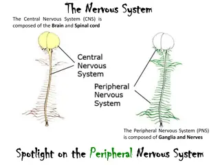
Nervous system The nervous system can be divided into the cerebrospinal and autonomic systems. The cerebrospinal system is composed of the central nervous system and the peripheral nervous system. The central nervous system is further subdivided into the brain and the spinal cord (Healey 1957; Bernstein 1970; Northcutt & Davis 1983).
Nervous system The peripheral system is composed of the cranial and spinal nerves and the associated sense organs (vision, smell, hearing, lateralis system, touch, taste, and electrical and temperature detection; see Chapter 6). The autonomic nervous system is composed of sympathetic and parasympathetic ganglia and fibers.
Fish brain Fish brains are on average only 1/15 the size of the brain of a bird or mammal of equal body size. Sharks have much larger brains relative to body size than teleosts and pelagic sharks have larger brains than pelagic teleosts (Linsey & Collins 2006). In pickerels (Esox), the brain is only 1/1305 of body weight. Elephantfishes (Mormyridae) have the largest brains among fishes, 1/52 to 1/82 of body weight.
The brain can be divided into fi ve parts from anterior to posterior (Fig. 4.11).
Five parts of fish brain The most anterior part is the telencephalon, or forebrain, which becomes the cerebrum of tetrapods. Its function in fishes is primarily associated with reception and passage of olfactory stimuli.
Five parts of fish brain The diencephalon, or tween brain, lies between the forebrain and the midbrain and is also known as the saccus dorsalis. It functions as a correlation center for incoming and outgoing messages regarding homeostasis and the endocrine system.
Five parts of fish brain The mesencephalon, or midbrain, is important in vision. The optic nerve (cranial nerve II) brings impulses from the eyes and enters the brain here. The midbrain is also a correlation center for messages coming from other sensory receptors. Fishes have two optic lobes, which are relatively large in sight-feeding species such as trouts and minnows
Five parts of fish brain The metencephalon, or hindbrain, functions in maintaining muscular tone and equilibrium in swimming. The cerebellum, a large single lobe, is the largest component of the fish brain. Cranial nerve IV (trochlear) runs from the metencephalon to the eye muscles. The metencephalon is small in lampreys (Petromyzontidae) and almost absent in hagfishes (Myxinidae). In elephantfishes (Mormyridae), the cerebellum is hypertrophied to form the valvula cerebelli (Fig. 4.11F), which extend over the dorsal surface of the telencephalon. This large cerebellum is related to reception of electrical impulses.
Five parts of fish brain The myelencephalon, brainstem, or medulla oblongata is the posterior portion of the brain and the enlarged anterior part of the spinal cord. Cranial nerves V through X arise here. The myelencephalon serves as the relay station for all the sensory systems except smell (cranial nerve I) and sight (cranial nerve II). It contains centers that control certain somatic and visceral functions. In bony fishes, it also contains respiratory and osmoregulatory centers
Peripheral nervous system Cranial nerveI, the olfactory nerve, is a sensory nerve that runs from the olfactory bulb to the olfactory lobes. The optic nerve (cranial nerve II) runs from the retina to the optic lobes. As in other vertebrates, cranial nerves III (oculomotor), IV (trochlear), and VI (abducens) are somatic motor nerves that innervate the six striated muscles of the eye: IV, the superior oblique; VI, the external rectus; and III, the other four eye muscles.
Peripheral nervous system Unlike in most other vertebrates, four cranial nerves (VII through X) innervate parts of the lateral line system. The trigeminal, V, is a mixed somatic sensory and motor nerve serving the anterior portion of the head. Cranial nerve VII, the facial, and VIII, the acoustic, usually join to form the acousticofacialis nerve, which then subdivides into four groups of mixed nerves serving the temporal and branchial regions of the head.
Peripheral nervous system Patterns of nerves, such as that of the ramus lateralis accessorius of the facial nerve (which innervates taste buds on the posterior head and body), have proved to be useful in assessing relationships of teleosts (Freihofer 1963). The glossopharyngeal, IX, is a mixed nerve that supplies the gill region. It often fuses with cranial nerve X, the anterior ramus of the vagus. The vagus is a mixed nerve connected to the body lateral line and viscera.
Summary The fish brain can be divided into five parts, from anterior to posterior: (i) the telencephalon, or forebrain, primarily associated with smell; (ii) the diencephalon, a correlation center for messages regarding homeostasis and the endocrine system; (iii) the mesencephalon, or midbrain, important in vision; (iv) the metencephalon, or hindbrain, which maintains muscle tone and equilibrium in swimming and has a large median lobe (cerebellum), which is the largest component of the fish brain; and (v) the myelencephalon, brainstem, or medulla oblongata, the posterior portion of the brain and enlarged anterior portion of the spinal cord that relays input for all sensory systems except smell and sight. Fishes have small brains but sharks have larger brains than teleosts. The largest brains occur in elephant fishes (Mormyridae), which have a large proportion of their brain devoted to electroreception.
HWK Homework 3.docx due on Oct 9 No late hand-in will be graded Read Chapter 6
Presentation for next week (Oct 9) Explain what are tuberous receptors, and how do they work Describe the three types of photoreceptive glycoprotein (opsin) and their difference Name two types of vision related structural adaptation
Presentation for next week Sent me your ppt on Sunday (Oct 9). Talk for 7 -10min
Have a nice week! Rhinogobius xiaoi
