Understanding the Reticuloendothelial System and Spleen
The lecture sheds light on the Reticuloendothelial System (RES) and spleen, explaining their cellular components, functions, and importance in the immune system. It details how macrophages in various organs form part of the RES, emphasizing their role in filtering and destroying foreign particles. The differentiation of macrophages based on their organ of residence and the process of monocytes maturing into macrophages are also discussed.
Download Presentation

Please find below an Image/Link to download the presentation.
The content on the website is provided AS IS for your information and personal use only. It may not be sold, licensed, or shared on other websites without obtaining consent from the author. Download presentation by click this link. If you encounter any issues during the download, it is possible that the publisher has removed the file from their server.
E N D
Presentation Transcript
Reticuloendothelial System & Spleen DR.MOHAMMED ALOTAIBI Assist. Professor of Physiology, College of Medicine King Khalid University Hospital King Saud University
At the end of this lecture the student is expected to be able to: 1. Define the term Reticuloendothelial system (RES) 2. Describe the cellular components of RES 3. Describe the functions of the RES 4. Define the structural function of the spleen 5. Describe the functions of the spleen 6. Understand the basic concept of the indication and risks of splenectomy
It is a network of connective tissue fibers inhabited (dwelled) by phagocytic cells such as macrophages ready to attack and ingest microbes and particles RES is an essential component of the immune system
Reticuloendothelial system is an older term for the mononuclear phagocyte system (MPS) Most endothelial cells are not macrophages.
1. Monocytes 2. Macrophage Located in all tissues, such as skin, liver (kupffer), spleen, bone marrow, lymph nodes, lung. 3. Endothelial cells: bone marrow, spleen, lymph node
Often remain fixed to their organs. They filter and destroy objects which are foreign to the body, such as bacteria, viruses. Some macrophages are mobile, and they can group together to become one big phagocytic cell in order to ingest larger foreign particles.
Macrophages differ depending on the organs in which they reside in. Kupffer cells ..in the liver. Microglia ..................in the CNS Reticular cells ..in the lymph nodes, bone marrow, spleen. Tissue histiocytes (fixed macrophages) .. ...in subcutaneous connective tissues Alveolar cells .in the lungs
Begin by Stem cell in Bone Marrow: monoblast maturing to promonocyte and mature monocytes released into blood Stay for 10-20 hours in circulation Then leave blood to tissues transforming into larger cells macrophage Macrophage life span is longer up to few months in tissues 1. 2. 3. 4.
Characterized by an increase in: Cell size Number and complexity of intracellular organelles Golgi, mitochondria, lysosomes Intracellular digestive enzymes
Phagocytosis: Bacterial, dead cells, foreign particles (direct) Immune function: processing antigen and antibodies production (indirect) Destruction of aging RBCs Storage and circulation of iron 1. 2. 3. 4.
Phagocytosis is a part of the natural, or innate immune process Macrophages are the powerful phagocytic cells: Ingest up to 100 bacteria Ingest larger particles such as old RBCs Get rid of waste products
File:Neutrophil with anthrax copy.jpg A scanning electron microscope image of a single neutrophil (yellow), engulfing anthrax bacteria (orange).
Indirectly immune function of RES: Ingest foreign body, process it, and present it to lymphocytes
1. Thymus: high rate of growth and activity until puberty, then begins to shrink; site of T-cell maturation 2. Lymph nodes: small, encapsulated, bean- shaped organs stationed along lymphatic channels and large blood vessels of the thoracic and abdominal cavities 3. Spleen: structurally similar to lymph node it filters circulating blood to remove worn out RBCs and pathogens. It is the largest system of the MPS.
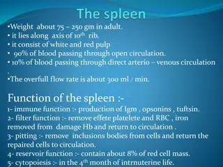
Is soft purple gray in color located in the left upper quadrant of the abdomen. It is a highly vascular lymphoid organ It plays important roles in: red blood cells integrity and has immune function. It holds a reserve of blood in case of hemorrhagic shock. It is one of the centers of activity of the RES and its absence leads to a predisposition toward certain infections Despite its importance, there are no tests specific to splenic function
White pulp: Thick sleeves of lymphoid tissue, that provides the immune function of the spleen Red pulp: surrounds white pulp, composed of Venous sinuses filled with whole blood and Splenic cords of reticular connective tissue rich in macrophages
1. Haematopoiesis (Hemopoiesis) (fetal life) 2. Spleen is a main site for destruction of RBCs specially old and abnormal e.g. spherocytosis 3. Blood is filtered through the spleen. 4. Reservoir of thrombocytes and immature erythrocytes 5. Recycles of iron
1. Because the organ is directly connected to blood circulation, it responds faster than other lymph nodes to blood-borne antigens 2. Reservoir of lymphocytes in white pulp 3. Destruction and processing of antigens 4. Site for Phagocytosis of bacteria and worn-out blood cells (Slow blood flow in the red pulp cords allows foreign particles to be phagocytosed )
5. Site of B cell maturation into plasma cells, which synthesize antibodies in its white pulp and initiates humoral response 6. Removes antibody-coated bacteria along with antibody-coated blood cells. 7. It contains (in its blood reserve) half of the body monocytes within the red pulp, upon moving to injured tissue (such as the heart), they turn into dendritic cells and macrophages that promote tissue healing.
Indications: 1. Hypersplenism: enlargement of the spleen (splenomegaly) with defects in the blood cells count. 2. primary spleen cancers 3. Sickle cell anaemia, Thalassemia,hereditary spherocytosis (HS) and elliptocytosis, 4. Idiopathic thrombocytopenic purpura (ITP). 5. Trauma. 6. Hodgkin's disease (lymphoma). 7. Autoimmune hemolytic disorders.
Overwhelming bacterial infection or post splenectomy sepsis. Patient prone to malaria Inflammation of the pancreas and collapse of the lungs. Excessive post-operative bleeding (surgical) Post-operative thrombocytosis and thrombosis
