Understanding the Nature and Composition of Human Bones
The skeletal system, composed of bones and cartilage, plays crucial roles in support, protection, mineral homeostasis, blood-cell formation, and triglyceride storage. Bones consist of organic collagen matrix, inorganic mineral salts, and water, with two main types of bone tissue - cortical and cancellous. The cortical bone provides dense outer protection, while the periosteum contributes to bone nourishment, protection, and remodelling. Understanding the structure and functions of bones enhances awareness of their importance in human anatomy.
Download Presentation

Please find below an Image/Link to download the presentation.
The content on the website is provided AS IS for your information and personal use only. It may not be sold, licensed, or shared on other websites without obtaining consent from the author. Download presentation by click this link. If you encounter any issues during the download, it is possible that the publisher has removed the file from their server.
E N D
Presentation Transcript
NATURE & FORMATION OF HUMAN BONES
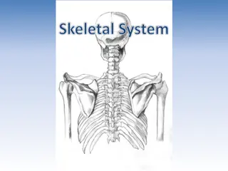
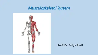
SKELETAL SYSTEM Composed of Bones & Cartilage connected by ligaments. Axial Skeleton : Bones along the axis of body i.e. skull, vertebral column and ribcage Appendicular skeleton : Appendages i.e. upper and lower libs, pelvic girdle and shoulder girdle.
FUNCTION The skeletal system has several key functions, including: Support and movement; Protection; Mineral homeostasis; Blood-cell formation; Triglyceride storage.
BONE COMPOSITION Bone matrix has three main components: 25% organic matrix (osteoid) : Collagen Fibres and other proteins such as glycoprotein, osteocalcin and proteoglycans. Hardens through the deposit of the calcium and other minerals around the fibers Flexibility and tensile strength of bone is derived from the collagen fibres. 50% inorganic mineral content (mineral salts) : Deposited between the gaps in the collagen layers with once these spaces are filled, minerals accumulate around the collagen fibres, crystallising and causing the tissue to harden; this process is called ossification. The hardness of the bone depends on the type and quantity of the minerals available for the body to use; hydroxyapatite is one of the main minerals present in bones. 25% water
STRUCTURE Bones are made up of two types of bone tissue : Cortical Bone Cancellous Bone
CORTICAL BONE Also known as compact bone. Dense outer layer. Provides support and protection for the inner cancellous structure. Cortical bone comprises three elements: Periosteum Intracortical area; Endosteum : Thin layer of connective tissue that lines the inside of the cortical surface.
PERIOSTEUM Tough, fibrous outer membrane. Highly vascular and almost completely covers the bone, except for the surfaces that form joints (covered by hyaline cartilage). Tendons and ligaments attach to the outer layer of the periosteum. Inner layer contains osteoblasts (bone-forming cells) and osteoclasts (bone-resorbing cells) responsible for bone remodelling.
FUNCTIONS : Protects the bone Help with remodelling Nourish bone tissue It also contains Volkmann s canals ( small channels running perpendicular to the diaphysis of the bone) which convey blood vessels, lymph vessels and nerves from the periosteal surface through to the intracortical layer. The periosteum has numerous sensory fibres, so bone injuries (such as fractures or tumours) can be extremely painful.
INTRACORTICAL BONE Organised into structural units, referred to as osteons or Haversian systems. These are cylindrical structures, composed of concentric layers of bone called lamellae, whose structure contributes to the strength of the cortical bone. Osteocytes (mature bone cells) sit in the small spaces between the concentric layers of lamellae, which are known as lacunae. Canaliculi are microscopic canals between the lacunae, in which the osteocytes are networked to each other by filamentous extensions. In the centre of each osteon is a central (Haversian) canal through which the blood vessels, lymph vessels and nerves pass. These central canals tend to run parallel to the axis of the bone. Volkmann s canals connect adjacent osteons and the blood vessels of the central canals with the periosteum.
ANATOMY OF CORTICAL BONE 1. Osteocyte (in lacuna) 2. Canaliculi ( extensions) 3. Lamellae ( Concentric layers of bone) 4. Haversian canal : Passage for blood, lymph vessels and nerves. 5. Connected by Volksmann canal
CANCELLOUS BONE Also known as spongy bone. It is formed of lamellae arranged in an irregular lattice structure of trabeculae, which gives a honeycomb appearance. The large gaps between the trabeculae help make the bones lighter, and so easier to mobilise. Trabeculae are characteristically oriented along the lines of stress to help resist forces and reduce the risk of fracture . The closer the trabecular structures are spaced, the greater the stability and structure of the bone . Red or yellow bone marrow exists in these spaces. Red bone marrow in adults is found in the ribs, sternum, vertebrae and ends of long bones.
BLOOD SUPPLY Bone and marrow are highly vascularised and account for approximately 10-20% of cardiac output. Blood vessels in bone are necessary for nearly all skeletal functions, including the delivery of oxygen and nutrients, homoeostasis and repair. The blood supply in long bones : derived from the nutrient artery and the periosteal, epiphyseal and metaphyseal arteries. Each artery is also accompanied by nerve fibres, which branch into the marrow cavities. Arteries are the main source of blood and nutrients for long bones, entering through the nutrient foramen, then dividing into ascending and descending branches. The ends of long bones are supplied by the metaphyseal and epiphyseal arteries, which arise from the arteries from the associated joint. If the blood supply to bone is disrupted, it can result in the death of bone tissue (osteonecrosis). A common example is following a fracture to the femoral neck, which disrupts the blood supply to the femoral head and causes the bone tissue to become necrotic. The femoral head structure then collapses, causing pain and dysfunction.
Bones are classified according to their shape Long bones typically longer than they are wide (such as humerus, radius, tibia, femur), they comprise a diaphysis (shaft) and epiphyses at the distal and proximal ends, joining at the metaphysis. In growing bone, this is the site where growth occurs and is known as the epiphyseal growth plate. Most long bones are located in the appendicular skeleton and function as levers to produce movement Short bones small and roughly cube-shaped, these contain mainly cancellous bone, with a thin outer layer of cortical bone (such as the bones in the hands and tarsal bones in the feet) Flat bones thin and usually slightly curved, typically containing a thin layer of cancellous bone surrounded by cortical bone (examples include the skull, ribs and scapula). Most are located in the axial skeleton and offer protection to underlying structures Irregular bones bones that do not fit in other categories because they have a range of different characteristics. They are formed of cancellous bone, with an outer layer of cortical bone (for example, the vertebrae and the pelvis) Sesamoid bones round or oval bones (such as the patella), which develop in tendons.
OSSIFICATION Bones begin to form in utero in the first eight weeks following fertilisation. The embryonic skeleton is first formed of mesenchyme (connective tissue) structures; this primitive skeleton is referred to as the skeletal template. These structures are then developed into bone, either through intramembranous ossification or endochondral ossification (replacing cartilage with bone).
OSSIFICATION / OSTEOGENESIS : INTRA MEMBRANOUS OSSIFICATION The term intra-membranous ossification describes the direct conversion of mesenchyme structures to bone, in which the fibrous tissues become ossified as the mesenchymal stem cells differentiate into osteoblasts. The osteoblasts then start to lay down bone matrix, which becomes ossified to form new bone.
*The stage of intramembranous ossification. The following stages are (a) Mesenchymal cells group into clusters, and ossification centers form. (b) Secreted osteoid traps osteoblasts, which then become osteocytes. (c) Trabecular matrix and periosteum form. (d) Compact bone develops superficial to the trabecular bone, and crowded blood vessels condense into red marrow. *Source: https://www.researchgate.net/figure/The-stage-of- intramembranous-ossification-The-following-stages-are-a- Mesenchymal-cells_fig3_330944846
ENDOCHONDRAL OSSIFICATION Long, short and irregular bones develop from an initial model of hyaline cartilage (cartilage models). Once the cartilage model has been formed, the osteoblasts gradually replace the cartilage with bone matrix through endochondral ossification Mineralisation starts at the centre of the cartilage structure, which is known as the primary ossification centre. Secondary ossification centres also form at the epiphyses (epiphyseal growth plates) . The epiphyseal growth plate is composed of hyaline cartilage and has four regions Resting or quiescent zone situated closest to the epiphysis, this is composed of small scattered chondrocytes with a low proliferation rate and anchors the growth plate to the epiphysis; Growth or proliferation zone this area has larger chondrocytes, arranged like stacks of coins, which divide and are responsible for the longitudinal growth of the bone; Hypertrophic zone this consists of large maturing chondrocytes, which migrate towards the metaphysis. There is no new growth at this layer; Calcification zone this final zone of the growth plate is only a few cells thick. Through the process of endochondral ossification, the cells in this zone become ossified and form part of the new diaphysis .
*The stage of endochondral ossification. The following stages are: (a) Mesenchymal cells differentiate into chondrocytes. (b) The cartilage model of the future bony skeleton and the perichondrium form. (c) Capillaries penetrate cartilage. Perichondrium transforms into periosteum. Periosteal collar develops. Primary ossification center develops. (d) Cartilage and chondrocytes continue to grow at ends of the bone. (e) Secondary ossification centers develop. ( f) Cartilage remains at epiphyseal (growth) plate and at joint surface as articular cartilage. *Source: https://www.researchgate.net/figure/The- stage-of-endochondral-ossification-The-following- stages-are-a-Mesenchymal-cells_fig4_330944846
Bones are not fully developed at birth, and continue to form until skeletal maturity is reached. By the end of adolescence around 90% of adult bone is formed and skeletal maturity occurs at around 20-25 years, although this can vary depending on geographical location and socio-economic conditions; for example, malnutrition may delay bone maturity. Genetic mutation can disrupt cartilage development, and therefore the development of bone : reduced growth and short stature - achondroplasia. The human growth hormone (somatotropin) is the main stimulus for growth at the epiphyseal growth plates. During puberty, levels of sex hormones (oestrogen and testosterone) increase, which stops cell division within the growth plate. As the chondrocytes in the proliferation zone stop dividing, the growth plate thins and eventually calcifies, and longitudinal bone growth stops. Males are on average taller than females because male puberty tends to occur later, so male bones have more time to grow .
REMODELLING Once bone has formed and matured, it undergoes constant remodelling by osteoclasts and osteoblasts, whereby old bone tissue is replaced by new bone tissue. Bone remodelling has several functions: Mobilisation of calcium and other minerals from the skeletal tissue to maintain serum homoeostasis, Replacing old tissue and repairing damaged bone, Helping the body adapt to different forces, loads and stress applied to the skeleton. Calcium plays a significant role in the body and is required for muscle contraction, nerve conduction, cell division and blood coagulation. As only 1% of the body s calcium is in the blood, the skeleton acts as storage facility, releasing calcium in response to the body s demands. Serum calcium levels are tightly regulated by two hormones, which work antagonistically to maintain homoeostasis. Calcitonin facilitates the deposition of calcium to bone, lowering the serum levels, whereas the parathyroid hormone stimulates the release of calcium from bone, raising the serum calcium levels.
Osteoclasts are large multinucleated cells typically found at sites where there is active bone growth, repair or remodelling, such as around the periosteum, within the endosteum and in the removal of calluses formed during fracture healing. Osteoclasts break down bone tissue by secreting lysosomal enzymes and acids into the space between the ruffled membrane . These enzymes dissolve the minerals and some of the bone matrix. The minerals are released from the bone matrix into the extracellular space and the rest of the matrix is phagocytosed and metabolised in the cytoplasm of the osteoclasts . Once the area of bone has been resorbed, the osteoclasts move on, while the osteoblasts move in to rebuild the bone matrix.
Osteoblasts synthesise collagen fibres and other organic components that make up the bone matrix. They also secrete alkaline phosphatase, which initiates calcification through the deposit of calcium and other minerals around the matrix. As the osteoblasts deposit new bone tissue around themselves, they become trapped in pockets of bone called lacunae. Once this happens, the cells differentiate into osteocytes, which are mature bone cells that no longer secrete bone matrix.
