Understanding Rinderpest: Symptoms, Transmission, and Host Range

Rinderpest is a viral disease characterized by high fever, lachrymal discharge, inflammation, erosions of the mouth, and diarrhea, leading to death. The disease affects cloven-hoofed animals, with cattle and water buffalo being most susceptible. Transmission occurs through aerosol vectors and ingestion. The virus is fragile and related to other viruses causing flu, distemper, measles, and Peste des Petits Ruminants. Eradication efforts target Rinderpest due to its lack of vertical transmission. The incubation period varies depending on the strain and exposure route.

Download Presentation

Please find below an Image/Link to download the presentation.
The content on the website is provided AS IS for your information and personal use only. It may not be sold, licensed, or shared on other websites without obtaining consent from the author. Download presentation by click this link. If you encounter any issues during the download, it is possible that the publisher has removed the file from their server.
E N D
Presentation Transcript
Rinderpest Rinderpest is characterized by high fever, lachrymal discharge, inflammation, Erosions of the epithelium of the mouth and of the digestive tract, profuse diarrhea, and death. The The four four D s D s of of Rinderpest Rinderpest: : Depression Depression Diarrhea Diarrhea Dehydration Dehydration Death Death hemorrhage, necrosis, Rinderpest
Rinderpest Rinderpest
Rinderpest The virus is relatively fragile and is immunologically related to viruses that cause - fluEM rpv2a Canine Distemper, Measles, and Peste des Petits Ruminants & Equine infuenza recently described in Austerlia. Rinderpest
Also known as cattle plague Rinderpest is a mucosal disease Rinderpest
Host Range All cloven-hoofed animals are susceptible (not all are clinical) Most clinical cases occur in cattle and water buffalo Rinderpest
Host Range Sheep, goats, and yak are mostly subclinical http://www.geo.arizona.edu/dgesl/research/regional/asian_monsoon_dynamics/yak.htm Rinderpest
Host Range Camels asymptomatic infections only Rinderpest
Host Range Wild Animals Most cloven-footed wild animals such as bison and deer Antelope Wildebeest Eland Giraffe Hippopotamus Gazelle Warthog Rinderpest
Transmission Aerosol Vectors Tabanids* Ingestion Fomites Krinsky, W.L. (1976) Animal disease agents transmitted by horse flies and deer flies (Diptera: Tabanidae). Journal of Medical Entomology 13(3): 225-275 Rinderpest
Transmission There is no vertical transmission, arthropod vector, or carrier state. This makes Rinderpest virus an ideal virus to be targeted for eradication. Rinderpest
Incubation period Varies with strain of RPV, dosage, and route of exposure (3-15 days) Normally a range of 3-9 days (can be as short as 3-4 days in experimental infection; also, can be as long as 10-15 days with virus of low virulence) Duration: 2 or more weeks Rinderpest
Pathogenesis Inhaled/ Ingestion Upper Respiratory infectio Vermia Target cell ( Lymphocyes & Alimentary Mucosa) Signs & Lesions.
*Virus is present in blood and secretions BEFORE symptoms appear Rinderpest
General Clinical signs Fever Depression Nasal & lachrymal secretion Congested mucosas Mucosal erosions Severe diarrhea Leukopenia Death Rinderpest
Clinical Signs in cattle The case definition of rinderpest is ocular and nasal discharges with any two of the additional signs: + fever + erosions in the mouth + diarrhea + dehydration + death Rinderpest
Clinical signs in cattle Two major forms of disease Acute or Classic form Peracute form Rinderpest
Clinical Signs in cattle (Peracute Form) Most often found in highly susceptible young and newborn animals No prodromal signs High fever (104-107 F) Congested mucous membranes Rinderpest
Clinical Signs in cattle (Acute Form) Acute (classic) form characterized by pyrexia, erosive stomatitis, gastroenteritis, dehydration, and death Four stages 1. Incubation period 2. Febrile period 3. Mucous membrane congestion 4. Gastrointestinal signs Rinderpest
Clinical Signs in cattle (Acute Form) Fever - 104 to 107 F (40-42 C) Serous oculo-nasal discharge Leukopenia Depression Anorexia Constipation followed by diarrhea Oral erosions Rinderpest
Clinical Signs in cattle (Acute Form) Decreases in fever and viral titer Diarrhea (may be watery or hemorrhagic) Dehydration, emaciation Prostration and death 6 to 12 days after onset of illness Rinderpest
Clinical Signs Shooting diarrhea Shooting diarrhea Rinderpest
Clinical Signs In Africa this also includes corneal opacity which has been associated with rinderpest in buffalos and lesser kudus but has also been noted in calves together with dermatitis. Rinderpest
Clinical Signs Early serous ocular discharge (Epiphora) Rinderpest
Clinical Signs Depression Diarrhea Dehydration Death Rinderpest
Clinical Signs Early focal mucosal erosions Rinderpest
Clinical Signs Early erosions rinderpest or trauma ? Rinderpest
Clinical Signs Inflammation and necrosis of cheek papillae Rinderpest
Clinical Signs Inflammation of cheek papillae Rinderpest
Clinical Signs Mucosal erosions cigarette burns Rinderpest
Clinical Signs Shallow erosions in the mouth Note how these have a sharp margin Rinderpest
Clinical Signs Erosion under the tongue Rinderpest
Convalescence muzzle skin sloughing Rinderpest
Convalescence eroded cheek papillae Rinderpest
Clinical Signs Profuse diarrhea and dysentery Rinderpest
Dehydration, emaciation and collapse Dehydration, emaciation and collapse Rinderpest
Dehydration and death Rinderpest
Lesions Eroded hard palate Rinderpest
Lesions Gastro-enteritis Rinderpest
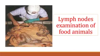
Lesions Hemorrhagic mesenteric lymph nodes The virus is lymphotropic as well as epitheliotropic. Lymph nodes throughout the body are at first swollen, oedematous and haemorrhagic, as seen here. Later they become pale and shrunken. The spleen is similarly affected. Rinderpest
Lesions Petaechial haemorrhages can be found on the mucosal surface throughout the intestines but in the large intestine they tend to be aligned along the surface of the longitudinal folds. Linear petaechial haemorrhages in colon Rinderpest
Lesions The petaechial haemorrhages enlarge and become confluent along the longitudinal folds. After death these stripes become blackened and give rise to the characteristic (but not pathognomic) zebra striping . Zebra striping in the colon Rinderpest
Lesions Virus replicates in intestinal epithelium and kills it dead, creating horrendous ulcers Rinderpest
Lesions Hemorrhagic Peyer s patches Oedema and necrosis of the Peyer s patches is a common feature. Later they are shrunken and may be frankly necrotic. Rinderpest
Diagnosis Samples: Conjunctival Fluid Intestinal contents or feces Whole blood Lymphoid tissue, lung, intestine Serum A definitive diagnosis require laboratory confirmation based on detecting viral antigens, the presence of microscopic lesions, and by isolating and identifying the virus. Laboratory confirmation depends on the collection of suitable samples from a group of animals, preferably in the febrile stage with oral lesions rather than from animals already dying, with profuse diarrhea. Rinderpest
Differential Diagnosis Bovine virus diarrhea Mucosal disease Infectious bovine rhinotracheaitis Malignant catarrhal fever Vesicular stomatitis Foot-and-mouth disease Rinderpest
Differential Diagnosis Salmonellosis Necrobacillosis paratuberculosis Bluetongue / EHD Mycotic Stomatitis Rinderpest
Diagnostic Tests Antigen Detection Antibody Detection Histopathology Blood should be collected in both serum and EDTA tubes. Swabs of clear tears, necrotic debris from gums and aspiration biopsies from superficial lymph nodes are also recommended. Virus isolation requires samples of spleen, lymph node and/or tonsils from a freshly euthanized animal Rinderpest
RINDERPEST Synonym : Cattle plaque Definition Acute, highly contagious viral disease of cattle characterized by high fever, necrotic stomatitis, gastroenteritis, diarrhoea and high mortality caused by morbillivirus In India "Hill Zebu cattle" are more susceptible than "Plain zebu cattle". This disease is characterized by necrosis, and erosions of the mucosa in the respiratory and digestive tracts. Etiology Morbillivirus The virus is antigenically closely related to the viruses of canine distemper, PPR of sheep and goats and measles of humans.
Incidence The disease has been the foremost cause of death in cattle in most African and Asian countries including India Rinderpest has not been reported since June 1995 in Indian suncontinent The seriousness of disease madeto start first Veterinary College in 1762 at Lyons, France. Susceptibility Mainly cattle and buffaloes, but also reported in sheep,goat and pigs Reported in deer, antelope, wild buffaloes, wild boars, bushbuck, warthogs and giraffe Mortality is 100 % in exotic breeds and 20-50 % mortality in indigenous breeds