Understanding Necrosis: Causes, Characteristics, and Differences from Autolysis
Necrosis is the death of cells and tissues in living animals, characterized by various causes such as poisons, toxins, circulatory disturbances, and mechanical injuries. It presents gross appearances like white, gray, or yellow areas with a cooked meat appearance and distinct demarcation from healthy tissue. Microscopically, necrosis manifests as eosinophilia, swelling, vacuolation, and changes in the nucleus. Autolysis, on the other hand, is post-mortem tissue breakdown and should be differentiated from necrosis.

Download Presentation

Please find below an Image/Link to download the presentation.
The content on the website is provided AS IS for your information and personal use only. It may not be sold, licensed, or shared on other websites without obtaining consent from the author. Download presentation by click this link. If you encounter any issues during the download, it is possible that the publisher has removed the file from their server.
E N D
Presentation Transcript
(Subject: Veterinary Pathology, Paper-I ; Unit:-I) Dr. Kaushal Kumar Assistant Professor & Head Department of Veterinary Pathology Bihar Veterinary College BASU Patna-14
Introduction There are two different ways by which a cells can die: Apoptosis (Programmed cell death) Necrosis (Unplanned cell death)
Necrosis Necrosis is death of cells and tissues in the living animal. Focal/ Multifocal necrosis- terms used for one or more, small, clearly defined areas of necrosis. Diffuse necrosis- term used when necrosis affects a large area or the entire tissue or organ.
General causes of necrosis: Poisons and toxins: 1. Chemical: Strong acids, alkalies, insecticides, mercury etc. 2. Infectious agents: Bacteria (Salmonella, Staphylococcus), viruses, 3. fungi, protozoa etc. Plant poisons - hepatotoxic alkaloids e.g. Senesce. Circulatory 4. disturbance: Anemia, congestion and ischemia. Mechanical injuries: Cutting, crushing and rubbing types. 5. Physical : Extreme temperature, electricity, free radical. 6.
Gross appearance: Affected areas white, gray or yellow in colour. 1. Have a cooked meat appearance. 2. Sharply demarcated (by red zone) from healthy tissue. 3. In case of gangrene the area is green, orange or black ( 4. iron sulphide)
Microscopic appearance: The microscopic changes of necrosis vary with the type of necrosis. Some general changes of necrosis in the cytoplasm are: Eosinophilia: The cytoplasm stains darker red in colour. Swelling and vacuolation: The cells are swollen and contain different types of vacuoles. Changes in the nucleus: The nucleus may show condensation (Pyknosis), fragmentation (karyorrhexis) and may disappear (karyolysis) 1. 2. 3.
Necrosis & Autolysis Autolysis: Autolysis is death of cells and tissues after the death of the animal (somatic death) and it should be distinguished from necrosis. The enzymatic digestion of cells by enzymes present within them. 1. The cells most susceptible to autolysis tend to be dying or dead cells. No sharp line of demarcation between affected and healthy tissue. 2. Circulatory changes like congestion and haemorrhage are not present. 3.
NECROSIS AUTOLYSIS No sharp line of demarcation between affected and healthy tissue Circulatory changes like congestion and haemorrhage are not present. A line of demarcation is usually present Circulatory changes are present Inflammatory changes like infiltration of leukocytes are present. Saprophytic growth not seen, but pathogenic bacteria maybe present. 1. 1. 2. 2. 3. Inflammatory changes are not present. 3. 4. Growth of saprophytic bacteria, bacillary rods in long chains are often present. 4.
Types of necrosis: Different types of necrosis are recognized according to the causes, pathogenesis and the tissue involved. These include Coagulative, liquefactive, caseous and fat necrosis.
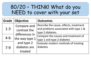
Coagulative necrosis: Most common type of necrosis. Architectural outlines persist but cellular details are lost. Type of tissue can be recognized. Denaturation (coagulation) of structural and enzymic proteins blocks proteolysis. Kidney Infarct (Source: Google)
Causes of Coagulative Necrosis Ischemia due to thrombosis/ embolism as in infarcts. Bacterial toxins e.g. Fusobacterium necrophorum in livers in cattle. Muscular dystrophy due to deficiency of selenium and Vit.-E in cattle and sheep. Necrosis of renal epithelium due to poisoning from mercuric salts.
Coagulative Necrosis Contd. Gross appearance: Necrotic area is firm, opaque with cooked meat appearance. It is sharply demarcated from the healthy areas. Microscopic appearance: Architectural outlines are present; cellular details are lacking. Result: Dead tissues remain in the body for a long period, ultimately removed by macrophages.
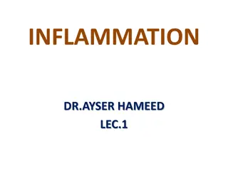
Liquefactive necrosis: There is digestion and liquefaction of necrotic tissue. Causes: Pyogenic bacterial infections attract neutrophils. Bacterial and leukocytic 1. enzymes liquefy dead cells and tissues. Some chemicals like turpentine oil also attract 2. neutrophils and cause pus formation and results Abcess in neck into liquefaction of the tissue. The necrosis in the nervous tissue is mostly liquefactive due to high 1. content of lipids and water.
Gross Appearance of Liquefactive Necrosis The necrotic tissue is liquefied and filled with semisolid creamy liquid called pus. Pus: It is a thick, white or yellow, creamy liquid consisting of exudate of leukocytes, tissue debris and microorganisms. Proteolytic enzymes released from neutrophils cause liquefaction of dead cells. 2. Abscess: It is a localized collection of pus, surrounded by fibrous capsule. 3. Empyema: It is accumulation of pus in a body cavity. 1.
Microscopic appearance of Liquefactive Necrosis No architectural or cellular details are visible in the area of necrosis. The necrotic area usually appears as a cavity containing a mass of necrotic neutrophils, bacteria and tissue debris. The entire necrotic mass is surrounded by a fibrous connective tissue capsule..
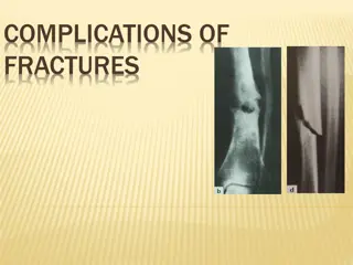
Caseous necrosis Dead tissue is converted into a homogenous, granular mass resembling cottage cheese. Cause: Associated with lesions of Mycobacterium tuberculosis and Arcanobacterium ovis, the cause of caseous lymphadinitis. Bovine tuberculosis ( Source Google.com)
Caseous necrosis Gross appearance: The area of necrosis is amorphous, granular, friable, white-gray resembling cottage cheese. The caseous mass is enclosed within a connective tissue capsule. Microscopic appearance: No architectural or cellular details are seen. Calcification commonly occurs in the necrotic areas. The necrotic tissue is amorphous, granular mass enclosed inside a zone of granulomatous inflammation, containing macrophages.
")