Understanding Choledochal Cysts: A Congenital Anomaly of the Biliary Tract

Choledochal cysts are congenital anomalies of the biliary tract characterized by cystic dilatation at various segments. They can lead to complications like biliary cirrhosis and recurrent pancreatitis. Clinical features include jaundice, abdominal pain, and right epigastric mass. Early detection is crucial for appropriate management.

Download Presentation

Please find below an Image/Link to download the presentation.
The content on the website is provided AS IS for your information and personal use only. It may not be sold, licensed, or shared on other websites without obtaining consent from the author. Download presentation by click this link. If you encounter any issues during the download, it is possible that the publisher has removed the file from their server.
E N D
Presentation Transcript
13 * 22 CBD . .
LAB CBC : WBC :7380 Hb : 12.8 PLT : 252000 AST :279 ALT:210 Total Bili : 6.12 Dirt Bili : 3.91 ALK : 645 ALB : 3 Total Pr : 5
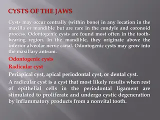
Choledochal cyst Choledochal cysts are considered to be congenital anomalies of the biliary tract characterized by varying degrees of cystic dilatation at various segments of the biliary tract (extrahepatic or intrahepatic). The frequency of choledochal cysts is about 1 in 15 000 live births in Western countries and as high as 1 in 1000 live births in Japan. There is a marked female predominance (4:1) regardless of the racial origin.
A choledochal cyst (or congenital bile duct cyst) may be detected at any age and in any portion of the bile duct. Choledochal cysts can be classified into five subtypes . Cysts are present in up to 2% of infants with obstructive jaundice.
Infants present in a manner simulating biliary atresia and, if unrecognized, may have progressive disease. Prolonged obstruction results in biliary cirrhosis, portal hypertension resulting from cirrhosis, and pressure on the portal vein by the distended cyst. Recurrent pancreatitis is an unusual complication of the malformation.
Clinical features The classic triad of intermittent abdominal pain, jaundice, and right epigastric mass varies in incidence; this triad is usually not present in infants and is uncommon in older children, occurring in about 20%. Jaundice (conjugated hyperbilirubinemia) is the common manifestation. Abdominal pain may be a presenting symptom, often with elevated serum amylase levels. The lesion may be detected at any age, with 18% appearing before 1 year of age. Older children may have mild chronic liver disease, which may reflect variable degrees of common bile duct obstruction.
In certain patients, the lesion appears to be a true congenital malformation and is associated with other anomalies of the biliary tree, such as double common duct, double gallbladder, and accessory hepatic ducts, as well as polycystic and hypoplastic kidneys. Adults with choledochal cyst disease commonly have acute biliary tract or pancreatic symptoms. It is possible that the variability in age and clinical course represents two distinct entities: congenital disease (in infants) versus acquired disease (in older children)
Spontaneous perforation of a choledochal cyst in infancy may occur. The cause of the perforation is postulated to be biliary epithelial irritation as a result of reflux of pancreatic juice caused by pancreaticobiliary malunion associated with mural immaturity, rather than an abnormal rise in ductal pressure or congenital mural weakness at a certain point.
Classification of bile duct cysts I Cystic dilatation of the common bile duct Ia Large saccular cystic dilatation Ib Small localized segmental dilatation Ic Diffuse (cylindric) fusiform dilatation II Diverticulum of the common bile duct and/or the gallbladder III Choledochocele IV Multiple cysts IVa Intrahepatic and extrahepatic (most common form) IVb Extrahepatic only V Fusiform intrahepatic dilatations (relation to Carol disease?)
Pathogenesis The pathogenesis of choledochal cysts is undetermined; there are several theories. Cysts may represent (1) anomalous union of the common bile duct and the pancreatic duct proximal to the sphincter of Oddi, which may permit reflux of pancreatic enzymes into the common bile duct with resultant inflammation, localized weakness, and dilatation (2) congenital segmental weakness of the common bile duct wall; or (3) obstruction of the distal common bile duct leading to dilatation.
Diagnosis In most infants, the diagnosis is suggested if non-invasive imaging studies are undertaken for vague right upper quadrant symptoms. Ultrasonography should be the initial procedure in the evaluation of suspected choledochal cyst. Radiographs of the upper gastrointestinal tract may outline the mass as it displaces the first and second portion of the duodenum but are unnecessary. Ultrasonography may be helpful in the preoperative differential diagnosis of choledochal cysts in neonates and infants. Cysts are larger, intrahepatic ducts are dilated, and gallbladders are not atretic in patients with choledochal cysts compared with patients with biliary atresia.
Treatment The goal is complete surgical excision of the cyst mucosa, with a Roux-en-Y choledochojejunostomy proximal to the most distal lesion.
Complications and outcome Cholangitis may occur in up to 15% of patients following surgery, even with the Roux-en-Y procedure, but is much less common than with direct anastomosis to the duodenum; the latter procedure is not advisable. The high rate of stricture (73%) after cyst enterostomy is also preventable by total cyst excision and Roux-en-Y reconstruction. Pancreatitis is uncommon but may occur secondary to proximal pancreatic duct or sphincter stenosis or stones.
Malignancy in choledochal cysts Carcinoma has been reported in residual cystic tissue in up to 26% of patients, an incidence that is 20 times greater than that in the general population. The typical malignancy is adenocarcinoma of the bile duct or gallbladder; less commonly squamous cell carcinoma and cholangiocarcinoma have been described. The risk of developing malignancy increases with age, making complete excision of the cyst and proximal bile duct mucosa an essential component of the operation in older patients. Malignant change also may occur in areas of the biliary tree remote from the cyst. The increased risk of malignant degeneration and the dismal prognosis once cancer has developed warrant complete cyst excision, even in asymptomatic patients, including those with prior cyst enterostomies.
Thanks for your attention


 may")