Understanding Cardiac MRI: Applications and Techniques
Learn about Cardiac MRI, its importance in assessing heart abnormalities, different imaging sequences, ECG gating, bright blood technique, and imaging planes in this comprehensive guide. Explore how CMR provides anatomical and functional information for acquired or congenital heart diseases, precise quantification of ventricular dimensions, and evaluation of myocardial viability and perfusion. Discover the essential role of ECG gating in acquiring motion-free heart images and the different pulse sequences used in cardiac MRI.

Download Presentation

Please find below an Image/Link to download the presentation.
The content on the website is provided AS IS for your information and personal use only. It may not be sold, licensed, or shared on other websites without obtaining consent from the author. If you encounter any issues during the download, it is possible that the publisher has removed the file from their server.
You are allowed to download the files provided on this website for personal or commercial use, subject to the condition that they are used lawfully. All files are the property of their respective owners.
The content on the website is provided AS IS for your information and personal use only. It may not be sold, licensed, or shared on other websites without obtaining consent from the author.
E N D
Presentation Transcript
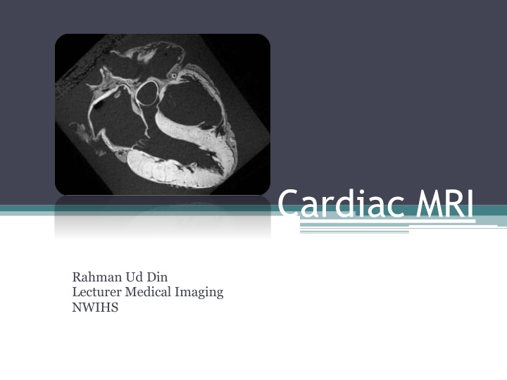
Cardiac MRI Rahman Ud Din Lecturer Medical Imaging NWIHS
Introduction CMRI Information to assess the abnormality of heart Anatomical and functional information Acquired or congenital heart diseases Modality of choice ARVD, Differentiation from constrictive pericarditis from restrictive pericarditis, aorticdissection Precise quantification of ventriculardimensions Myocardial viability and perfusion ARVD
ECG Gating ECG/EKG gating essential for motion-free image of heart Images are acquired in a particular phase of cardiac cycle in every cardiac cycle To avoid image blur and motion artifacts The image is decided by ECG gating Usually R-wave is used to trigger the acquisition Such data acquired in the diastolic phase Peripheral pulse also used for gating But less effective than ECG gating
Imaging Sequences Pulse sequences used Dark-blood sequences Bright-blood sequences Dark-blood tech Spin-echo seq; that show the flowing blood as flow void Seqs; include breath-hold turbo or FSE (TSE, FSE) Single shot FSE Double inversion recovery FSE (Double-IR-FSE)
Bright blood technique GRE seq: based shows blood bright Seqs; include spoiled GRE (turboFLASH/SPGR/T1-FSE) Balanced SSFP (TrueFISP/FIESTA/balanced TFE) Balanced TFE is mainstay seq; in CMRI Motion loop is obtained rapid cine imaging (RCI) Used RCI used ventricular function To calculate EF, SV as well as valvular & RWM Phase contrast useful in velocity & flow direction Initial CMRI starts from black blood than bright blood techniques used to assess functions
Imaging planes Orthogonal planes (axial, sagittal and coronal) Used for chest imaging and not suitable in CMRI Because cardiac axes are not parallel to body axes Three axes images are taken and used as localiser
1. Vertical long-axis plan (two chamber view) Axial image Large oblique diameter image of LV Chambers; LA and LV
2. Horizontal long-axis(Four Chamber view) Planned on two chamber view By a drawing linepassing through LA, MV & LV All four chambers, MV and TV assessed Cine GRE images obtained on this plane to assess function of MV, TV, AV and LV, RV wall motion
3. Short-axis plane Multiple cross sections are obtained perpendicular to LV long axis as seen on a two-chamber view Sections are taken from the base to the apex of the heart Cine GRE images allow visualization and quantification of systolic myocardial wall thickening Images in this plane are used for calculating ventricle volume, mass and EF by postprocessing
4. Five-Chamber view View obtained parallel to the line passing through the LV apex and aortic outflow tract on coronal images Apart from all four chambers, this view also shows aortic root This describe MV and AV
5. RVOT Plane passing through the RV outflow tract
Clinical Applications of CMRI 1. Congenital Heart Disease (CHD) Complex information about heart anatomy When echocardiography fail ASD, VSD detected with high sensitivity & specificity Calculate shunt size Conditions like TGA (transposition of Greater Arteries) Truncus arteriosus Many anomalies CMRI in children are having limitation
ASD Truncus arteriosus
2. Valvular Heart Disease (VHD) Arrhythmogenic right ventricular dysphagia Enlargement and dilatation of RV Thinning of wall, area of dyskinesia, focal bulging of free wall in systole Decreased EF and impair ventricular filling during diastole HCM (Hypertrophied) Echo detect but RV involvement is checkedCMRI RCM (Restrictive) vs Constrictive Constrictive calcified pericardium, WT +4mm Hemochromatosis Myocardial iron deposition i.e. Thalasemia Quantified by T2*-w seq;
4. Ventricular Function CMRI more accurate than Echo It measure EF, EDV & ESV (end systolic vol) Done on short axis image using software Balanced SSFP, good contrast b/w blood pool and myocardium
5. Coronary artery assessment Not good enough for visualisation of coronary arteries and its branches Presently it is used for such purpose, to find anomalies, aneurysms and bypass grafting patency GRE seq; balanced SSFP C+ or C-
6. Myocardial perfusion and viability IV Gd T1-weighted GRE seq; turboFLASH Low signal areas of underperfusion on these images correspond with regions of ischemia or infarct Myocardial viability Viability seq; run after 10-15 minutes after Gd contrast T1-w GRE or inversion recovery balanced SSFP seq IR pulse used to suppress myocardium to get LV info Proper TI is important Infracted area on viability imaging shows enhancement This imaging is Bright is dead Answer feasibility revascularisation procedure like angioplasty and bypass or not CMRI viability is superior compared to the PET
7. Cardiac and Pericardial masses CMRI accurate method for cardiac and pericardial masses evaluation Thrombus the most common filling defect in cardiac chamber Gd enhancement differentiate b/w mass or thrombus Most cardiac neoplasm are metastatic Primary neoplasm are rare and 80% benign
8. Pericardial disease Visualised with spin echo or GRE images Normal pericardium seen on SE images As a line of low signal intensity located b/w high signals of pericardial and epicardial fat Normal thickness 1-2 mm; more than 4 mm is considered thickening


")