Cardiac Action Potential: Ionic Basis and Excitability in Cells
CARDIAC ACTION
POTENTIAL: IONIC
BASIS
References
1.Clinical arrhythmology and electrophysiology, a companion to
braunwald’s heart disease, third edition
2.Harrison’s principles of internal medicine,21
st
edition
3. Ganong’s review of medical physiology, 26
th
edition
4.Cardiac Ion Channels,Augustus O. Grant
https://doi.org/10.1161/CIRCEP.108.789081
Circulation: Arrhythmia
and Electrophysiology. 2009;2:185–194
5.Cardiac transmembrane ion channels and action potentials: cellular
physiology and arrhythmogenic behavior
András Varró, Jakub Tomek,
Norbert Nagy, László Virág, Elisa Passini, Blanca Rodriguez, and István
Baczkó,
András Varró
https://doi.org/10.1152/physrev.00024.2019
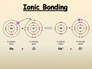
IONIC EQUILIBRIUM
Cell membrane is resistant to hydrophilic ion movement
Ions use specialized pores called channels to move across
membrane
ion movement is driven by
1. Electrical gradient
2. Chemical gradient
Movement of ion is from higher gradient to lower gradient.
Most of the movements occur passively
•
electrical gradient becomes equal and opposite to the
chemical gradient, the ion is said to be in
electrochemical
equilibrium
•
the electrical potential is called the
equilibrium potential
(E
ion
) (reversal potential or Nernst potential)
of that individual
ion
•
No ion movement occurs at this potential
•
E
ion
of an ion depends on its concentration on either side of
the membrane
RESTING MEMBRANE POTENTIAL
It is the potential difference across the cell membrane at rest.
It is negative inside with respect to outside the membrane.
Cells in their resting state are said to be polarised
RMP is not same in all cardiac cells
-90mV in atrium, ventricles
-60mV in pacemaker cells
THRESHOLD POTENTIAL
is the critical level to which the membrane
potential must be depolarized to initiate an action potential
EXCITABILITY
Ease with which a cell respond to a stimulus with a
regenerative action potential
In cardiac cells excitability depends on number of available
Na
+
channels
Sodium channels are more open at negative E
m
Cardiac cells with more negative Em(ventricles and atrium)
are more excitable than SA node
REFRACTORINESS
Inability to initiate another action potential in response to
stimulus of threshold intensity
Absolute refractory period
: no stimulus, regardless of
strength can re-excite the cell
Relative refractory period
: suprathreshold stimulus can
initiate action potential
Refractoriness in cardiac tissue is a function of Na
+
channels
CARDIAC ACTION POTENTIAL
Action Potential is a sudden reversal of membrane polarity
when a stimulus strikes the cell membrane.
Action potential in cardiac muscle is different from that of
other tissues such as skeletal muscles and nerve tissues.
Duration of action potential in cardiac muscle is 250 to 350ms
(0.25 to 0.35s)
CARDIAC ACTION POTENTIAL
Cardiac action potential is unique in itself
Not only it is different from action potential seen in other
excitable tissues but different in different part of the heart
This heterogeneity is brought by differential distribution of
various ionic channels
Cardiac Action potential are of two types
1.Fast response action potential : seen in atrium, ventricles
2. Slow response action potential: seen in SA node, AVN
Phases of action potential
There are 5 phases
Phase 0
: rapid depolarisation
Phase 1
: early repolarisation
Phase 2
: plateau phase
Phase 3
: rapid repolarisation
Phase 4
: resting membrane potential
FAST RESPONSE
ACTION POTENTIAL
Phase 4: The Resting Membrane
Potential
Caused by the different ionic concentration across the
membrane and selective membrane permeability
At RMP, membrane is most permeable to K
+
ions.
Hence E
m
tends to be close to E
k
(-94mV)
K
ir
plays a major role in maintaining E
m
Resting E
m
is also maintained by Na
+-
K
+
ATPase pump
Intracellular Ca
2+
also plays role via Na
+
-Ca
2+
pump
Phase 0: The Upstroke rapid
repolarisation
Depolarisation activates Na
+
channels
Rapid influx of Na
+
ions
depolarises the membrane leading to more influx of Na
+
I
Na
is generated which lowers the E
m
to lesser negative till the
E
m
reaches threshold for Ca
2+
channel opening
Phase 1 : Early repolarisation
Membrane repolarise rapidly and transiently to almost 0mV
Due to inactivation of Na
+
channels
Transient outward K
+
current (I
to
)
Na
+
-Ca
2+
exchanger also plays a role
Phase 2: The plateau
Delicate balance between
a) the depolarizing
inward currents (I
CaL
and a
small residual component of inward I
NaL
)
b) the repolarizing outward currents (ultrarapidly [I
Kur
], rapidly
[I
Kr
], and slowly [I
Ks
] activating delayed outward rectifying
current
longest phase of the action potential
Unique among excitable tissues
I
CaL
is activated by membrane depolarization, is largely
responsible for the action potential plateau
Na
+
channels also make a minor contribution in the form of
late I
Na
I
kr
and I
ks
play their part in maintaining steady E
m
While I
Kr
is active during early phase 2, I
Ks
is more active
during later half of phase 2
I
kur
since is present only in atria plays role in phase 2 of atrium
alone
Na
+
-K
+
ATPase pump and Na
+
-Ca
2+
exchanger also plays a
minor role
Phase 3: Final rapid
repolarisation
restores the E
m
to its resting value
mediated by
1.increasing conductance of the delayed outward rectifying
currents (I
Kr
and I
Ks
)
2. the inwardly rectifying K+ currents (IK1 and acetylcholine-
activated K+ current [I
KACh
])
3.inactivation of I
CaL
Slow response action potential
Seen in SA node and AV node
More depolarised E
m
at the onset of phase 4(-50 to -65mV)
Characterised by slow upstroke phase 0
Mediated by I
CaL
instead of I
Na
Phase 4 : Diastolic
depolarisation
SA node and AV node exhibits variable E
m
E
m
progressively decline during diastole
Once it reaches -40mV, action potential generated
Due to funny current, I
f
which is a inwardly directed current
Funny current is mainly driven by Na
+
ions and K
+
channel to a
lesser extent
Funny current deactivate during action potential
Ca
2+
channels are also thought to play role in diastolic
depolarisation
Phase 0: the upstroke-slow
depolarisation
Mainly driven by I
CaL
.
I
Na
is mostly inactive at phase 0 in SA and AV node
I
CaL
is a slow peaking channel
SA node shows slowly peaked upstroke
What does this
magic?
Cardiac ion
channels
Cardiac action potential generation and propagation depends
on presence and activity of various ion channels
This ion channels are characterised by their variable
distribution throughout cardiac tissue
This variability gives cardiac action potential its heterogeneity
Cardiac ion channels are differentiated on the basis of their
permeability to different ions and their gating pathways
Movement of ion is guided by its concentration difference
across the cell membrane
Ions channels switches between different state that
determines their permeability to an ion
The ion channels opens and closes by the mechanism of
gating
According to gating mechanism cardiac ion channels are
classified as
Major cardiac on channels
1.
Sodium channels
2.
Potassium channels
3.
Calcium channels
Sodium Channels
typical example of voltage-gated ion channels
The I
Na
determines excitability and conduction in atrial, His-
Purkinje system (HPS), and ventricular myocardium
Na
+
entry also modulates intracellular Na
+
levels, intracellular
Ca
2+
concentration and cell contraction.
contributes in the plateau phase (phase 2)
determine the duration of the action potential
determine the frequency of action potential firing
Pharmacological aspect
targets for the action of class I antiarrhythmic drugs.
blockade decreases tissue excitability and conductance
velocity
Class IC drugs (flecainide and propafenone) block both the
open and inactivated state Na
+
channels
The class IB agents (lidocaine, mexiletine, and tocainide) block
both open and inactivated Na
+
channels
class IB drugs exhibit minimal or no effects on the Na
+
channels in normal tissue
causes significant conduction slowing in depolarized tissue,
especially at faster depolarization rates.
Class IA drugs (quinidine, procainamide, and disopyramide)
exhibit open state block, have intermediate effects on Na
+
channels
Clinical aspect
congenital LQTS (LQT3)
, caused by gain-of-function mutations on
the Na
+
channel gene, SCN5A
accounts for approximately 8% of congenital LQTS cases
QT prolongation and the risk of developing arrhythmia are more
pronounced at slow heart rates
LQT9
is caused by gain-of-function mutations on the CAV3 gene
LOF mutation of SCN5A gene : type 1 brugada syndrome
LQT10
-
loss-of-function mutations on the SCN4B gene,
Loss-of-function SCN5A mutation
s-
familial forms of progressive
cardiac conduction disease
characterized by
a) slowing of electrical conduction through the atria, AVN, His
bundle, Purkinje fibers, and ventricles
b) age-related degenerative process and fibrosis of the cardiac
conduction system, in the absence of structural or systemic disease
Gain-of-function mutations in SCN5A : the most prevalent
genetic cause of SIDS
Potassium channels
K
+
channels represent the most diverse class of cardiac ion
channels
categorized as voltage-gated (K
v
) and ligand-gated channels.
regulate the resting E
m
the frequency of pacemaker cells
shape and duration of the cardiac action potential
Transient Outward Potassium
Current (I
to
)
I
to
is a prominent repolarizing current
shapes the rapid (phase 1) repolarization
sets the height of the initial plateau (phase 2)
2 phenotypes
1.I
to
fast (I
to,f
)
2.I
to
slow (I
to,s
)
I
to
density are much higher in the epicardium and mid-myocardium
than in the endocardium
Transient outward channels are subject to α- and β-adrenergic
regulation.
Pharmacological aspect
Quinidine, 4-aminopyridine, flecainide, and propafenone blocks
the channel and accelerate I
to
inactivation
I
to
blockers can potentially prolong the action potential duration
in the atrial and in ischemic ventricular myocardium
Clinical aspect
myocardial ischemia, MI, dilated cardiomyopathy, and end-stage
heart failure cause downregulation of I
to
The reduction in I
to
results in prolongation and increased
heterogeneity of action potential duration
development of a marked dispersion of repolarization
which provides the substrate for reentrant arrhythmias
predisposes to ventricular arrhythmias and SCD
I
to
is reduced in
a)
Chronic AF
b)
Hypothyroidism
c)
Diabetes
Ultrarapidly Activating Delayed
Outward Rectifying Current
(I
Kur
)
I
Kur
activates rapidly on depolarization in the plateau range and
displays outward rectification
inactivates very slowly
detected only in human atria and not in the ventricles
basis for the much shorter duration of the action potential in
the atrium.
Lead to the less positive plateau phase in atrial compared with
ventricular cardiomyocytes
I
Kur
is highly sensitive to 4-aminopyridine
relatively insensitive to class III anti-arrhythmics
Vernakalant
is a I
kur
channel blocker and is atrium specific
Hence terminates AF without affecting ventricles
Rapidly Activating Delayed
Outward Rectifying Current (I
Kr
)
I
Kr
is the principal repolarizing current at the end of the plateau
phase
Governs the cardiac action potential duration and refractoriness.
I
Kr
progressively increase in phases 2 and 3
maximal current before the final rapid declining phase of the action
potential.
Beta adrenergic stimulation enhances while alpha adrenergic
stimulation diminishes I
kr
Pharmacological aspect
K
r
is the target of class III antiarrhythmic drugs of the
methanesulfonanilide group (almokalant, dofetilide, d-sotalol,
ibutilide)
I
Kr
blockers
a)
prolong atrial and ventricular action potential duration
b)
increases refractoriness
c)
no significant changes in conduction velocity
I
Kr
is blocked by variety of drugs and is the major cause for drug
induced long QT syndromes
Clinical aspect
Hyperkalemia enhances while hypokalemia diminishes I
kr
LQT2(second most prevalent type of LQTS) caused by KCNH2 [HERG]
loss-of-function mutations)
LQT6 (caused by KCNE2 [MiRP1] mutations)
Gain of function mutation in I
kr
is associated with short QT syndrome
Both hyperglycemia and hypoglycemia depress I
Kr
I
Kr
amplitude increases on elevation of extracellular K
+
concentrations
Slowly Activating Delayed
Outward Rectifying Current (I
Ks
)
I
Ks
contributes to human atrial and ventricular repolarization
I
Ks
contributes most during late plateau phase.
important role in determining the rate-dependent shortening
of the cardiac action potential
As heart rate increases, I
Ks
increases
safeguard against loss of repolarizing power
•
Pharmacological aspect
selectively blocked by indapamide, thiopentone, propofol,
and benzodiazepines.
I
Ks
accumulates at fast driving rates because of its slow
deactivation
I
Ks
blockers can be expected to be more effective in prolonging
action potential duration and refractoriness at fast rates
•
Clinical aspect
LQT1(
most common type of LQTS)
, is caused by autosomal
dominant loss-of-function mutations on the KCNQ1 gene
Romano Ward syndrome
(AD)
Lange- Nielson syndrome
(AR)
LQT11 is caused by loss-of-function mutations on the AKAP9
gene
SQT2 is caused by mutations on the KCNQ1 gene (KvLQT1)
Heart failure reduces I
Ks
in atrial, ventricular, and sinus node
myocytes
account for the prolonged action potential duration in heart
failure.
Inward Rectifying Current (I
K1
)
I
K1
sets and stabilizes the resting E
m
regulates cellular excitability of atrial and ventricular myocytes
during phase 4
I
K1
channels close on depolarization.
I
K1
limits the outward current during phases 0, 1, and 2
allows membrane depolarization , slows membrane
repolarization, and prolongs cardiac action potential
I
K1
is almost absent in sinus node and AVN cells
relatively more depolarized resting diastolic potentials
•
Clinical aspect
loss-of-function mutations of KCNJ2 gene encoding Kir2
Andersen-Tawil syndrome
(LQT7), a rare autosomal dominant
disorder characterized by the triad of
skeletal developmental abnormalities,
periodic paralysis
ventricular arrhythmias
Catecholaminergic polymorphic VT
due to LOF mutation
Characterized by prominent U Wave, ventricular ectopy,
polymorphic VT
A gain-of-function mutation of KCNJ2
SQTS type 3 (SQT3)
gain-of-function KCNJ2 mutation
familial AF.
I
K1
is downregulated in patients with severe heart failure and
cardiomyopathy
Acetylcholine-Activated
Potassium Current (I
KACh
)
I
KACh
is more prominent in atrial tissue, as well as in the sinus
node and AVN
absent in the ventricles
I
KACh
mediates vagal influences on sinus rate and atrial
repolarization, as well as AVN conduction
hyperpolarizes the E
m
and shortens action potential duration.
slowing of phase 4 depolarization,
reduction in the spontaneous firing rate of the pacemaker cells of
the sinus node and AVN, and slowing of AVN conduction
LQT13 is caused by loss-of-function mutations on the KCNJ5
gene.
I
KACh
is constitutively active in chronic AF
ATP dependent potassium
current(IK
ATP
)
Ligand gated K
+
channel
Regulated by ATP/ADP ratio
Inhibited by increased intracellular ATP
Couples cellular metabolism to Em
Gets activated during the time of metabolic stress
abbreviates action potential duration and reduces Ca
2+
entry
depress muscle contractility and conserve energy
Plays role in ischemic preconditioning
K
ATP
channel opener like Nicorandil shortens action potential
duration
Suppresses EAD and DAD in patients with LQTS
K
ATP
channel blocker inhibits hypoxia induced shortening of AP and
prevents development of VT
L-Type Calcium Current
(I
CaL
)
Found in all types of cardiac cells
largely responsible for the action potential plateau
major determinant of the duration of the plateau phase and hence
of action potential duration and refractoriness.
also links membrane depolarization to myocardial contraction
I
CaL
is responsible for the upstroke (phase 0) of slow response action
potentials
Target of class IV antiarhythmics
•
Clinical aspect
Gain-of-function mutations of the CACNA1C gene leads to
inappropriate continuation of I
CaL
(
Timothy syndrome
)
Prolongs AP duration
EADs and DADs
Approximately 12% of cases of the Brugada syndrome
loss-
of-function mutations in the cardiac Ca
2+
channel ,
Brugada syndrome type 3-- CACNA1C gene, which encodes
the pore-forming α1 subunit (Cav1.2).
Brugada syndrome type 4 is caused by mutations in the
CACNB2 gene
SQT4 is caused by mutations on the CACNA1C gene
I
CaL
is downregulated in AF
T-Type Calcium Current (I
CaT
)
T-type channels are abundant in sinus node pacemaker cells
and Purkinje fibers of many species
important for maintenance of pacemaker activity by setting
the frequency of action potential firing
it has been suggested that T-type Ca
2+
channels play a role in
generating pacemaker depolarization and contribute to
automaticity
Cardiac Pacemaker Current (I
f
)
Hyperpolarization-activated cyclic nucleotide-gated (HCN)
channels.
I
f
is a mixed Na
+
-K
+
current, with a threefold higher selectivity for
Na+ than for K
+
HCN channels are activated on hyperpolarization
I
f
conducts an inward current during phases 3 and 4 of the
action potential and underlie slow membrane depolarization in
cells with pacemaker activity
major player in both generation of spontaneous activity and rate
control of cardiac pacemaker cells(pacemaker current)
begin to activate at the end of the action potential
HCN2/HCN4 expression is upregulated in
1)
AF
2)
ventricular hypertrophy
3)
congestive heart failure
Ivabradine
, a selective I
f
blocker is used for the treatment of IHD,
Systolic HF, inappropiate sinus tachycardia
Heterogeneity of action potential within
cardiac cells
Cardiac chanellopathies
Summary
Cardiac action potential is different from other action
potentials
There is heterogeneity of action potential within cardiac tissue
Ion channels are the show runner for the generation and
propagation of action potential
There are 5 phases in a cardiac action potential
These phases are due to variable activity of cardiac ion
channels
Cardiac ion channels are the site of defect in various
congenital and acquired heart disease
Ion channels are the target of various drugs used to treat
arrhythmias and heart diseases.
Explore the complex processes underlying cardiac action potential, from ionic equilibrium to resting membrane potential and excitability in cardiac cells. Learn about the critical thresholds, equilibrium potentials, and gradients that regulate the electrical activity of the heart. Discover the intricate interplay of ion movements and channels that drive the generation and propagation of action potentials in the cardiac system.

Uploaded on Sep 18, 2024 | 0 Views
Download Presentation

Please find below an Image/Link to download the presentation.
The content on the website is provided AS IS for your information and personal use only. It may not be sold, licensed, or shared on other websites without obtaining consent from the author. Download presentation by click this link. If you encounter any issues during the download, it is possible that the publisher has removed the file from their server.
E N D
Presentation Transcript
CARDIAC ACTION POTENTIAL: IONIC BASIS
References 1.Clinical arrhythmology and electrophysiology, a companion to braunwald s heart disease, third edition 2.Harrison s principles of internal medicine,21stedition 3. Ganong s review of medical physiology, 26thedition 4.Cardiac Ion Channels,Augustus O. Grant https://doi.org/10.1161/CIRCEP.108.789081Circulation: Arrhythmia and Electrophysiology. 2009;2:185 194 5.Cardiac transmembrane ion channels and action potentials: cellular physiology and arrhythmogenic behaviorAndr s Varr , Jakub Tomek, Norbert Nagy, L szl Vir g, Elisa Passini, Blanca Rodriguez, and Istv n Baczk ,Andr s Varr https://doi.org/10.1152/physrev.00024.2019
IONIC EQUILIBRIUM Cell membrane is resistant to hydrophilic ion movement Ions use specialized pores called channels to move across membrane ion movement is driven by 1. Electrical gradient 2. Chemical gradient Movement of ion is from higher gradient to lower gradient. Most of the movements occur passively
electrical gradient becomes equal and opposite to the chemical gradient, the ion is said to be in electrochemical equilibrium the electrical potential is called the equilibrium potential (Eion) (reversal potential or Nernst potential) of that individual ion No ion movement occurs at this potential Eionof an ion depends on its concentration on either side of the membrane
RESTING MEMBRANE POTENTIAL It is the potential difference across the cell membrane at rest. It is negative inside with respect to outside the membrane. Cells in their resting state are said to be polarised RMP is not same in all cardiac cells -90mV in atrium, ventricles -60mV in pacemaker cells THRESHOLD POTENTIAL is the critical level to which the membrane potential must be depolarized to initiate an action potential
EXCITABILITY Ease with which a cell respond to a stimulus with a regenerative action potential In cardiac cells excitability depends on number of available Na+channels Sodium channels are more open at negative Em Cardiac cells with more negative Em(ventricles and atrium) are more excitable than SA node
REFRACTORINESS Inability to initiate another action potential in response to stimulus of threshold intensity Absolute refractory period: no stimulus, regardless of strength can re-excite the cell Relative refractory period: suprathreshold stimulus can initiate action potential Refractoriness in cardiac tissue is a function of Na+ channels
CARDIAC ACTION POTENTIAL Action Potential is a sudden reversal of membrane polarity when a stimulus strikes the cell membrane. Action potential in cardiac muscle is different from that of other tissues such as skeletal muscles and nerve tissues. Duration of action potential in cardiac muscle is 250 to 350ms (0.25 to 0.35s)
CARDIAC ACTION POTENTIAL Cardiac action potential is unique in itself Not only it is different from action potential seen in other excitable tissues but different in different part of the heart This heterogeneity is brought by differential distribution of various ionic channels Cardiac Action potential are of two types 1.Fast response action potential : seen in atrium, ventricles 2. Slow response action potential: seen in SA node, AVN
Phases of action potential There are 5 phases Phase 0: rapid depolarisation Phase 1: early repolarisation Phase 2 : plateau phase Phase 3 : rapid repolarisation Phase 4: resting membrane potential
FAST RESPONSE ACTION POTENTIAL
Phase 4: The Resting Membrane Potential Caused by the different ionic concentration across the membrane and selective membrane permeability At RMP, membrane is most permeable to K+ions. Hence Emtends to be close to Ek(-94mV) Kirplays a major role in maintaining Em Resting Emis also maintained by Na+-K+ATPase pump Intracellular Ca2+also plays role via Na+-Ca2+pump
Phase 0: The Upstroke rapid repolarisation Depolarisation activates Na+channels Rapid influx of Na+ions depolarises the membrane leading to more influx of Na+ INais generated which lowers the Emto lesser negative till the Emreaches threshold for Ca2+channel opening
Phase 1 : Early repolarisation Membrane repolarise rapidly and transiently to almost 0mV Due to inactivation of Na+channels Transient outward K+current (Ito) Na+-Ca2+exchanger also plays a role
Phase 2: The plateau Delicate balance between a) the depolarizing inward currents (ICaLand a small residual component of inward INaL) b) the repolarizing outward currents (ultrarapidly [IKur], rapidly [IKr], and slowly [IKs] activating delayed outward rectifying current longest phase of the action potential Unique among excitable tissues
ICaLis activated by membrane depolarization, is largely responsible for the action potential plateau Na+channels also make a minor contribution in the form of late INa Ikrand Iksplay their part in maintaining steady Em While IKris active during early phase 2, IKsis more active during later half of phase 2 Ikursince is present only in atria plays role in phase 2 of atrium alone Na+-K+ATPase pump and Na+-Ca2+exchanger also plays a minor role
Phase 3: Final rapid repolarisation restores the Emto its resting value mediated by 1.increasing conductance of the delayed outward rectifying currents (IKrand IKs) 2. the inwardly rectifying K+ currents (IK1 and acetylcholine- activated K+ current [IKACh]) 3.inactivation of ICaL
Slow response action potential Seen in SA node and AV node More depolarised Emat the onset of phase 4(-50 to -65mV) Characterised by slow upstroke phase 0 Mediated by ICaLinstead of INa
Phase 4 : Diastolic depolarisation SA node and AV node exhibits variable Em Emprogressively decline during diastole Once it reaches -40mV, action potential generated Due to funny current, Ifwhich is a inwardly directed current Funny current is mainly driven by Na+ions and K+channel to a lesser extent Funny current deactivate during action potential Ca2+channels are also thought to play role in diastolic depolarisation
Phase 0: the upstroke-slow depolarisation Mainly driven by ICaL. INais mostly inactive at phase 0 in SA and AV node ICaLis a slow peaking channel SA node shows slowly peaked upstroke
What does this magic?
Cardiac ion channels
Cardiac action potential generation and propagation depends on presence and activity of various ion channels This ion channels are characterised by their variable distribution throughout cardiac tissue This variability gives cardiac action potential its heterogeneity
Cardiac ion channels are differentiated on the basis of their permeability to different ions and their gating pathways Movement of ion is guided by its concentration difference across the cell membrane
Ions channels switches between different state that determines their permeability to an ion
The ion channels opens and closes by the mechanism of gating According to gating mechanism cardiac ion channels are classified as
Major cardiac on channels 1. Sodium channels 2. Potassium channels 3. Calcium channels
Sodium Channels typical example of voltage-gated ion channels The INadetermines excitability and conduction in atrial, His- Purkinje system (HPS), and ventricular myocardium Na+entry also modulates intracellular Na+levels, intracellular Ca2+concentration and cell contraction.
contributes in the plateau phase (phase 2) determine the duration of the action potential determine the frequency of action potential firing Pharmacological aspect targets for the action of class I antiarrhythmic drugs. blockade decreases tissue excitability and conductance velocity Class IC drugs (flecainide and propafenone) block both the open and inactivated state Na+channels
The class IB agents (lidocaine, mexiletine, and tocainide) block both open and inactivated Na+channels class IB drugs exhibit minimal or no effects on the Na+ channels in normal tissue causes significant conduction slowing in depolarized tissue, especially at faster depolarization rates. Class IA drugs (quinidine, procainamide, and disopyramide) exhibit open state block, have intermediate effects on Na+ channels
Clinical aspect congenital LQTS (LQT3), caused by gain-of-function mutations on the Na+channel gene, SCN5A accounts for approximately 8% of congenital LQTS cases QT prolongation and the risk of developing arrhythmia are more pronounced at slow heart rates LQT9 is caused by gain-of-function mutations on the CAV3 gene LOF mutation of SCN5A gene : type 1 brugada syndrome
LQT10 - loss-of-function mutations on the SCN4B gene, Loss-of-function SCN5A mutations-familial forms of progressive cardiac conduction disease characterized by a) slowing of electrical conduction through the atria, AVN, His bundle, Purkinje fibers, and ventricles b) age-related degenerative process and fibrosis of the cardiac conduction system, in the absence of structural or systemic disease Gain-of-function mutations in SCN5A : the most prevalent genetic cause of SIDS
Potassium channels K+channels represent the most diverse class of cardiac ion channels categorized as voltage-gated (Kv) and ligand-gated channels. regulate the resting Em the frequency of pacemaker cells shape and duration of the cardiac action potential
Transient Outward Potassium Current (Ito) Itois a prominent repolarizing current shapes the rapid (phase 1) repolarization sets the height of the initial plateau (phase 2) 2 phenotypes 1.Itofast (Ito,f) 2.Itoslow (Ito,s) Itodensity are much higher in the epicardium and mid-myocardium than in the endocardium Transient outward channels are subject to - and -adrenergic regulation.
Pharmacological aspect Quinidine, 4-aminopyridine, flecainide, and propafenone blocks the channel and accelerate Itoinactivation Itoblockers can potentially prolong the action potential duration in the atrial and in ischemic ventricular myocardium Clinical aspect myocardial ischemia, MI, dilated cardiomyopathy, and end-stage heart failure cause downregulation of Ito The reduction in Itoresults in prolongation and increased heterogeneity of action potential duration
development of a marked dispersion of repolarization which provides the substrate for reentrant arrhythmias predisposes to ventricular arrhythmias and SCD Itois reduced in a) Chronic AF b) Hypothyroidism c) Diabetes
UltrarapidlyActivating Delayed Outward Rectifying Current (IKur) IKuractivates rapidly on depolarization in the plateau range and displays outward rectification inactivates very slowly detected only in human atria and not in the ventricles basis for the much shorter duration of the action potential in the atrium. Lead to the less positive plateau phase in atrial compared with ventricular cardiomyocytes
IKuris highly sensitive to 4-aminopyridine relatively insensitive to class III anti-arrhythmics Vernakalant is a Ikurchannel blocker and is atrium specific Hence terminates AF without affecting ventricles
Rapidly Activating Delayed Outward Rectifying Current (IKr) IKris the principal repolarizing current at the end of the plateau phase Governs the cardiac action potential duration and refractoriness. IKrprogressively increase in phases 2 and 3 maximal current before the final rapid declining phase of the action potential. Beta adrenergic stimulation enhances while alpha adrenergic stimulation diminishes Ikr
Pharmacological aspect Kris the target of class III antiarrhythmic drugs of the methanesulfonanilide group (almokalant, dofetilide, d-sotalol, ibutilide) IKrblockers a) b) c) prolong atrial and ventricular action potential duration increases refractoriness no significant changes in conduction velocity IKris blocked by variety of drugs and is the major cause for drug induced long QT syndromes
Clinical aspect Hyperkalemia enhances while hypokalemia diminishes Ikr LQT2(second most prevalent type of LQTS) caused by KCNH2 [HERG] loss-of-function mutations) LQT6 (caused by KCNE2 [MiRP1] mutations) Gain of function mutation in Ikris associated with short QT syndrome
Both hyperglycemia and hypoglycemia depress IKr IKramplitude increases on elevation of extracellular K+ concentrations
Slowly Activating Delayed Outward Rectifying Current (IKs) IKscontributes to human atrial and ventricular repolarization IKscontributes most during late plateau phase. important role in determining the rate-dependent shortening of the cardiac action potential As heart rate increases, IKsincreases safeguard against loss of repolarizing power
Pharmacological aspect selectively blocked by indapamide, thiopentone, propofol, and benzodiazepines. IKsaccumulates at fast driving rates because of its slow deactivation IKsblockers can be expected to be more effective in prolonging action potential duration and refractoriness at fast rates
Clinical aspect LQT1(most common type of LQTS), is caused by autosomal dominant loss-of-function mutations on the KCNQ1 gene Romano Ward syndrome(AD) Lange- Nielson syndrome(AR) LQT11 is caused by loss-of-function mutations on the AKAP9 gene SQT2 is caused by mutations on the KCNQ1 gene (KvLQT1) Heart failure reduces IKsin atrial, ventricular, and sinus node myocytes account for the prolonged action potential duration in heart failure.


")