Understanding Barium Meal Radiological Study
Barium meal is a radiological study of the upper gastrointestinal tract, including the esophagus, stomach, duodenum, and proximal jejunum. It helps diagnose various conditions such as peptic ulcers, cancers, and gastrointestinal obstructions. The procedure involves oral administration of barium contrast, with indications, contraindications, preparation steps, and the use of different contrast media like single and double contrast techniques explained in detail.
Download Presentation

Please find below an Image/Link to download the presentation.
The content on the website is provided AS IS for your information and personal use only. It may not be sold, licensed, or shared on other websites without obtaining consent from the author. Download presentation by click this link. If you encounter any issues during the download, it is possible that the publisher has removed the file from their server.
E N D
Presentation Transcript
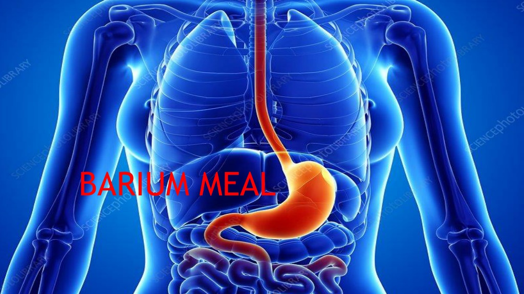
Barium meal BARIUM MEAL
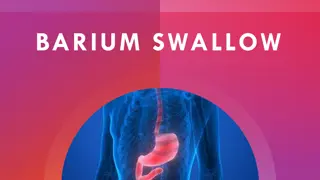
INTRODUCTION Barium meal is the radiological study of oesophagus, stomach, duodenum and proximal jejunum. It is done by oral administration of BARIUM CONTRAST.
INDICATIONS Epigastric pain suggestive of peptic ulceration. Anorexia. Weight loss. Vomiting. Anaemia. Heart burn. Dyspepsia. Upper abdominal mass. Gastro-intestinal haemorrhage. Gastric or duodenal obstruction. Malignancies of oesophago-gastric junction, stomach and duodenum. Systemic diseases like Tuberculosis affecting the upper gastrointestinal tract.
CONTRAINDICATIONS Suspected cases of gastro-duodenal perforation History or suspicion of aspiration, where alternative contrast medium should be considered. Large bowel obstruction (Barium inspissation occurs in these cases) Fistulous communication with any organs other than parts of G.I.T Recent biopsy from GIT, as barium granuloma may form at biopsy site.
PREPARATION Over night fasting or atleast 6 hours fasting Low residue diet. Avoiding smoking and chewing gum. Diabetic patient should be given early appointment. In patients with gastric outlet obstruction, prolonged fasting or intravenous Metaclopramide and sometimes nasogastric intubation and aspiration of the contents may be necessary.
CONTRAST MEDIA SINGLE CONTRAST STUDY: Low density barium suspension (80-100% w /v) is used. 30% w /v suspension is used for high kV single contrast study. Water soluble contrast media are indicated when a gastro-duodenal perforation is suspected. Use of newer non-ionic water soluble contrast media have to advocated for the detection of upper GI perforation, when there is risk of aspiration.
DOUBLE CONTRAST A high density (approximately 250% w /v), low viscosity barium suspension produces best mucosal coating and hence detail. Between 100 and 150 ml of barium suspension is usually necessary to achieve adequate double contrast studies.
CONVENTIONAL SINGLE CONTRAST STUDY 10-15 ml of 80-100% w/v barium suspension is given. Patient lying supine is rotated with the right side going up in a continuous clockwise manner for obtaining of the entire stomach mucosa. 100-250 ml of barium is given. Spot films of the filled fundus in varying obliquity maybe taken. Patient is turned prone oblique right side dependent as barium enters into the duodenum through pylorus . Spot films for duodenal bulb and C loop can be taken and in RAO.
DOUBLE CONTRAST Mass screening of the gastric tumors for early detection. This technique relies much less on fluoroscopy and more on filming which is done over couch for better image quality. This was subsequently found very useful for small mucosal lesions like polyps, mucosal erosions and ulcers, recurrent tumors and post operative studies. About 100-150 ml of high density low viscosity barium is given. Injection Buscopan is given as described before. Gas forming agents are given. Then patient is rotated slowly for mucosal coating, beginning from supine to right lateral to prone to left lateral and back to supine. Filming for various parts of stomach and duodenum is done with standard views as stated before. The table may have to be tilted 30 head up /head low to attain maximum distension of the part to be filmed. The study can be done without buscopan, rest of the procedure remains the same.
AFTER CARE The patient should be warned that his bowel motion will be white for few days after the examination and to keep his bowel open with laxative to avoid barium impaction which can be painful. The patient must not leave the department until any blurring of vision produced by Buscopan has resolved.
COMPLICATIONS Leakage of barium from an unsuspected perforation-peritonitis. Aspiration pneumonia. Barium impaction-converts a partial large bowel obstruction into a complete obstruction. Side effects from the pharmacological agents used along with barium. Acute gastric dilatation. Barium embolization if a bleeding ulcer is present.
