Understanding Bacterial Form and Function: Structures, Shapes, and Arrangements
Explore the diverse structures common to bacterial cells, including membranes, ribosomes, and chromosomes. Learn about the shapes and arrangements of bacteria, such as cocci, bacilli, and spirilla. Discover external appendages that provide motility and aid in attachment and mating.
Download Presentation

Please find below an Image/Link to download the presentation.
The content on the website is provided AS IS for your information and personal use only. It may not be sold, licensed, or shared on other websites without obtaining consent from the author. Download presentation by click this link. If you encounter any issues during the download, it is possible that the publisher has removed the file from their server.
E N D
Presentation Transcript
Bacterial Form and Function Bacterial Form and Function A. Structures common to all bacterial cells 1. Cell membrane 2. Cytoplasm 3. Ribosomes 4. One (or a few) chromosomes
Bacterial Form and Function B. Structures found in most bacterial cells 1. Cell wall 2. Surface coating or glycocalyx C. Structures found in some bacterial cells 1. Flagella 2. Pili
Bacterial Form and Function 3. Fimbriae 4. Capsules 5. Slime layers 6. Inclusions 7. Actin cytoskeleton 8. Endospores
Shapes & Arrangements D. Bacterial Arrangements and Sizes 1. Three general shapes A) Coccus roughly spherical B) Bacillus rod-shaped 1) Coccobacillus short and plump 2) Vibrio gently curved C) Spirillum curviform or spiral-shaped
Shapes & Arrangements 2. Pleomorphism when cells of a single species vary to some extent in shape and size 3. Arrangements & Groupings A) Cocci greatest variety in arrangement 1) Singles 2) Pairs (diplococci)
Shapes & Arrangements 3) Tetrads 4) Irregular clusters (staphylococci) 5) Chains (streptococci) 6) Cubical packet (sarcina)
Shapes & Arrangements B) Bacilli less varied 1) Singles 2) Pairs (diplobacilli) 3) Chains (streptobacilli) 4) Row of cells oriented side-by-side (palisades)
Shapes & Arrangements C) Spirilla 1) Usually singles 2) Occasionally found in short chains
External Structures External Structures A. Appendages: Cell extensions 1. Common but not present on all species 2. Can provide motility (flagella, pili and axial filaments) 3. Can be used for attachment and mating (pili and fimbriae)
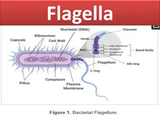
External Structures 4. Flagella A) Three parts: Filament, hook (sheath), and basal body 1) Filament a) Whip-like, helical structure 2) Hook a) Holds the filament b) Attached to the rod portion of the basal body
External Structures 3) Basal body a) A complex structure consisting of a rod, 4 rings and a motor contained within the cell envelope b) Activation of the motor causes the hook (and therefore, the filament) to swivel
External Structures B) Vary in both number and arrangement 1) Monotrichous single flagellum 2) Lophotrichous small bunches or tufts of flagella emerging from the same site 3) Amphitrichous flagella attached at both ends of the cell 4) Peritrichous dispersed randomly over the structure of the cell
External Structures C) Flagellar Function 1) Chemotaxis movement of the cell in response to a chemical signal a) 2 types i) Positive chemotaxis ii) Negative chemotaxis
External Structures b) Some photosynthetic bacteria exhibit phototaxis c) Move is accomplished through a series of runs and tumbles i) Run linear movement (a) Created by counterclockwise flagellar rotation
External Structures ii) Tumble cell stops and reverses directions or spins in place (a) Created by clockwise flagellar rotation
External Structures 5. Axial Filaments A) Also known as a periplasmic flagella B) Seen in a special group of bacteria known as spirochetes C) Consists of a filament and hook but the entire structure is located between the cell wall and membrane (the periplasmic space) D) Creates movement through twisting and flexing actions
External Structures 6. Pili A) Elongated, rigid hollow structures B) Found on some gram-negative bacteria C) Involved in attachment, movement and conjugation 7. Fimbriae A) Small, bristle-like fibers B) Tend to stick to each other and to surfaces
External Structures 8. Glycocalyx A) Develops as a coating of repeating polysaccharide units, protein, or both B) Differ among bacteria in thickness, organization, and chemical composition
External Structures 1) Slime layer a loose shield that protects some bacteria from loss of water and nutrients 2) Capsule when the glycocalyx is bound more tightly to the cell and is denser and thicker
External Structures C) Functions of the Glycocalyx 1) Protects the cell a) Formed by many pathogenic bacteria to protect the bacteria against phagocytes 2) Sometimes helps the cell adhere to the environment a) Important in formation of biofilms 3) Helps prevent the loss of water and nutrients
The Cell Envelope The Cell Envelope: The Boundary layer of Bacteria A. Majority of bacteria have a cell envelope B. Composed of two or three basic layers 1. Cell wall 2. Cell membrane 3. In some bacteria, the outer membrane
The Cell Envelope C. Differences in Cell Envelope Structure 1. The differences between gram-positive and gram-negative bacteria lie in the cell envelope 2. Gram-positive A) Two layers cell wall and cell membrane 3. Gram-negative A) Three layers outer membrane, cell wall, and cell membrane
The Cell Envelope D. Structure of the Cell Wall 1. Helps determine the shape of a bacterium 2. Provides strong structural support 3. Most are rigid because of peptidoglycan content 4. Keeps cells from rupturing because of changes in pressure due to osmosis
The Cell Envelope A) Target of many antibiotics disrupt the cell wall, and cells have little protection from lysis 5. Gram-positive cell wall A) A thick sheath of peptidoglycan B) There is little space between the cell wall and membrane (periplasmic space)
The Cell Envelope C) 2 molecules (besides peptidoglycan) are commonly found 1) Teichoic acid binds together layers of peptidoglycan 2) Lipoteichoic acid link the peptidoglycan layers to the cell membrane D) Gram positive cells walls are less susceptible to lysis (stronger) but more permeable than gram negative bacteria
The Cell Envelope 6. Gram-negative cell wall A) Single, thin sheet of peptidoglycan B) A wide periplasmic space surrounds the peptidoglycan C) Unlike gram positive bacteria, it possesses an outer membrane (a.k.a. LPS layer) 1) Similar to the cell membrane, except it contains specialized polysaccharides and proteins
The Cell Envelope 2) Innermost layer phospholipid layer anchored by lipoproteins to the peptidoglycan layer below 3) Outermost layer contains lipopolysaccharide: 2 important components a) Lipid A found within the bilayer; recognized by our immune systems
The Cell Envelope b) O-specific polysaccharide found externally; used to identify certain strains/species of bacteria (E. coli O157:H7) 4) Outer membrane serves as a partial chemical sieve a) Only relatively small molecules can penetrate
The Cell Envelope D) Gram negative bacteria are less permeable (because of the LPS) but more susceptible to lysis than gram positive bacteria E. Cell Membrane Structure 1. Also known as the cytoplasmic membrane or plasma membrane 2. Contain primarily phospholipids and proteins
The Cell Envelope 3. Functions A) Provides a site for functions such as energy reactions, nutrient processing, and synthesis B) Regulates transport (selectively permeable membrane) C) Secretion
Internal Structures Bacterial Internal Structure A. Contents of the Cell Cytoplasm 1. Gelatinous solution 2. Site for many biochemical and synthetic activities 3. 70%-80% water 4. Also contains larger, discrete cell masses (chromosome/nucleoid, plasmids, ribosomes, inclusions, and actin strands)
Internal Structures 5. Bacterial Chromosome A) Single circular strand of essential DNA B) Aggregated in a dense area of the cell the nucleoid 6. Plasmids A) Extra, nonessential pieces of DNA B) May be found floating freely in the cytoplasm or attached to the chromosome
Internal Structures C) Often confer protective traits such as drug resistance or the production of toxins and enzymes D) Can be transferred from one bacterium to another naturally or artificially, thereby transferring the traits it carries
Internal Structures 7. 70S Ribosomes A) Made of rRNA and protein B) The site of protein production in the cell
Internal Structures 8. Inclusions also known as inclusion bodies A) Some bacteria lay down nutrients in these inclusions during periods of nutrient abundance B) Serve as a storehouse when nutrients become depleted 1) Some enclose condensed, energy-rich organic substances C) Some aquatic bacterial inclusions include gas vesicles to provide buoyancy and flotation
Internal Structures 9. Actin Cytoskeleton A) Long polymers of actin B) Contribute to cell shape
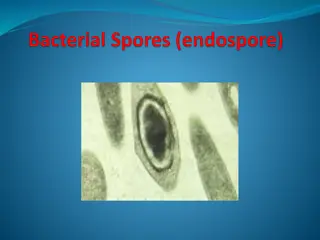
Internal Structures B. Bacterial Endospores: An Extremely Resistant Stage 1. Dormant bodies produced by Bacillus, Clostridium, and Sporosarcina 2. These bacteria have a two-phase life cycle A) Phase One: Vegetative cell 1) Metabolically active and growing
Internal Structures 2) Can be induced by the environment to undergo spore formation (sporulation) B) Phase Two: Endospore 1) Stimulus for sporulation the depletion of nutrients a) Process takes 6-8 hours 2) Vegetative cell undergoes a conversion to a sporangium 3) The DNA of the cell is duplicated
Internal Structures 4) A septum forms dividing the cell into unequal parts each with its own DNA 5) The larger portion engulfs the smaller portion resulting in a forespore 6) A thick peptidoglycan coat forms around the forespore making it impervious to other substances and heat resistant; it is now an endospore
Internal Structures 7) The endospore is released as the sporangium deteriorates 8) The endospore remains dormant until conditions improve around it
Internal Structures C) Endospores are the hardiest of all life forms 1) Withstand extremes in heat, drying, freezing, radiation, and chemicals a) Resist ordinary cleaning methods 2) Some viable endospores have been found that were more than 250 million years old
