Tutorial on Standardized Evaluation of Tumor-Infiltrating Lymphocytes in Breast Cancer
This tutorial, prepared by the International Working Group for TIL in breast cancer, provides guidelines for pathologists on the standardized evaluation of tumor-infiltrating lymphocytes (TILs) in breast cancer based on H&E slides of core biopsies or tumor resections. It details steps such as defining TIL evaluation areas, focusing on stromal TILs, and scanning tumors for evaluation. These standardized practices are essential for daily clinical practice and research in breast cancer.
Download Presentation

Please find below an Image/Link to download the presentation.
The content on the website is provided AS IS for your information and personal use only. It may not be sold, licensed, or shared on other websites without obtaining consent from the author.If you encounter any issues during the download, it is possible that the publisher has removed the file from their server.
You are allowed to download the files provided on this website for personal or commercial use, subject to the condition that they are used lawfully. All files are the property of their respective owners.
The content on the website is provided AS IS for your information and personal use only. It may not be sold, licensed, or shared on other websites without obtaining consent from the author.
E N D
Presentation Transcript
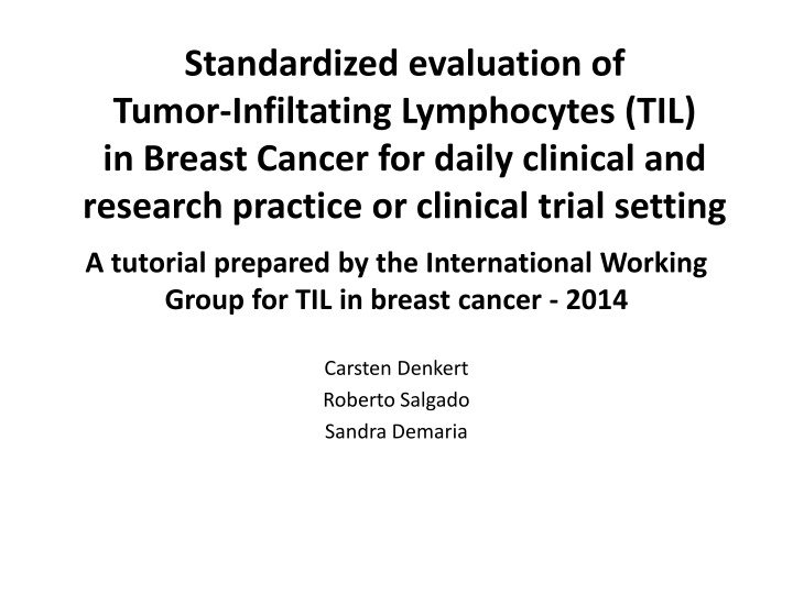
Standardized evaluation of Tumor-Infiltating Lymphocytes (TIL) in Breast Cancer for daily clinical and research practice or clinical trial setting A tutorial prepared by the International Working Group for TIL in breast cancer - 2014 Carsten Denkert Roberto Salgado Sandra Demaria
Aim of this tutorial To provide a guideline to pathologists for the standardized evaluation of tumor-infiltrating lymphocytes based on H&E slides of core biopsies or tumor resections. Please consult the manuscript for more specific details.
Step 1: Define area for TIL evaluation Only TILs within the borders of the invasive tumors are evaluated The invasive edge is included in the evaluation, but not reported separately Immune infiltrates outside of the tumor borders, e.g. in adjacent normal tissue or DCIS are not included do not include immune infiltrate outside of the tumor TLS area within tumor borders area within tumor borders Example 1 Example 2
Step 1: Define area for TIL evaluation Large areas of central necrosis or fibrosis are not included in the evaluation area for TIL evaluation do not include in evaluation Example 3
Step 2: Focus on stromal TIL In the diagnostic setting, only stromal TILs are relevant Do not include TILs in this area Include only TILs in this area = stromal TILs Example 4
Step 2: Focus on stromal TIL in the diagnostic setting, only stromal TIL are relevant Do not include TIL in this area Include only TIL in this area = stromal TIL Do not include TIL in this area Do not include TIL in this area Example 5
Step 2: Scan tumor at low magnification focus on the tumor stroma Stroma contains predominantly collagenous tissue, few round cells Stroma contains predominantly round cell infiltrate, collagenous tissue difficult to recognize Example 6 Example 6
Step 2: Scan tumor at low magnification focus on the tumor stroma Stroma contains predominantly collagenous tissue, few round cells Stroma contains predominantly round cell infiltrate, collagenous tissue difficult to recognize Example 8 Example 9
Step 2: Scan tumor at low magnification focus on the tumor stroma Stroma contains predominantly collagenous tissue, few round cells Stroma contains predominantly round cell infiltrate, collagenous tissue difficult to recognize Example 10 Example 11
Step 3: Determine type of inflammatory infiltrate Include only mononuclear infiltrate (lymphocytes & plasma cells) Do not include granulocytic infiltrate in areas of tumor necrosis do not include granulocytes in necrotic areas mononuclear stromal TIL infiltrate Example 12
Step 3: Determine type of inflammatory infiltrate Include only mononuclear infiltrate (lymphocytes & plasma cells) do not include granulocytic infiltrate in areas of tumor necrosis Example 13 Example 14
Step 4: As a first approach, include tumor in one of three groups based on low magnification and assess % stromal TILs (continue with Step 5 for percentage) Group A: tumor with no/minimal immune cells Group B: tumor with intermediate / heterogeneous infiltrate Group C: tumor with high immune infiltrate 0-10% stromal TILs 10-40% stromal TILs 40-90% stromal TILs For this intermediate group evaluate different areas at higher magnification. Example 15 Example 16
Step 5: Report percentage of stromal lymphocytes 1% 5% Report the average of the stromal area, do not focus on hot spots. For intermediate group evaluate different areas at higher magnification. Please note that lymphocytes to not form solid aggregates, therefore even with 90-100% stromal TILs there will still be some space between the individual lymphocytes. 10% 20% 60% 70% 80% 90%
Please send any questions or comments to: Carsten Denkert: carsten.denkert@charite.de Roberto Salgado: roberto@salgado.be Sandra Demaria: szd3005@med.cornell.edu
