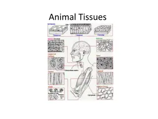
Tissue Processing and Common Fixatives Overview

undefined
TISSUE
 
PROCESSING
1.
Fixation
2.
Dehydration
3.
Clearing
4.
Impregnation
5.
Embedding
 
and
 
blocking
6.
Section
 
cutting
7.
Routine
 
staining
undefined
COMMONLY
 
USED
 
FIXATIVES
Formalin
 
 
MC
 
 
routine
Glutaraldehyde
 
 
electron
 
microscopy
Picric
 
acid(Bouin’s
 
solution)
 
 
renal
 
&
 
testicular
 
tissue
Alcohol(Carnoy’s
 
fixative)
 
 
cytologic
 
smears,
 
endometrial
 
sampling
Osmium
 
tetraoxide
 
 
CNS
 
tissues
 
&
 
electron
 
microscopy
undefined
undefined
undefined
undefined
undefined
Pathophysiology
 
of
Endocrine
 
System
Dr.
Athmar mohamed
Pituitary
 
disorders
Causes
 
:
tumor
hemorrhage
trauma
irradiation
Pituitary
 
disorders
G
r
o
w
t
h
 
h
o
r
m
o
n
e
d
e
f
i
c
i
e
n
c
y
 
children
 
-
 
short
 
stature
(
p
i
t
u
i
t
a
r
y
 
d
w
a
r
f
i
s
m
)
,
n
o
r
m
a
l
 
i
n
t
e
l
l
i
g
e
n
c
e
,
o
b
e
s
i
t
y
 
adults
 
-
 
central
 
obesity,
reduced
 
muscle
 
mass
and
 
exercise
 
capacity.
(pituitary
dwarfism),
Pituitary
 
disorders
E
x
c
e
s
s
 
o
f
 
G
H
 
i
n
 
c
h
i
l
d
h
o
o
d
P
i
t
u
i
t
a
r
y
 
g
i
g
a
n
t
i
s
m
 
 
growth
 
velocity
 
proportional
 
enlargement
 
of
skeleton
 
and
 
inner
 
organs
 
enlargement
 
of
 
the
peripheral
 
nerves
 
delayed
 
puberty
 
and
hypogonadism
Pituitary
 
gigantism
Pituitary
 
disorders
E
x
c
e
s
s
 
o
f
 
G
H
 
i
n
 
a
d
u
l
t
s
A
c
r
o
m
e
g
a
l
y
 
reason
 
–somatotrope
 
adenoma
 
hyperplasia
 
and
 
hypertrophy
 
of
 
soft
tissues
  
 
size
 
of
 
hands,
 
feet,
 
nose,
 
ears
 
cartilaginous
 
proliferation
 
of
 
the
larynx
  
coarsening
 
of
 
the
 
facial
 
features.
 
enlarged
 
tongue
 
and
 
increase
 
of
inner
 
organs
 
thick
 
and
 
dark
 
skin.
 
degenerative
 
arthritis.
Acromegaly
Thyroid
 
disorders
.
G
o
i
t
e
r
 
-
 
 
s
i
z
e
 
o
f
 
t
h
e
 
t
h
y
r
o
i
d
 
g
l
a
n
d
(not
 
related
 
of
 
TH
 level)
 
Complications
 
of
 
goiter:
 
difficulty
 
in
 
swallowing,
 
distention
 
of
 
the
 
veins
 
of
 
the
neck
 
and
 
upper
 
extremities,
 
edema
 
of
 
the
 
eyelids
 and
conjunctiva,
 
syncope
 
with
 
coughing.
Goit
e
r
undefined
Thyroid
 
disorders
C
o
n
g
e
n
i
t
a
l
 
h
y
p
o
t
h
y
r
o
i
d
i
s
m
 
-
c
r
e
t
i
n
i
s
m
 
C
a
u
s
e
s
 
congenital
 
absence
 
of
 
the
thyroid
 
gland
 
abnormal
 
biosynthesis
 
of
thyroid
 
hormone
 
deficient
 
TSH
 
secretion.
 
Clinics:
 
mental
 
retardation,
impaired
 
growth.
undefined
Thyroid
 
disorders
A
c
q
u
i
r
e
d
 
h
y
p
o
t
h
y
r
o
i
d
i
s
m
 
m
y
x
e
d
e
m
a
 
accumulation
 
of
 
a
mucopolysaccharide
substance
 
in
 
the
 
connective
tissues.
 
Causes
 
destruction
 
or
 
dysfunction
of
 
the
 
thyroid
 
gland,
 
impaired
 
pituitary
 
function
 
hypothalamic
 
dysfunction.
Disorders
 
of
 
gall
 
bladder
By
Dr.
Athmar mohamed
2
 
main
 
types
 
of
 
gall
 
stones
 
:
Cholesterol
 
stones
Pigment
 
stones
In
 
the
 
West,
 
about
 
90%
 
are
 
cholesterol
 
stones.
Pigment
 
gall
 
stone
 
is
 
predominant
 
in
 
non-
Western
 
population
 
 
associated
 
with
bacterial
 
infection
 
of
 
biliary
 
tree
 
and
 
parasitic
infestations.
Cholesterol
 
stone
Content
 
:
 Crystalline
 
cholesterol
monohydrate
 
is
 
predominant
Pigment
 
stone
Bilirubin
 
calcium
 
salt
 
is
predominant
Pigment
 
stones
 
Black
 
pigment
 
,
hemolytic
 
anemia
(hemoglobinopathy,
 
red
 
cell
disorders)
 
Brown
 
pigment
 
 
Asian
patients
 
(infection)
Pathology
Cholesterol
 
stones
 
:
Gross
 
:
 
pale
 
yellow,
 
ovoid,
 
firm,
 
single
 
to
 
multiple
 
with
 
faceted
 
surfaces
Mostly
 
radiolucent,
 
20%
 
is
radio
 
opaque
 
due
 
to
 
the
presence
 
of
 
calcium
 
carbonate
content.
:
Pigment
 
stones
Black
 
stone
 
(in
 
sterile
 
gall
 
bladder
bile)-
 
small
 
size,
 
fragile
 
to
 
touch,
numerous,
 
50-
70%
 
are
 
radioopaque
Brown
 
stone
 
(in
 
infected
 
intrahepatic
or
 
extrahepatic
 
ducts)-
 
single
 
to
 
a
few,
 
soft,
 greasy,
 
soaplike
 
consistency
due
 
to
 
presence
 
of
 
retained
 
fatty
acids
 
released
 
by
 
bacterial
phospholipases
 
on
 
biliary
 
lecithins,
radiolucent.
Stone
 
content
 
:
 
calcium
 
salts
 of
unconjugated
 bilirubin,
 
lesser
amounts
 
of
 
other
 
calcium
 
salts,
mucin
 
glycoproteins
 
and
 
cholesterol.
Summary
 
:
Complications
 
:
Inflammation
 
of
 
gall
bladder
 
(cholecystitis)
Empyema
Perforation,
Fistulas
Complications-
cont
Inflammation
 
of
 
biliary
 
tree
 
(cholangitis)
Obstructive
 
cholestasis
Pancreatitis
Erode
 
adjacent
 
bowel
 
and
 
cause
 
intestinal
 
obstruction
 
(gallstone
 
ileus)
Summary:
Pathology
 
of
 
acute
 
cholecystitis
Gross
 
:
Enlarged,
 
tense,
 
edematous,
 
red
 
or
violaceous
 
colour
 
(subserosal
haemorrhage)
Fibrinous
 
/fibrinopurulent
 
exudate
covering
 
the
 
serosa
 
+-
 
stones
 
obstructing
 
the
 
neck
 
or
cystic
 
duct
Lumen
 
contains
 
blood
 
and
 
pus
(empyema)
Green
 
black
 
necrotic
Microscopic
 
:
acute
 
inflammation
 
in
 
the
 
wall
mucosal
 
ulceration.
May
 
be
 
associated
 with
 
abscess
formation
 
or
 
gangrenous
 
necrosis.
Clinical
 
features
 
:
recurrent
 
attacks
 
of
 
epigastric
 
or
right
 
upper
 
quadrant
 
pain
Nausea,
 
vomiting
 
and
 
intolerance
 
to
 
fatty
 
foods.
Pathology:
Gross
 
:
smooth
 
and
 
glistening
 
to
 
dull
 
serosa
 
(subserosal
fibrosis)
thickened
 
wall,
 
opaque
 
gray-
white
 
appearance
Uncomplicated 
cases,
 
lumen
 
contains
 
clear, 
green,
mucoid
 
bile
 
and
 
stones
 
with
 
normal
 
mucosa
Microscopic
 
:
Reactive
 
proliferation
 
of
 
mucosa
Inflammation
 
(lymphocytes,
 
plasma
 
cells,
 
and
 
macrophages
 
in
 
the
 
mucosa
 
and
 
in
 
the
 
subserosal
 
fibrous
 
tissue).
 
May
 
be
 
minimal.
Prominent
 
outpouching 
of
 
the
 
mucosal
 
epithelium
 
through
 
the
 
wall
 
(Rokitansky
 
Aschoff
 
sinuses)
Marked
 
subepithelial
 and
 
subserosal
 
fibrosis
 
+-
Superimposed
 
acute
 
inflammation
 
+-
Extensive
 
calcification
 
within
 
the
 
wall
→porcelain
 
gall
 
bladder
 
→increase
 
risk
 
of
 
cancer
Xanthogranulomatous
cholecystitis: 
massively
thickened
 
wall
 
with
shrunken,
 
nodular,
chronically
 
inflamed
with
 
foci
 
of
 
necrosis
 
and
haemorrhage.
Hydrops
 
of
 
the
 
gall
bladder
 
:
 
atrophic,
chronically
 
obstructed
gall
 
bladder
 
containing
only
 
clear
 
secretion
Complications
 
of
 
cholecystitis
Bacterial
 
superinfection
 
with
cholangitis
 
or
 
sepsis
Gall
 
bladder
 
perforation
 
and
 
local
abscess
 
formation
Gall
 
bladder
 
rupture
 
with
 
diffuse
peritonitis
Biliary
 
enteric
 
(cholecystenteric)
fistula,
 
with
 
drainage
 
of
 
bile
 
into
adjacent
 
organs,
 
entry
 
of
 
air
 
and
bacteria
 
into
 
biliary
 
tree
 
and
potentially
 
gallstone-
induced
intestinal
 
obstruction
 
(ileus)
Aggravating
 
of
 
preexisting
medical
 
illness,
 
with
 
cardiac,
pulmonary,
 
renal
 
or
 liver
decompensation
Porcelain
 
gall
 
bladder
 
with
increased
 
risk
 
of
 
cancer
Treatment
 
:
 
Cholecystectomy
Slide Note
Embed
Share

In tissue processing, the steps include fixation, dehydration, clearing, impregnation, embedding, section cutting, and routine staining. Common fixatives like Formalin, Glutaraldehyde, and Picric acid are used for different tissues. Understand the pathophysiology of the endocrine system and pituitary disorders such as growth hormone deficiency and excess of growth hormone in children and adults. Explore thyroid disorders like goiter and their complications.

  • Tissue Processing
  • Fixatives
  • Pathophysiology
  • Endocrine System
  • Pituitary Disorders

Uploaded on Feb 24, 2025 | 0 Views


Download Presentation

Please find below an Image/Link to download the presentation.

The content on the website is provided AS IS for your information and personal use only. It may not be sold, licensed, or shared on other websites without obtaining consent from the author. Download presentation by click this link. If you encounter any issues during the download, it is possible that the publisher has removed the file from their server.

E N D

Presentation Transcript


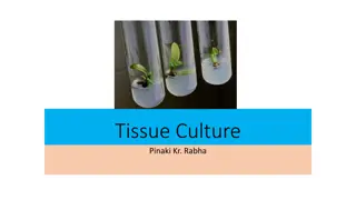
  1. TISSUE PROCESSING Fixation 1. Dehydration 2. Clearing 3. Impregnation 4. Embedding and blocking 5. Section cutting 6. Routine staining 7.

  2. COMMONLY USED FIXATIVES Formalin MC routine Glutaraldehyde electron microscopy Picric acid(Bouin s solution) renal & testicular tissue Alcohol(Carnoy s fixative) cytologic smears, endometrial sampling Osmium tetraoxide CNS tissues & electron microscopy

  3. Pathophysiology of Endocrine System Dr.Athmar mohamed

  4. Pituitary disorders Causes : tumor hemorrhage trauma irradiation

  5. Pituitary disorders Growth hormone deficiency children - short stature (pituitary dwarfism), normal intelligence, obesity adults - central obesity, reduced muscle mass and exercise capacity. (pituitary dwarfism),

  6. Pituitary disorders Excess of GH in childhood Pituitary gigantism growth velocity proportional enlargement of skeleton and inner organs enlargement of the peripheral nerves delayed puberty and hypogonadism Pituitary gigantism

  7. Pituitary disorders Excess of GH in adults Acromegaly reason somatotrope adenoma hyperplasia and hypertrophy of soft tissues size of hands, feet, nose, ears cartilaginous proliferation of the larynx coarsening of the facial features. enlarged tongue and increase of inner organs thick and dark skin. degenerative arthritis. Acromegaly

  8. Thyroid disorders Goiter Goiter - size of the thyroid gland (not related of TH level) Complications of goiter: difficulty in swallowing, distention of the veins of the neck and upper extremities, edema of the eyelids and conjunctiva, syncope with coughing. .

  9. Thyroid disorders Congenital hypothyroidism - cretinism Causes congenital absence of the thyroid gland abnormal biosynthesis of thyroid hormone deficient TSH secretion. Clinics: mental retardation, impaired growth.

  10. Thyroid disorders Acquired hypothyroidism myxedema accumulation of a mucopolysaccharide substance in the connective tissues. Causes destruction or dysfunction of the thyroid gland, impaired pituitary function hypothalamic dysfunction.

  11. Disorders of gall bladder By Dr.Athmar mohamed

  12. 2 main types of gall stones : Cholesterol stones Pigment stones In the West, about 90% are cholesterol stones. Pigment gall stone is predominant in non- Western population associated with bacterial infection of biliary tree and parasitic infestations.

  13. Pigment stone Bilirubin calcium salt is predominant Pigment stones Black pigment , hemolytic anemia (hemoglobinopathy, red cell disorders) Brown pigment Asian patients (infection) Cholesterol stone Content : Crystalline cholesterol monohydrate is predominant

  14. Pathology Pigment stones Cholesterol stones : : Black stone (in sterile gall bladder bile)- small size, fragile to touch, numerous, 50-70% are radioopaque Brown stone (in infected intrahepatic or extrahepatic ducts)- single to a few, soft, greasy, soaplike consistency due to presence of retained fatty acids released by bacterial phospholipases on biliary lecithins, radiolucent. Stone content : calcium salts of unconjugated bilirubin, lesser amounts of other calcium salts, mucin glycoproteins and cholesterol. Gross : pale yellow, ovoid, firm, single to multiple with faceted surfaces Mostly radiolucent, 20% is radio opaque due to the presence of calcium carbonate content.

  15. Summary :

  16. Complications : Inflammation of gall bladder (cholecystitis) Empyema Perforation, Fistulas

  17. Complications-cont Inflammation of biliary tree (cholangitis) Obstructive cholestasis Pancreatitis Erode adjacent bowel and cause intestinal obstruction (gallstone ileus)

  18. Summary:

  19. Pathology of acute cholecystitis Gross : Enlarged, tense, edematous, red or violaceous colour (subserosal haemorrhage) Fibrinous /fibrinopurulent exudate covering the serosa +- stones obstructing the neck or cystic duct Lumen contains blood and pus (empyema) Green black necrotic Microscopic : acute inflammation in the wall mucosal ulceration. May be associated with abscess formation or gangrenous necrosis.

  20. Clinical features : recurrent attacks of epigastric or right upper quadrant pain Nausea, vomiting and intolerance to fatty foods. Pathology: Gross : smooth and glistening to dull serosa (subserosal fibrosis) thickened wall, opaque gray-white appearance Uncomplicated cases, lumen contains clear, green, mucoid bile and stones with normal mucosa

  21. Microscopic : Reactive proliferation of mucosa Inflammation (lymphocytes, plasma cells, and macrophages in the mucosa and in the subserosal fibrous tissue). May be minimal.

  22. Prominent outpouching of the mucosal epithelium through the wall (Rokitansky Aschoff sinuses) Marked subepithelial and subserosal fibrosis +-Superimposed acute inflammation +-Extensive calcification within the wall porcelain gall bladder increase risk of cancer

  23. Xanthogranulomatous cholecystitis: massively thickened wall with shrunken, nodular, chronically inflamed with foci of necrosis and haemorrhage. Hydrops of the gall bladder : atrophic, chronically obstructed gall bladder containing only clear secretion

  24. Complications of cholecystitis Bacterial superinfection with cholangitis or sepsis Gall bladder perforation and local abscess formation Gall bladder rupture with diffuse peritonitis Biliary enteric (cholecystenteric) fistula, with drainage of bile into adjacent organs, entry of air and bacteria into biliary tree and potentially gallstone-induced intestinal obstruction (ileus) Aggravating of preexisting medical illness, with cardiac, pulmonary, renal or liver decompensation Porcelain gall bladder with increased risk of cancer

  25. Treatment : Cholecystectomy

More Related Content

giItT1WQy@!-/#giItT1WQy@!-/#giItT1WQy@!-/#giItT1WQy@!-/#giItT1WQy@!-/#giItT1WQy@!-/#giItT1WQy@!-/#giItT1WQy@!-/#giItT1WQy@!-/#giItT1WQy@!-/#giItT1WQy@!-/#