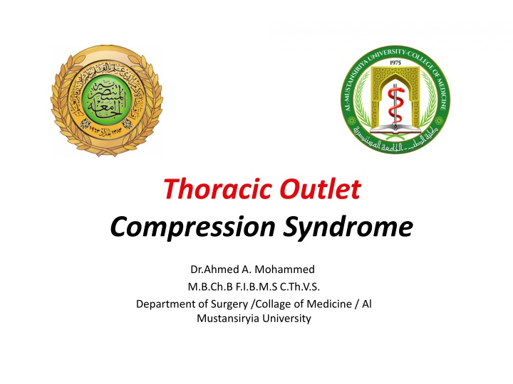
Thoracic Outlet Compression Syndrome
Thoracic Outlet Compression Syndrome is characterized by upper extremity pain and paresthesias due to vascular or neurologic compression at the thoracic outlet. Learn about its anatomical features, incidence, etiology, pathophysiology, clinical manifestations, and diagnosis.

Download Presentation

Please find below an Image/Link to download the presentation.
The content on the website is provided AS IS for your information and personal use only. It may not be sold, licensed, or shared on other websites without obtaining consent from the author.If you encounter any issues during the download, it is possible that the publisher has removed the file from their server.
You are allowed to download the files provided on this website for personal or commercial use, subject to the condition that they are used lawfully. All files are the property of their respective owners.
The content on the website is provided AS IS for your information and personal use only. It may not be sold, licensed, or shared on other websites without obtaining consent from the author.
E N D
Presentation Transcript
Thoracic Outlet Compression Syndrome Dr.Ahmed A. Mohammed M.B.Ch.B F.I.B.M.S C.Th.V.S. Department of Surgery /Collage of Medicine / Al Mustansiryia University
T Thoracic horacic O Outlet C Compression ompression S Syndrome utlet yndrome Is a symptoms complex of upper Is a symptoms complex of upper - - extremity pain and parasthesias resulting from vascular or neurologic parasthesias resulting from vascular or neurologic compression of the subclavian vessels or brachial compression of the subclavian vessels or brachial plexus as they pass through the thoracic outlet. plexus as they pass through the thoracic outlet. extremity pain and
Anatomic Features The Thoracic outlet is bounded by the 1stdorsal vertebra post- eriorly, the upper border of the manubrium sterni anteriorly & the 1strib on each side crossed by clavicles posterolaterally in a scissor-like fashion. The subclavian vessels and the brachial plexus cross the 1st rib through the cervicoaxillary canal in order to reach the upper limb.
Anatomic Features Neurovascular Compression occurs in 3 anatomic areas:- 1. Scalene Triangle. 2. Costoclavicular Space. 3. Subcoracoid Space.
Incidence Normal Frequency Incidence of Cervical Rib Female:Male Ratio 0.12-1% 0.5 % 1.76:1
Etiology 1. Bony Abnormalities. 2. Soft Tissue Abnormalities. 3. Traumatic Causes. 4. Rare Causes.
Clinical Manifestations Neurological Vascular Mixed
Diagnosis 1. Physical Evaluation 2. Radiographic Evaluation 3. Doppler Study 4. MRI Study 5. Electrodiagnostic Study
History and Clinical examination History: Trauma Occupation Exercise Part of general disease Spinal cord tumor Osteoarthritis Conn.tissue disease Periphral emboli Carpal tunnel synd. atheroscelrosis Physical exam. Neurological Arterial venous
Diagnostic tests for TOS Adson(scalene test) Deep breath Extend neck Turn chin toward the exam side Radial a. ve pulse Halsted(costoclavicular test): Exaggerated military position Shoulder back Subclavian art. Between clavicle & 1strib.
Diagnostic tests for TOS Hyperabduction test: Move hands passively to hyper abduction-artery compression by pectoralis minor m. 3 min. elevated arm stress test(roos test): Both arms abducted Externally rotated Elbow flexed in 90 degree. Close and open hands
Investigations for TOS Chest & neck radiograph: Bony deformity Tumor Vertebral abnormality Ct scan Electromyography EMG & NCS SSEP Arteriography Plethysmography Colour Doppler U/S MRI & MRA Myelogram: Cervical disc Other causes of cord compression
Treatment of TOS Conservative( non operative): Wt. reduction Exercise programs Chang the occupation Results: Improve symp. 50%-70% of patients More successful in: Obese Middle age Poor posture
Surgical Treatment Surgical Treatment Surgical Treatment : Indication: 1. 1. No response to No response to 4 4 ms. 2. 2. Progression of motor or sensory Progression of motor or sensory symp 3. 3. Excessive Excessive prolonge prolongeulnar or median NCS velocities. 4. 4. Narrowing or occlusion of subclavian A. Narrowing or occlusion of subclavian A. 5. 5. Thrombosis of axillary or subclavian veins Thrombosis of axillary or subclavian veins ms. Or more trials of conservative Rx. Or more trials of conservative Rx. symp. . ulnar or median NCS velocities. Approaches: Transaxillary TransaxillaryApproach Supraclavicular Supraclavicular Approach Parascapular ParascapularApproach Double Double- -Route Route Approach Approach Approach Approach Approach
S Staging of patient with TOS taging of patient with TOS Advantage: more objective surgical evaluation Priciple: Clinical evaluation Patient history Stage 0 : asymptomatic Stage I : appearance of symptoms with provocative tests Stage II : symptoms in daily life. Stage III : deprivational symptoms that hinder performance of daily work
Differential Dx. Neurological Cervical spine Spinal cord tumor Osteoarthritis Traumatic arthritis Musculofascial Myofascitis Tendinites capsulitis Vascular Arterial: Atherosclerosis Embolism Raynoid disease Vasculitis Collagen vascular diseases Venous: Thrombophlebitis Mediastinal venous obstruction Others Cardiac diseases Esophageal diseases Pulmonary diseases
Conclusion TOCS more common in females than males TOCS more common on the right than left TOCS more common in the 3rddecade TOCS most commonly caused by cervical rib TOCS not necessarily caused by cervical rib TOCS even when bilateral, one side predominate TOCS ; neurological symptoms > vascular symptoms Physical tests are useful but not confirm diagnosis
CXR & EMG are important in all cases Doppler & Angiography are effective in vascular TOCS CT scan & MRI are very effective Conservative measures have to be started in all cases but are usually of little value in our patients Para scapular approach is preferred by most surgeons Scalenotomy, fibrous band division & 1strib removal are important points in surgical treatment