The Nervous System
NERVOUS SYSTEM
Prof. Dr. Dalya Basil
Nervous System
◦
The
nervous system
is a complex collection
of
nerves
and specialized cells known as
neurons
that transmit signals between different parts of
the body.
◦
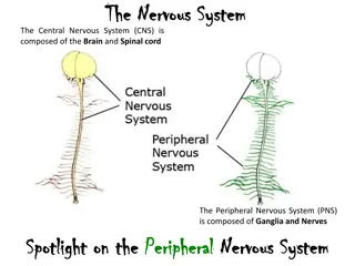
Anatomically, the nervous system is divided into
central nervous system (CNS) & peripheral
nervous system (PNS).
Further divisions of each system are
shown in the below figure.
Some important definitions
◦
Neuron:
(the nerve cell): a highly specialized cell that makes
the nervous tissue & does its functions. Neurons have different
sizes, shapes & functions. However, they all have a cell body,
from which cytoplasmic processes (called the dendrites) emerge,
one of them (called the axon) is long & usually has branches
(axon terminals) at its end. Neurons receive nerve signals
(electrical signals = action potentials) from other cells via the
dendrites, & send those signals to other cells via the axon (in one
direction: cell body axon).
Some important definitions
◦
Neurotransmitter:
a chemical material secreted from the axon terminals
of a neuron upon the arrival of nerve signal. They reach the other cell (another
neuron, muscle cell, gland cell, etc) causing certain effect.
◦
Nervous tissue:
a specialized tissue made of nerve cells (neurons) &
supportive cells (glial cells) or (neuroglia). Nervous tissue is very soft & delicate,
& needs to be protected by harder tissues (eg: bones).
◦
Nerve:
a collection of large number of axons bundled together in the PNS.
◦
Tract:
a collection of large number of axons bundled together in the CNS.
Some important definitions
◦
Sensory nerve:
a nerve that transmits signals from the body tissues to the CNS
◦
Motor nerve:
a nerve that transmits signals from the CNS to the body tissues
Note: some nerves contain both sensory & motor fibers.
◦
Myelinated nerve fibers:
axons surrounded by a myelin sheath (an
insulating lipid layer)
◦
Non-myelinated nerve fibers:
axons not surrounded by a myelin sheath
◦
Nucleus:
a collection of nerve cell bodies in the CNS.
◦
Ganglion:
a collection of nerve cell bodies in the PNS. Each ganglion receives
pre-ganglionic (afferent) fibers & sends post-ganglionic (efferent) fibers. Ganglia
are present in: (1) some sensory nerves (sensory ganglia), (2) sympathetic &
parasympathetic nervous systems (sympathetic & parasympathetic ganglia,
respectively).
Functional Anatomy of the Nervous
System
◦
The CNS is made of the brain & the spinal cord. The brain is the
higher center that receives information (stimuli) from the external
environment & internal body tissues, analyzes them & makes the
proper response.
◦
The brain is also responsible for consciousness, thinking, emotions,
& many other functions. The spinal cord is the part of CNS that
connects the brain to the spinal nerves (so, all information
received by the spinal nerves reach the brain through the spinal
cord, & all commands from the brain reach the spinal nerves
through the spinal cord). In addition, the spinal cord is responsible
for many nervous "reflexes".
Functional Anatomy of the Nervous
System
◦
The PNS is made of the cranial nerves & the spinal nerves. The
cranial nerves are connected to the brain directly, & are
distributed mainly in the head & neck. The spinal nerves are
connected to the brain indirectly via the spinal cord, & are
distributed in the neck & the rest of the body.
◦
The autonomic nervous system (ANS) is part of the PNS. However,
its function is controlled by the CNS. It is divided into sympathetic,
parasympathetic, & enteric nervous systems. The fibers of the ANS
are distributed through the PNS (both spinal & cranial nerves). The
ANS regulates the involuntary body activities (eg: heart beats,
respiration, secretion & movement of the GIT, etc).
Anatomy of the Central Nervous
System (CNS)
◦
THE BRAIN
◦
The brain is the organ situated in the cranial cavity of the skull, that
ensures an ideal protection to its soft structure. The brain is -simply- a
collection of billions of neurons, & a larger number of glial cells,
arranged in a specific way: the nerve cell bodies are gathered in the
outer zone of the brain, forming the "gray matter" or "cortex", while
the myelinated axons of those neurons occupy the interior of the
brain forming the "white matter".
Anatomy of the Central Nervous
System (CNS)
◦
This arrangement is inverted in the "brainstem", in which the gray
matter lies inside the white matter. The axons forming the white
matter are grouped in bundles or "tracts" that originate in certain
areas of the cortex & terminate in other regions in the brain or
spinal cord. Note the following regarding the brain:
◦
The largest parts of the brain are the 2 cerebral hemispheres
(cerebrum). Each hemisphere is divided into 4 lobes.
◦
The frontal lobe: the anterior part of cerebrum, responsible for
voluntary movement, speaking, thinking, emotions, & other
functions.
Anatomy of the Central Nervous
System (CNS)
◦
The parietal lobe: the supero-lateral part of cerebrum,
responsible for sensation, spatial orientation, understanding
language, & other functions.
◦
The temporal lobe: the infero-lateral part of cerebrum,
responsible for memory, hearing, balance, feeling of hunger, &
other functions.
◦
The occipital lobe: the posterior part of cerebrum, responsible for
vision & other functions.
◦
The right hemisphere of the brain controls the left side of the
body, & vice versa.
Anatomy of the Central Nervous
System (CNS)
◦
The "cerebellum" is the postero-inferior part of the brain. It is
responsible for maintaining muscle tone, involuntary planning &
control of complex movements, & balance.
◦
The "brainstem" is a large midline mass of white matter that
connects the cerebrum & cerebellum to the spinal cord. Inside it,
the brainstem contains many "nuclei", which are islets of gray
matter (collections of nerve cell bodies) that serve the following
functions:
◦
Sleep/wakefulness control
◦
Control of the cranial nerves
◦
Function of the respiratory system
◦
Function of the cardiovascular system
THE SPINAL CORD
◦
The spinal cord is the elongated part of the CNS located inside the vertebral
column. Like the brainstem, the spinal cord has white matter outside & gray
matter inside. The spinal cord white matter consists of ascending &
descending bundles of nerve fibers (tracts) that transport signals from & to the
brain. The gray matter of the spinal cord is aggregation of nerve cell bodies
arranged as an inner column with H-shape cross section, having 2 anterior
horns & 2 posterior horns & a transverse part.
THE SPINAL CORD
◦
Note the following regarding the spinal cord:
◦
The spinal cord gives rise to 31 spinal nerves on each side.
◦
The anterior horns contain the cell bodies of the large motor
neurons.
◦
The posterior horns contain the cell bodies of the smaller sensory
neurons.
◦
The spinal cord mediates some "reflexes". A reflex is a sudden
involuntary motor action ( = muscles contraction) in response to
sudden sensory stimuli. This action is completed at the spinal cord
level, without involving the brain.
Anatomy of the Peripheral Nervous
System (PNS)
◦
CRANIAL NERVES:
These are 12 pairs of nerves that are connected directly
to the brain, & are responsible for supplying motor & sensory fibers to the head
& neck region, & the viscera of the neck, thorax & abdomen.
◦
Olfactory nerves (I): small nerve fibers in the upper part of nasal cavity,
responsible for olfaction (smelling)
◦
Optic nerve (II): the nerve responsible for vision. It gathers signals from the
retina of the eye & sends them to the visual cortex in the occipital lobe.
◦
Oculomotor nerve (III): the motor supply of the extra-ocular muscles (that
move the eyeball) plus the sphincter muscle of the pupil.
◦
Trochlear nerve (IV): the motor supply of one extra-ocular muscle (the
trochlear muscle)
Anatomy of the Peripheral Nervous
System (PNS)
◦
Trigeminal nerve (V): the biggest cranial nerve, responsible for the sensory
supply of the head. It also gives motor supply to the muscles of mastication
(moving the mandible)
◦
Abducent nerve (VI): the motor supply of one extra-ocular muscle (the lateral
rectus muscle)
◦
Facial nerve (VII): the motor supply of facial muscles (of expression). It also
supplies the anterior 2/3rd of the tongue with taste sensory fibers.
◦
Vestibulo-choclear nerve (VIII): the nerve of hearing & balance, connected to
the inner ear.
◦
Glosopharyngeal nerve (IX): the motor supply of the pharyngeal muscles, it
also supplies sensory & taste sensory fibers to the posterior 1/3rd of the tongue.
Anatomy of the Peripheral Nervous
System (PNS)
◦
Vagus nerve (X): the longest cranial nerve. It has motor fibers (to the larynx &
pharynx), sensory fibers, taste sensory fibers, & preganglionic parasympathetic
fibers to the viscera of the thorax & abdomen.
◦
Accessory nerve (XI): the motor supply of sternocleidomastoid & trapezius
muscles.
◦
Hypoglossal nerve (XII): the motor supply of the tongue muscles.
SPINAL NERVES
◦
These are 31 pairs of nerves connected to the lateral aspect of the spinal cord.
They include: 8 cervical, 12 thoracic, 5 lumbar, 5 sacral, & 1 coccygeal nerves.
Note the following:
◦
Each spinal nerve, after exiting the vertebral canal, divides into ventral &
dorsal branches.
◦
Lower cervical & upper thoracic spinal nerves form a plexus (the brachial
plexus), which gives rise to the nerves of the upper limb. The main nerves of the
upper limb are the median nerve (anteriorly), the ulnar nerve (medially) & the
radial nerve (posteriorly).
SPINAL NERVES
◦
Lumbar & sacral spinal nerves form a plexus (the lumbosacral plexus), which
gives rise to the nerves of the lower limb. The main nerves of the lower limb are
the femoral nerve (anteriorly), the obturator nerve (medially) & the sciatic
nerve (posteriorly).
◦
Dermatomes: a dermatome is the area of skin supplied by a specific spinal
nerve. In general, dermatomes are arranged as follows:
◦
The cervical spinal nerves supply the skin of the back of the head, the whole
neck, shoulders, lateral sides of the arm & forearm, & the whole hand.
◦
The thoracic spinal nerves supply the skin of medial sides of the arm & forearm,
axilla, & the trunk to the level of pelvic bones.
SPINAL NERVES
◦
The lumbar spinal nerves supply the skin of the pelvis (except the buttocks) &
the whole lower limb (except the posterior aspect & the sole of the foot)
◦
The sacral spinal nerves supply the skin of the genitalia, buttocks & the
posterior aspect of the lower limb & the sole of the foot.
◦
The coccygeal spinal nerve supplies the skin around the anus.
The Autonomic Nervous System
(ANS)
◦
The autonomic nervous system is a group of nerves & ganglia
distributed throughout the body, supplying mainly the smooth
muscles, glands, & the viscera of the body. This system controls
our involuntary activities related to the different functions of the
body systems, eg: heart rate, blood pressure, secretion of the
glands, respiration, GIT movements, & many other functions. The
autonomic nervous system is divided into the sympathetic &
parasympathetic divisions, with each division having the opposite
function of the other.
SYMPATHETIC NERVOUS SYSTEM
◦
Anatomically, the sympathetic system is made of sympathetic ganglia
connected to sympathetic nerves. Note the following:
◦
Sympathetic ganglia are arranged as 2 sympathetic chains, one on each side
of the vertebral column. In addition, there are few sympathetic ganglia in the
abdomen. Functionally, the medulla of the adrenal gland is also considered as
a large sympathetic ganglion.
◦
Sympathetic ganglia receive preganglionic fibers from the spinal cord.
Preganglionic neurons secret acetylcholine (ACh) in the ganglion (so they are
"cholinergic" neurons)
SYMPATHETIC NERVOUS SYSTEM
◦
Sympathetic ganglia give postganglionic fibers to the body tissues & organs.
Postganglionic neurons secret noradrenaline (NA) at the target organ (so they
are "adrenergic" neurons)
◦
Actions mediated by the sympathetic system: increasing heart rate, blood
pressure, cardiac output, sweating, relaxation of bronchial smooth muscles
(bronchodilation), etc.
PARASYMPATHETIC NERVOUS SYSTEM
◦
Anatomically, the parasympathetic system is made of
parasympathetic ganglia connected to parasympathetic nerves.
Note the following:
◦
Parasympathetic ganglia are located near the target organs.
◦
Actions mediated by the parasympathetic system: decreasing
heart rate, blood pressure, cardiac output, increasing GIT
secretion & movement, contraction of bronchial smooth muscles
(bronchoconstriction), etc.
Enteric Nervous System (ENS)
◦
This is a complex, extensive network of nerve fibers &
ganglia spread throughout the gastrointestinal tract
(GIT), specifically in the submucosa & muscularis layers
of the tract. This system receives innervation from the
sympathetic & parasympathetic systems, & controls the
sensation, secretion, & movements of the GIT.
The nervous system, a complex network of neurons transmitting signals throughout the body, is divided into the central and peripheral nervous systems. Neurons, neurotransmitters, and nerve structures play vital roles in this system. Explore the details and divisions within each system, learn about neurons, neurotransmitters, nerves, ganglia, and more.

Download Presentation

Please find below an Image/Link to download the presentation.
The content on the website is provided AS IS for your information and personal use only. It may not be sold, licensed, or shared on other websites without obtaining consent from the author.If you encounter any issues during the download, it is possible that the publisher has removed the file from their server.
You are allowed to download the files provided on this website for personal or commercial use, subject to the condition that they are used lawfully. All files are the property of their respective owners.
The content on the website is provided AS IS for your information and personal use only. It may not be sold, licensed, or shared on other websites without obtaining consent from the author.
E N D
Presentation Transcript
NERVOUS SYSTEM Prof. Dr. Dalya Basil
Nervous System The nervous system is a complex collection of nerves and specialized cells known as neurons that transmit signals between different parts of the body. Anatomically, the nervous system is divided into central nervous system nervous system (PNS). (CNS) & peripheral
Further divisions of each system are shown in the below figure.
Some important definitions Neuron: (the nerve cell): a highly specialized cell that makes the nervous tissue & does its functions. Neurons have different sizes, shapes & functions. However, they all have a cell body, from which cytoplasmic processes (called the dendrites) emerge, one of them (called the axon) is long & usually has branches (axon terminals) at its end. Neurons receive nerve signals (electrical signals = action potentials) from other cells via the dendrites, & send those signals to other cells via the axon (in one direction: cell body axon).
Some important definitions Neurotransmitter: a chemical material secreted from the axon terminals of a neuron upon the arrival of nerve signal. They reach the other cell (another neuron, muscle cell, gland cell, etc) causing certain effect. Nervous tissue: a specialized tissue made of nerve cells (neurons) & supportive cells (glial cells) or (neuroglia). Nervous tissue is very soft & delicate, & needs to be protected by harder tissues (eg: bones). Nerve: a collection of large number of axons bundled together in the PNS. Tract: a collection of large number of axons bundled together in the CNS.
Some important definitions Sensory nerve: a nerve that transmits signals from the body tissues to the CNS Motor nerve: a nerve that transmits signals from the CNS to the body tissues Note: some nerves contain both sensory & motor fibers. Myelinated nerve fibers: axons surrounded by a myelin sheath (an insulating lipid layer) Non-myelinated nerve fibers: axons not surrounded by a myelin sheath Nucleus: a collection of nerve cell bodies in the CNS. Ganglion: a collection of nerve cell bodies in the PNS. Each ganglion receives pre-ganglionic (afferent) fibers & sends post-ganglionic (efferent) fibers. Ganglia are present in: (1) some sensory nerves (sensory ganglia), (2) sympathetic & parasympathetic nervous systems (sympathetic & parasympathetic ganglia, respectively).
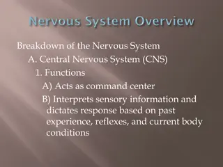
Functional Anatomy of the Nervous System The CNS is made of the brain & the spinal cord. The brain is the higher center that receives information (stimuli) from the external environment & internal body tissues, analyzes them & makes the proper response. The brain is also responsible for consciousness, thinking, emotions, & many other functions. The spinal cord is the part of CNS that connects the brain to the spinal nerves (so, all information received by the spinal nerves reach the brain through the spinal cord, & all commands from the brain reach the spinal nerves through the spinal cord). In addition, the spinal cord is responsible for many nervous "reflexes".
Functional Anatomy of the Nervous System The PNS is made of the cranial nerves & the spinal nerves. The cranial nerves are connected to the brain directly, & are distributed mainly in the head & neck. The spinal nerves are connected to the brain indirectly via the spinal cord, & are distributed in the neck & the rest of the body. The autonomic nervous system (ANS) is part of the PNS. However, its function is controlled by the CNS. It is divided into sympathetic, parasympathetic, & enteric nervous systems. The fibers of the ANS are distributed through the PNS (both spinal & cranial nerves). The ANS regulates the involuntary body activities (eg: heart beats, respiration, secretion & movement of the GIT, etc).
Anatomy of the Central Nervous System (CNS) THE BRAIN The brain is the organ situated in the cranial cavity of the skull, that ensures an ideal protection to its soft structure. The brain is -simply- a collection of billions of neurons, & a larger number of glial cells, arranged in a specific way: the nerve cell bodies are gathered in the outer zone of the brain, forming the "gray matter" or "cortex", while the myelinated axons of those neurons occupy the interior of the brain forming the "white matter".
Anatomy of the Central Nervous System (CNS) This arrangement is inverted in the "brainstem", in which the gray matter lies inside the white matter. The axons forming the white matter are grouped in bundles or "tracts" that originate in certain areas of the cortex & terminate in other regions in the brain or spinal cord. Note the following regarding the brain: The largest parts of the brain are the 2 cerebral hemispheres (cerebrum). Each hemisphere is divided into 4 lobes. The frontal lobe: the anterior part of cerebrum, responsible for voluntary movement, speaking, thinking, emotions, & other functions.
Anatomy of the Central Nervous System (CNS) The responsible language, & other functions. The temporal responsible for memory, hearing, balance, feeling of hunger, & other functions. The occipital lobe: the posterior part of cerebrum, responsible for vision & other functions. The right hemisphere of the brain controls the left side of the body, & vice versa. parietal lobe: sensation, the supero-lateral spatial part of understanding cerebrum, for orientation, lobe: the infero-lateral part of cerebrum,
Anatomy of the Central Nervous System (CNS) The "cerebellum" is the postero-inferior part of the brain. It is responsible for maintaining muscle tone, involuntary planning & control of complex movements, & balance. The "brainstem" is a large midline mass of white matter that connects the cerebrum & cerebellum to the spinal cord. Inside it, the brainstem contains many "nuclei", which are islets of gray matter (collections of nerve cell bodies) that serve the following functions: Sleep/wakefulness control Control of the cranial nerves Function of the respiratory system Function of the cardiovascular system
THE SPINAL CORD The spinal cord is the elongated part of the CNS located inside the vertebral column. Like the brainstem, the spinal cord has white matter outside & gray matter inside. The spinal cord white matter consists of ascending & descending bundles of nerve fibers (tracts) that transport signals from & to the brain. The gray matter of the spinal cord is aggregation of nerve cell bodies arranged as an inner column with H-shape cross section, having 2 anterior horns & 2 posterior horns & a transverse part.
THE SPINAL CORD Note the following regarding the spinal cord: The spinal cord gives rise to 31 spinal nerves on each side. The anterior horns contain the cell bodies of the large motor neurons. The posterior horns contain the cell bodies of the smaller sensory neurons. The spinal cord mediates some "reflexes". A reflex is a sudden involuntary motor action ( = muscles contraction) in response to sudden sensory stimuli. This action is completed at the spinal cord level, without involving the brain.
Anatomy of the Peripheral Nervous System (PNS) CRANIAL NERVES: These are 12 pairs of nerves that are connected directly to the brain, & are responsible for supplying motor & sensory fibers to the head & neck region, & the viscera of the neck, thorax & abdomen. Olfactory nerves (I): small nerve fibers in the upper part of nasal cavity, responsible for olfaction (smelling) Optic nerve (II): the nerve responsible for vision. It gathers signals from the retina of the eye & sends them to the visual cortex in the occipital lobe. Oculomotor nerve (III): the motor supply of the extra-ocular muscles (that move the eyeball) plus the sphincter muscle of the pupil. Trochlear nerve (IV): the motor supply of one extra-ocular muscle (the trochlear muscle)
Anatomy of the Peripheral Nervous System (PNS) Trigeminal nerve (V): the biggest cranial nerve, responsible for the sensory supply of the head. It also gives motor supply to the muscles of mastication (moving the mandible) Abducent nerve (VI): the motor supply of one extra-ocular muscle (the lateral rectus muscle) Facial nerve (VII): the motor supply of facial muscles (of expression). It also supplies the anterior 2/3rd of the tongue with taste sensory fibers. Vestibulo-choclear nerve (VIII): the nerve of hearing & balance, connected to the inner ear. Glosopharyngeal nerve (IX): the motor supply of the pharyngeal muscles, it also supplies sensory & taste sensory fibers to the posterior 1/3rd of the tongue.
Anatomy of the Peripheral Nervous System (PNS) Vagus nerve (X): the longest cranial nerve. It has motor fibers (to the larynx & pharynx), sensory fibers, taste sensory fibers, & preganglionic parasympathetic fibers to the viscera of the thorax & abdomen. Accessory nerve (XI): the motor supply of sternocleidomastoid & trapezius muscles. Hypoglossal nerve (XII): the motor supply of the tongue muscles.
SPINAL NERVES These are 31 pairs of nerves connected to the lateral aspect of the spinal cord. They include: 8 cervical, 12 thoracic, 5 lumbar, 5 sacral, & 1 coccygeal nerves. Note the following: Each spinal nerve, after exiting the vertebral canal, divides into ventral & dorsal branches. Lower cervical & upper thoracic spinal nerves form a plexus (the brachial plexus), which gives rise to the nerves of the upper limb. The main nerves of the upper limb are the median nerve (anteriorly), the ulnar nerve (medially) & the radial nerve (posteriorly).
SPINAL NERVES Lumbar & sacral spinal nerves form a plexus (the lumbosacral plexus), which gives rise to the nerves of the lower limb. The main nerves of the lower limb are the femoral nerve (anteriorly), the obturator nerve (medially) & the sciatic nerve (posteriorly). Dermatomes: a dermatome is the area of skin supplied by a specific spinal nerve. In general, dermatomes are arranged as follows: The cervical spinal nerves supply the skin of the back of the head, the whole neck, shoulders, lateral sides of the arm & forearm, & the whole hand. The thoracic spinal nerves supply the skin of medial sides of the arm & forearm, axilla, & the trunk to the level of pelvic bones.
SPINAL NERVES The lumbar spinal nerves supply the skin of the pelvis (except the buttocks) & the whole lower limb (except the posterior aspect & the sole of the foot) The sacral spinal nerves supply the skin of the genitalia, buttocks & the posterior aspect of the lower limb & the sole of the foot. The coccygeal spinal nerve supplies the skin around the anus.
The Autonomic Nervous System (ANS) The autonomic nervous system is a group of nerves & ganglia distributed throughout the body, supplying mainly the smooth muscles, glands, & the viscera of the body. This system controls our involuntary activities related to the different functions of the body systems, eg: heart rate, blood pressure, secretion of the glands, respiration, GIT movements, & many other functions. The autonomic nervous system is divided into the sympathetic & parasympathetic divisions, with each division having the opposite function of the other.
SYMPATHETIC NERVOUS SYSTEM Anatomically, the sympathetic system is made of sympathetic ganglia connected to sympathetic nerves. Note the following: Sympathetic ganglia are arranged as 2 sympathetic chains, one on each side of the vertebral column. In addition, there are few sympathetic ganglia in the abdomen. Functionally, the medulla of the adrenal gland is also considered as a large sympathetic ganglion. Sympathetic ganglia receive preganglionic fibers from the spinal cord. Preganglionic neurons secret acetylcholine (ACh) in the ganglion (so they are "cholinergic" neurons)
SYMPATHETIC NERVOUS SYSTEM Sympathetic ganglia give postganglionic fibers to the body tissues & organs. Postganglionic neurons secret noradrenaline (NA) at the target organ (so they are "adrenergic" neurons) Actions mediated by the sympathetic system: increasing heart rate, blood pressure, cardiac output, sweating, relaxation of bronchial smooth muscles (bronchodilation), etc.
PARASYMPATHETIC NERVOUS SYSTEM Anatomically, parasympathetic ganglia connected to parasympathetic nerves. Note the following: Parasympathetic ganglia are located near the target organs. Actions mediated by the parasympathetic system: decreasing heart rate, blood pressure, cardiac output, increasing GIT secretion & movement, contraction of bronchial smooth muscles (bronchoconstriction), etc. the parasympathetic system is made of
Enteric Nervous System (ENS) This is a complex, extensive network of nerve fibers & ganglia spread throughout the gastrointestinal tract (GIT), specifically in the submucosa & muscularis layers of the tract. This system receives innervation from the sympathetic & parasympathetic systems, & controls the sensation, secretion, & movements of the GIT.


")
")