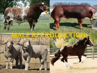
The Male Reproductive System


The Reproductive System
Male reproductive system

•
The Reproductive system includes the
following:
•
Gonads: or reproductive organs that produce
gametes & hormones.
•
Ducts: that receive and transport the gametes.
•
Accessory glands & organs that secrete fluids
(into the same glands or other excretory
ducts).
•
External genitalia.
•
The male and female reproductive systems are
functionally deferent:
•
*in an adult male, the testes or male gonads secrete
sex hormones called androgens (
testosterone
) &
produce
½ billion sperm each day
.(mature sperms
travel along a lengthy duct system, where they are
mixed with secretions of the accessory glands the
mixture is known as semen.
•
*in an adult female the ovaries or female gonads,
typically release
only one immature gamete
(called
oocyte)
per month
, this gamete travels along short
uterine tubes (oviducts) that end in the muscular
uterus. & hormones are
Estrogen & Progesterone
.
The Male reproductive system:
•
In the testes the sperm cells or spermatozoa,
travel within the epididymis→ the ductus
deferens or vas deferens→ the ejaculatory
duct→ &the urethra.
•
Accessory organs: the seminal vesicles, the
prostate gland & the bulbourethral glands
secrete into the ejaculatory ducts & urethra.
The external genitalia consist of the scrotum
(which encloses the testes) & the penis.
Histology of the testes:
•
The testes are subdivided by the
septa
to series of
lobules, and
the seminiferous tubules
(which are tightly
coiled) are distributed among the lobules. Typical
testes contain nearly ½ mile of seminiferous tubules &
the sperm production occurs within these tubules.
•
Each seminiferous tubule is surrounded by delicate
capsule, & loss connective tissue that fills the spaces
between the tubules. Within those spaces are
numerous blood vessels and large
interstitial cells (or
Leydig cells)
, interstitial cells are responsible for the
production of androgens (most important is
testosterone).
•
Sperm cells (or
spermatozoa
) are produced by the
Spermatogenesis
begins at the
outer layer of cells in
the seminiferous tubules & proceed towards the
lumen.
•
Stem cells called
spermatogonia
divided by
mitosis
to
produce generations of daughter cells, some of which
differentiate into spermatocytes, while
meiosis
is a
specialized form of cell division involved only in the
production of gametes (sperms & ovaries).
•
The spermatocytes give rise to the
spermatids
.
•
(at each step in this process, the
daughter cells move
closer to the lumen of the tubule
. The spermatids
differentiate into spermatozoa, this differentiation
process is called
Spermiogenesis
& ends as a physically
mature spermatozoa & spermoigenesis is the last step
in spermatogenesis).
Spermatogenesis
involves three
integrated processes:
1.
Mitosis
2.
Meiosis
3.
Speriogenesis
1.
Mitosis
:
is a process that separates the
duplicated chromosomes
of the original cells
into
two identical nuclei
. The cell division
produces daughter cells which are pushed
toward the lumen of the tubule. These cells
differentiate into spermatocytes that prepare to
begin meiosis.
Mitosis is a part of the process of
somatic cell
division
, which produces two daughter cells
containing
46 chromosomes (23 pairs)
, called the
diploid cells
(because the daughter cells contain
both members of each chromosome pair).
2.
Meiosis
:
is a special form of cell division involved
in
gamete production
(the
gametes contain half
the normal chromosomes
), so the fusion of the
nuclei of a sperm & an ovum produces a cell that
has a normal number of chromosomes (46).
In the seminiferous tubules, the meiotic
divisions of spermatocytes produce spermatids
(
undifferentiated male gametes
).
Meiosis involves two cycles of cell division
(meiosis
I & II
), and produce
four cells
, each
contain
23 individual chromosomes
called
haploid cells
(because these cells contain only one
member of each chromosome). These are the
same in the formation of sperm &ova.
3.
Spermiogenesis
:
spermatids are small & unspecialized
cells
in spermiogenesis the spermatids differentiate
into a
physically mature spermatozoa
. (spermiogenesis
involves major changes in spermatids internal &
external structure). Each spermatid matures into a
single sperm cell or (spermatozoon).
-developing spermatids undergoing spermiogenesis are
not free in the seminiferous tubules but they are
surrounded by the cytoplasm of the
Sustentacular
cells
.
-as spermiogenesis proceeds the spermatids develop the
appearance of mature spermatozoa, then it loses its
attachment to the sustentacular cells & enters the
lumen of the seminiferous tubules by Spermation. The
process from spermatogonial division to Spermation,
takes approximately 9 weeks.
Spermatogenesis & Sustentacular
cells:
•
Sustentacular cells play an important role in
spermatogenesis, these cells have 6 functions
that directly or indirectly affect mitosis,
meiosis & spermiogenesis with in the
seminiferous tubules:
a- Maintenance of the blood – testes
barrier
:
•
the seminiferous tubules are isolated from the general
circulation by a blood – testes barrier.
The sustentacular
cells are joined by tight junctions forming a layer that
divides the seminiferous tubules into an
outer
basal
compartment (contains spermatogonia) and an
inner
luminal compartment (meiosis & spermiogenesis occurs).
•
(transport across the sustentacular cells is
tightly regulated
so that conditions in the luminal compartment remain very
stable
. And the fluid within the seminiferous tubules is
produced by the sustentacular cells which also regulate the
fluids composition(tubular fluid is very different from the
surrounding interstitial fluid).
•
- the blood – testes barrier is essential for
preserving the differences
between the
tubular fluid & interstitial fluid.
•
- developing spermatozoa contain
sperm-
specific antigens
in their cell membrane &
these antigens are not found in the somatic
cell membrane, so they would be attacked by
the immune system if blood – testes barrier
did not prevent being detected.
b- Support of mitosis & meiosis:
•
spermatogenesis depends on the stimulation
of sustentacular cells by circulating follicle-
stimulating hormone (FSH) & testosterone.
•
-stimulated sustentacular cells then promote
the division of spermatogonia & the meiotic
divisions of spermatocytes.
c- Support of spermiogenesis:
•
the sustentacular cells surround & enfold the
spermatids providing nutrients & chemical
stimulation that spermatids providing
nutrients & chemical stimulation that promote
development.
d- Secretion of inhibin:
•
inhibin is a peptide hormone secreted in
response to factors released by the developing
sperm.
•
-inhibin depresses the pituitary production of
(FSH) from pituitary & gonadotropin-releasing
hormone (GnRH) from the hypothalamus.
•
-the faster the rate of sperm production, the
greater the amount of inhibin secreted. So
sustentacular cells provide a feed back control of
spermatogenesis.
e- Secretion of androgen-binding
protein:
•
androgen-binding protein(ABP) binds
androgen(testosterone) in the fluid contents
of the seminiferous tubules, this protein is
important in elevating the concentration of
androgens within the tubules & stimulated
spermiogenesis(the production of ABP is
stimulated by FSH).
f- Secretion of Mullerian-inhibiting
factor:
•
MIF is secreted by sustentacular cells in the
developing tests (this hormone causes
regression of the fetal mullerian duct).
Inadequate MIF production leads to failure of
the testes to descend into the scrotum.
The Reproductive Tract:
•
The testes produce physically mature
spermatozoa (but they are incapable of
successful fertilization). The other proteins of
the male reproductive system are responsible
for the functional maturation (nourishment,
storage & transport of spermatozoa).
The Epididymis:
•
It lies along the posterior border of the testis,
the epididymis has a head, body & a tail.
•
The head receives spermatozoa coming
from the seminiferous tubules.
•
The body is coiled & twisted so to take
very little space.
•
The tail, here the sperms are stored
primarily.
The epididymis has three functions:
1-
it monitors & adjusts the composition of the
tubular fluid (epididymis is lined by a pseudo
stratified columnar epithelium so the cilia
increase the surface area available for
absorption & secretion into the fluid in
tubule).
2-
it acts as a recycling center for damaged
spermatozoa (cellular debris & damaged
spermatozoa are absorbed in the epididymis).
Epididymis functions..cont..
3-
it stores spermatozoa & facilitates their functional
maturation: it takes about 2 weeks for a spermatozoon to
pass through the epididymis, during this period the
spermatozoon completes its functional maturation.
Spermatozoa leaving the epididymis are mature but
they remain immobile. So to become active, motile & fully
functional they must undergo Capacitation which occurs in
two steps:
•
Spermatozoa become motile when mixed with secretions
of seminal vesicles.
•
Spermatozoa become capable of fertilization when exposed
to conditions inside the female reproductive system.
Epididymis functions..cont..
4-
the epididymis secretes a substance that
prevents premature capacitation.
5-
transport along the epididymis involves some
combination of fluid movement & peristaltic
contractions of smooth muscles.
The Ductus Deferens:
•
or vas deferens 40-45cm long begins at the tail of the
epididymis. The vas deferens is part of the spermatic
cord & it inters the abdominal cavity moves along the
lateral surface of the urinary bladder & before the vas
deference reaches the prostate gland & seminal
vesicles its lumen enlarges & called the
ampulla
.
•
The vas deferens contain a thick layer of smooth
muscle which
function in transporting the sperms by
the peristaltic contractions which propel spermatozoa
& fluid along the duct, also the vas deference can store
spermatozoa for several months.
The Accessory Glands:
•
Fluids of the seminiferous tubules & the
epididymis are only 5% of the volume of
semen, so the fluid components of semen is a
mixture of the secretions of many different
glands.
Seminal vesicles, prostate gland & the
bulbourethral glands.
Major functions of male accessory
glands:
1-Activating spermatozoa.
2-Providing the nutrients spermatozoa needs for
motility.
3-Propelling spermatozoa & fluids along the
reproductive tract by peristaltic contractions.
4-Producing buffers that counteract the acidity
of urethral & vaginal contents.
The Seminal Vesicles:
•
The vas deferens ends on each side at the junction
between the ampulla & the duct that drains the
seminal vesicle. The seminal vesicles are extremely
active secretary glands with an epithelial lining that
contains extensive folds. And the seminal vesicles
contribute about
60 %
of the volume of semen. The
composition of the secretion contains:
1- High concentrations of fructose (which is easily
metabolized by spermatozoa).
2- Fibrinogen (which form a temporary clot after
ejaculation).
•
Secretion of the seminal vesicles are slightly
alkaline which helps neutralize acids in the
prostatic secretions & within the vagina.
Inactive but functional spermatozoa begin
beating their flagella when mixed with the
secretions of the seminal vesicles & become
highly mobile.
The Prostate Glands:
•
The prostate gland is a small, muscular, rounded
organ & it encloses the proximal portion of the urethra
as it leaves the urinary bladder. Prostate glands
produce
prostatic fluid a slightly acidic solution
that
contributes
20-30 %
of the volume of semen.
•
The prostatic secretions contain
seminalplasmin
an
antibiotic that may help prevent urinary tract infections
in males. The secretions are ejected into the prostatic
urethra by peristaltic contractions of the muscular wall.
The Bulbourethral Glands:
(or Cowper’s Glands)
•
Are stimulated at the base of the penis, they
secrete a thick,
alkaline mucus
. the secretion
helps neutralization any urinary acids that may
remain in the urethra.
Hormones of the Male Reproductive
Function:
•
The anterior pituitary release Follicle-
Stimulating Hormone(FSH) & Luteinizing
hormone(LH), under the effect of
Gonadotropein Releasing Hormone(GnRH).
=FSH and Spermatogenesis=
•
In males, FSH targets primarily the sustentacular cells in the
seminiferous tubules.
•
Under FSH stimulation & in the presence of testosterone from
interstitial cells, the sustentacular cells:
•
-promote spermatogenesis & spermiogenesis.
•
-secrete androgen-binding protein (ABP).
•
The rate of spermatogenesis is regulated by a negative feed back
mechanism involving GnRH, FSH & inhibin.
•
-FSH levels elevated→ inhibin production ↑es→ FSH levels to
normal.
•
-FSH levels decline → inhibin production falls→ FSH production
accelerate.
=LH & Androgen production=
LH causes the secretion of testosterone by interstitial cells in
testes.
Testosterone
has many functions:
•
Stimulating spermatogenesis & promoting the functional
maturation of spermatozoa.
•
Affecting CNS function.
•
Stimulating metabolism throughout the body specially
protein synthesis & muscle growth
•
Establishing & maintaining the secondary sex
characteristics (facial hair, increased muscle mass).
•
Maintaining the accessory glands of the mail reproductive
tract.
The male reproductive system is a complex network involving gonads, ducts, and accessory glands that function to produce and transport sperm. Testes, the primary male gonads, play a crucial role in sperm production through a process called spermatogenesis. This system functions differently from the female reproductive system, with testosterone and semen production being key components. Understanding the structure and function of the male reproductive system is essential for comprehending the process of human reproduction.

Download Presentation

Please find below an Image/Link to download the presentation.
The content on the website is provided AS IS for your information and personal use only. It may not be sold, licensed, or shared on other websites without obtaining consent from the author.If you encounter any issues during the download, it is possible that the publisher has removed the file from their server.
You are allowed to download the files provided on this website for personal or commercial use, subject to the condition that they are used lawfully. All files are the property of their respective owners.
The content on the website is provided AS IS for your information and personal use only. It may not be sold, licensed, or shared on other websites without obtaining consent from the author.
E N D
Presentation Transcript
The Reproductive System Male reproductive system
The Reproductive system includes the following: Gonads: or reproductive organs that produce gametes & hormones. Ducts: that receive and transport the gametes. Accessory glands & organs that secrete fluids (into the same glands or other excretory ducts). External genitalia.
The male and female reproductive systems are functionally deferent: *in an adult male, the testes or male gonads secrete sex hormones called androgens (testosterone) & produce billion sperm each day.(mature sperms travel along a lengthy duct system, where they are mixed with secretions of the accessory glands the mixture is known as semen. *in an adult female the ovaries or female gonads, typically release only one immature gamete (called oocyte) per month, this gamete travels along short uterine tubes (oviducts) that end in the muscular uterus. & hormones are Estrogen & Progesterone.
The Male reproductive system: In the testes the sperm cells or spermatozoa, travel within the epididymis the ductus deferens or vas deferens the ejaculatory duct &the urethra. Accessory organs: the seminal vesicles, the prostate gland & the bulbourethral glands secrete into the ejaculatory ducts & urethra. The external genitalia consist of the scrotum (which encloses the testes) & the penis.
Histology of the testes: The testes are subdivided by the septa to series of lobules, and the seminiferous tubules (which are tightly coiled) are distributed among the lobules. Typical testes contain nearly mile of seminiferous tubules & the sperm production occurs within these tubules. Each seminiferous tubule is surrounded by delicate capsule, & loss connective tissue that fills the spaces between the tubules. Within those spaces are numerous blood vessels and large interstitial cells (or Leydig cells), interstitial cells are responsible for the production of androgens (most important is testosterone).
Sperm cells (or spermatozoa) are produced by the Spermatogenesis begins at the outer layer of cells in the seminiferous tubules & proceed towards the lumen. Stem cells called spermatogonia divided by mitosis to produce generations of daughter cells, some of which differentiate into spermatocytes, while meiosis is a specialized form of cell division involved only in the production of gametes (sperms & ovaries). The spermatocytes give rise to the spermatids. (at each step in this process, the daughter cells move closer to the lumen of the tubule. The spermatids differentiate into spermatozoa, this differentiation process is called Spermiogenesis & ends as a physically mature spermatozoa & spermoigenesis is the last step in spermatogenesis).
Spermatogenesis involves three integrated processes: 1. Mitosis 2. Meiosis 3. Speriogenesis
1. Mitosis: is a process that separates the duplicated chromosomes of the original cells into two identical nuclei. The cell division produces daughter cells which are pushed toward the lumen of the tubule. These cells differentiate into spermatocytes that prepare to begin meiosis. Mitosis is a part of the process of somatic cell division, which produces two daughter cells containing 46 chromosomes (23 pairs), called the diploid cells (because the daughter cells contain both members of each chromosome pair).
2. Meiosis: is a special form of cell division involved in gamete production (the gametes contain half the normal chromosomes), so the fusion of the nuclei of a sperm & an ovum produces a cell that has a normal number of chromosomes (46). In the seminiferous tubules, the meiotic divisions of spermatocytes produce spermatids (undifferentiated male gametes). Meiosis involves two cycles of cell division (meiosis I & II ), and produce four cells, each contain 23 individual chromosomes called haploid cells(because these cells contain only one member of each chromosome). These are the same in the formation of sperm &ova.
3. Spermiogenesis : spermatids are small & unspecialized cells in spermiogenesis the spermatids differentiate into a physically mature spermatozoa. (spermiogenesis involves major changes in spermatids internal & external structure). Each spermatid matures into a single sperm cell or (spermatozoon). -developing spermatids undergoing spermiogenesis are not free in the seminiferous tubules but they are surrounded by the cytoplasm of the Sustentacular cells. -as spermiogenesis proceeds the spermatids develop the appearance of mature spermatozoa, then it loses its attachment to the sustentacular cells & enters the lumen of the seminiferous tubules by Spermation. The process from spermatogonial division to Spermation, takes approximately 9 weeks.
Spermatogenesis & Sustentacular cells: Sustentacular cells play an important role in spermatogenesis, these cells have 6 functions that directly or indirectly affect mitosis, meiosis & spermiogenesis with in the seminiferous tubules:
a- Maintenance of the blood testes barrier: the seminiferous tubules are isolated from the general circulation by a blood testes barrier. The sustentacular cells are joined by tight junctions forming a layer that divides the seminiferous tubules into an outer basal compartment (contains spermatogonia) and an inner luminal compartment (meiosis & spermiogenesis occurs). (transport across the sustentacular cells is tightly regulated so that conditions in the luminal compartment remain very stable. And the fluid within the seminiferous tubules is produced by the sustentacular cells which also regulate the fluids composition(tubular fluid is very different from the surrounding interstitial fluid).
- the blood testes barrier is essential for preserving the differences between the tubular fluid & interstitial fluid. - developing spermatozoa contain sperm- specific antigens in their cell membrane & these antigens are not found in the somatic cell membrane, so they would be attacked by the immune system if blood testes barrier did not prevent being detected.
b- Support of mitosis & meiosis: spermatogenesis depends on the stimulation of sustentacular cells by circulating follicle- stimulating hormone (FSH) & testosterone. -stimulated sustentacular cells then promote the division of spermatogonia & the meiotic divisions of spermatocytes.
c- Support of spermiogenesis: the sustentacular cells surround & enfold the spermatids providing nutrients & chemical stimulation that spermatids providing nutrients & chemical stimulation that promote development.
d- Secretion of inhibin: inhibin is a peptide hormone secreted in response to factors released by the developing sperm. -inhibin depresses the pituitary production of (FSH) from pituitary & gonadotropin-releasing hormone (GnRH) from the hypothalamus. -the faster the rate of sperm production, the greater the amount of inhibin secreted. So sustentacular cells provide a feed back control of spermatogenesis.
e- Secretion of androgen-binding protein: androgen-binding protein(ABP) binds androgen(testosterone) in the fluid contents of the seminiferous tubules, this protein is important in elevating the concentration of androgens within the tubules & stimulated spermiogenesis(the production of ABP is stimulated by FSH).
f- Secretion of Mullerian-inhibiting factor: MIF is secreted by sustentacular cells in the developing tests (this hormone causes regression of the fetal mullerian duct). Inadequate MIF production leads to failure of the testes to descend into the scrotum.
The Reproductive Tract: The testes produce physically mature spermatozoa (but they are incapable of successful fertilization). The other proteins of the male reproductive system are responsible for the functional maturation (nourishment, storage & transport of spermatozoa).
The Epididymis: It lies along the posterior border of the testis, the epididymis has a head, body & a tail. The head receives spermatozoa coming from the seminiferous tubules. The body is coiled & twisted so to take very little space. The tail, here the sperms are stored primarily.
The epididymis has three functions: 1-it monitors & adjusts the composition of the tubular fluid (epididymis is lined by a pseudo stratified columnar epithelium so the cilia increase the surface area available for absorption & secretion into the fluid in tubule). 2-it acts as a recycling center for damaged spermatozoa (cellular debris & damaged spermatozoa are absorbed in the epididymis).
Epididymis functions..cont.. 3-it stores spermatozoa & facilitates their functional maturation: it takes about 2 weeks for a spermatozoon to pass through the epididymis, during this period the spermatozoon completes its functional maturation. Spermatozoa leaving the epididymis are mature but they remain immobile. So to become active, motile & fully functional they must undergo Capacitation which occurs in two steps: Spermatozoa become motile when mixed with secretions of seminal vesicles. Spermatozoa become capable of fertilization when exposed to conditions inside the female reproductive system.
Epididymis functions..cont.. 4-the epididymis secretes a substance that prevents premature capacitation. 5-transport along the epididymis involves some combination of fluid movement & peristaltic contractions of smooth muscles.
The Ductus Deferens: or vas deferens 40-45cm long begins at the tail of the epididymis. The vas deferens is part of the spermatic cord & it inters the abdominal cavity moves along the lateral surface of the urinary bladder & before the vas deference reaches the prostate gland & seminal vesicles its lumen enlarges & called the ampulla. The vas deferens contain a thick layer of smooth muscle which function in transporting the sperms by the peristaltic contractions which propel spermatozoa & fluid along the duct, also the vas deference can store spermatozoa for several months.
The Accessory Glands: Fluids of the seminiferous tubules & the epididymis are only 5% of the volume of semen, so the fluid components of semen is a mixture of the secretions of many different glands. Seminal vesicles, prostate gland & the bulbourethral glands.
Major functions of male accessory glands: 1-Activating spermatozoa. 2-Providing the nutrients spermatozoa needs for motility. 3-Propelling spermatozoa & fluids along the reproductive tract by peristaltic contractions. 4-Producing buffers that counteract the acidity of urethral & vaginal contents.
The Seminal Vesicles: The vas deferens ends on each side at the junction between the ampulla & the duct that drains the seminal vesicle. The seminal vesicles are extremely active secretary glands with an epithelial lining that contains extensive folds. And the seminal vesicles contribute about 60 % of the volume of semen. The composition of the secretion contains: 1- High concentrations of fructose (which is easily metabolized by spermatozoa). 2- Fibrinogen (which form a temporary clot after ejaculation).
Secretion of the seminal vesicles are slightly alkaline which helps neutralize acids in the prostatic secretions & within the vagina. Inactive but functional spermatozoa begin beating their flagella when mixed with the secretions of the seminal vesicles & become highly mobile.
The Prostate Glands: The prostate gland is a small, muscular, rounded organ & it encloses the proximal portion of the urethra as it leaves the urinary bladder. Prostate glands produce prostatic fluid a slightly acidic solution that contributes 20-30 % of the volume of semen. The prostatic secretions contain seminalplasmin an antibiotic that may help prevent urinary tract infections in males. The secretions are ejected into the prostatic urethra by peristaltic contractions of the muscular wall.
The Bulbourethral Glands: (or Cowper s Glands) Are stimulated at the base of the penis, they secrete a thick, alkaline mucus. the secretion helps neutralization any urinary acids that may remain in the urethra.
Hormones of the Male Reproductive Function: The anterior pituitary release Follicle- Stimulating Hormone(FSH) & Luteinizing hormone(LH), under the effect of Gonadotropein Releasing Hormone(GnRH).
=FSH and Spermatogenesis= In males, FSH targets primarily the sustentacular cells in the seminiferous tubules. Under FSH stimulation & in the presence of testosterone from interstitial cells, the sustentacular cells: -promote spermatogenesis & spermiogenesis. -secrete androgen-binding protein (ABP). The rate of spermatogenesis is regulated by a negative feed back mechanism involving GnRH, FSH & inhibin. -FSH levels elevated inhibin production es FSH levels to normal. -FSH levels decline inhibin production falls FSH production accelerate.
=LH & Androgen production= LH causes the secretion of testosterone by interstitial cells in testes. Testosterone has many functions: Stimulating spermatogenesis & promoting the functional maturation of spermatozoa. Affecting CNS function. Stimulating metabolism throughout the body specially protein synthesis & muscle growth Establishing & maintaining the secondary sex characteristics (facial hair, increased muscle mass). Maintaining the accessory glands of the mail reproductive tract.


 are produced by the")
 are produced by the")