Teat Canal Disorders in Dairy Cows: Causes and Procedures
Teat canal disorders such as contract sphincter, enlarged teat orifice, calculus, polyps, and teat orifice occlusion can affect milk flow in dairy cows. Procedures for addressing these issues include local anesthesia, orifice cleaning, enlargement, and removal of obstructions. Images provided depict the conditions and procedures for better understanding.
Download Presentation

Please find below an Image/Link to download the presentation.
The content on the website is provided AS IS for your information and personal use only. It may not be sold, licensed, or shared on other websites without obtaining consent from the author.If you encounter any issues during the download, it is possible that the publisher has removed the file from their server.
You are allowed to download the files provided on this website for personal or commercial use, subject to the condition that they are used lawfully. All files are the property of their respective owners.
The content on the website is provided AS IS for your information and personal use only. It may not be sold, licensed, or shared on other websites without obtaining consent from the author.
E N D
Presentation Transcript
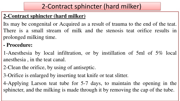
2-Contract sphincter (hard milker) 2-Contract sphincter (hard milker) Its may be congenital or Acquired as a result of trauma to the end of the teat. There is a small stream of milk and the stenosis teat orifice results in prolonged milking time. - Procedure: 1-Anesthesia by local infiltration, or by instillation of 5ml of 5% local anesthesia , in the teat canal. 2-Clean the orifice, by using of antiseptic. 3-Orifice is enlarged by inserting teat knife or teat slitter. 4-Applying Larson teat tube for 5-7 days, to maintain the opening in the sphincter, and the milking is made through it by removing the cap of the tube.
3-Enlarged teat orifice 3-Enlarged teat orifice (free milker or leaker). This occurs due to relaxed or traumatized sphincter, in which the milk is leaks from the teat at times other than milking time and result in milk loss. Procedure: By inject of few drops of sterile mineral oil or lugol's solution around the orifice using 22-26 gauge needle. This it may be repeated more than one time to obtain narrowing and stenosis of the sphincter.
4-calculus 4-lactolith (milk stone, or calculus of the teat canal). Mineral deposition form on a nucleus made the lactolith. lactolith may lodged at the orifice causing obstruction and difficulty in obtaining milk, there may be intermittent free movement of milk flow and obstruction due to the object moving up and down the teat canal during the milking process. Lactolith usually not attached and moved freely and by careful palpation of the teat will reveal a firm, round movable object. Procedure: It may be removed by milking through the orifice when it is small. Or can be crushed with small forceps (mosquito or alligator) and milk out. If it is hard and too large, slit the sphincter as for contract sphincter and removed and after care is as for contract sphincter.
5-Polyps of the teat canal. These found as a pea-sized enlargement protruding from the wall of the teat canal. It may interfere with milk flow, and can be palpated by careful examination, after removing of milk. Procedure: 1-prepare the area of the teat orifice for aseptic surgery. 2-Anesthesia by injection of 5 ml local anesthetic into the teat canal. 3- Insert a tumor extractor or curette through the orifice and remove the polyps from the wall. 4- Milk it out or retrieve it with alligator forceps.
6-Occlusion of teat orifice. 6-Occlusion of teat orifice. It may be congenital or acquired due to trauma to the end of the teat that result in healing with occlusion. Clinical sings: the teat fills with milk at the time of lactation but the teat orifice is absent and no milk is obtained. Procedure: 1-Anesthesia local anesthesia injection into the area. 2-Prepare the area for aseptic surgery. 3-Insert a sterile 15 or 16 gauge hypodermic needle where the opening should be, insert the needle into the teat canal until milk flow is obtained, then without the needle and enlarge the opening as described for contracted sphincter.
