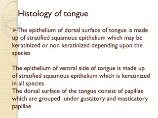
Taste: Gustation and Taste Receptors
Taste
A. Taste (gustation)
1. Taste buds – contain the taste receptors
A) Most are located within the papillae of
the tongue
B) A few are located in soft palate, cheeks,
pharynx, and epiglottis
C) Contain 3 cell types
Taste
1) Gustatory cells – receptor cells; have long
microvilli projections
A) Bind to chemical particles dissolved in
saliva to stimulate taste sensation
2) Supporting cells – most abundant; insulate the
receptors cells from each other
3) Basal cells – at the base of the taste bud
Taste
2. Papillae
A) Raised bumps on the tongue that house the
taste buds & aid in the handling of food
B) 3 types
1) Circumvallate papillae
a) 7 to 12 large, round papillae found on
the back of the tongue in a V pattern
b) Taste buds along the sides of each
papillae
Taste
2) Fungiform papillae
a) Mushroom-shaped papillae scattered over the
surface of the tongue
b) Primarily have taste buds on the tops of each
papillae
3) Filiform papillae – most common
a) Cone-shaped papillae scattered over the entire
tongue surface
b) Have touch receptors rather than taste buds
Taste
3. Taste sensations
A) Salty
1) Stimulated by NaCl and other inorganic
salts
B) Sweet
1) Stimulated by sugars, alcohols,
saccharin, and some amino acids
Taste
C) Sour
1) Stimulated by acids
D) Bitter
1) Stimulated by alkaloids such as nicotine and
caffeine
E) Umami
1) Responsible for the “beef taste” of steak, tang
in aging cheese, and the flavor of MSG
2) Stimulated by the chemical glutamate
Taste
4. Impulse pathway
A) Taste bud (gustatory receptor cell)
1) Gustatory hair
a) Binds to chemical particles dissolved
in saliva
b) Generates impulse
B) Facial (VII), glossopharyngeal (IX) or
vagus (X) nerves
Taste
C) Medulla oblongata
D) Thalamus
E) Primary gustatory area in parietal lobe
5. Taste is 80% smell; inhibited by blocked
olfactory receptors
Smell
B. Smell (olfaction)
1. Olfactory epithelium – patch of receptors
located on the superior portion of the nasal
cavity; 3 cell types
A) Olfactory receptor cells
1) Bowling-pin shaped cells
2) 10-100 million
Smell
3)
Contain olfactory cilia that detect
chemical particles dissolved in air
4) Synapse with olfactory nerves (I)
B) Supporting cells – surround & insulate the
receptor cells
C) Basal cells – line base of olfactory epithelium
Smell
2. Pathway
A) Olfactory receptors
1) Olfactory cilia – on dendritic end of
receptor cells
a) Binds with chemical particles dissolved
in air
b) Generates impulse
Smell
B) Olfactory nerves (I)
1) Olfactory bulbs
a) Cell bodies & dendrites of olfactory
neurons
2) Olfactory tracts
a) Axons of olfactory neurons
Smell
C) Thalamus
D) Lateral olfactory area – temporal lobe
1) Conscious awareness of smell
E) Orbitofrontal area – frontal lobe
1) Identification & discrimination
Vision
C. Eye
1. About 2.5cm (1 inch) in diameter; lies in
the orbit surrounded by protective fat layer
2. External wall is made up of 3 tunics
(layers)
A) Fibrous tunic – outermost layer made
up of dense CT; 2 regions
1) Sclera – posterior portion; white
portion of the eye
Vision
a) Protects & shapes eyeball; provides
sturdy anchoring site for extrinsic eye
muscles
2) Cornea – anterior portion
a) Translucent to allow light to pass
through
B) Vascular tunic – middle layer of the eyeball; 3
regions
Vision
1) Choroid – highly vascular region
2) Ciliary body – thickened ring of tissue
surrounding the lens
a) Ciliary muscle – alters shape of the lens to
focus light
3) Iris – visible, colored portion of the eye
a) Controls amount of light entering the eye
b) Pupil – opening in the iris
Vision
C) Sensory Tunic (retina) – innermost layer
1) Contains photoreceptor cells
a) Rod cells – sensitive to light and permit
vision in dim light; do not provide sharp
images or color vision
b) Cone cells – allow for color vision
i) Blue cones – 16%
ii) Green cones – 10%
iii) Red cones – 74%
Vision
3. Lens – structure that focuses light on the
photoreceptors; divides the eye into anterior &
posterior segments
A) Aqueous humor – clear liquid similar to
plasma that fills the anterior segment
B) Vitreous humor – clear, jelly-like
substance that fills the posterior segment
Vision
4. Accessory structures
A) Eyebrows – coarse hairs above the eye that
protect the eye from sunlight and perspiration
B) Eyelids – mobile, skin-covered folds that
protect the eye
C) Conjunctiva – transparent mucus membrane
that covers the inner surface of the eyelids and
the anterior surface of the eye
Vision
D) Lacrimal apparatus – glands and ducts that
drain lacrimal fluid (tears) on the surface of
the eye to keep it moist and also to flush the
eye when necessary
5. Extrinsic Eye Muscles
A) Superior rectus – elevates eye (III)
B) Inferior rectus – depresses eye (III)
Vision
C) Lateral rectus – moves eye laterally (VI)
D) Medial rectus – moves eye medially (III)
E) Inferior oblique – elevates eye and turns it
laterally (III)
F) Superior oblique – depresses eye and turns it
laterally (IV)
Vision
6. Impulse pathway
A) Photoreceptors
1) Generate impulse
B) Optic nerve (II)
C) Optic chiasma
D) Optic tract
E) Thalamus
F) Primary visual area – occipital lobe
Vision
7. Physiology of Vision
A) Refraction (bending) of light rays by the
cornea and lens causes the light rays to
come into exact focus onto the retina
B) Once the light stimulates the rods and
cones of the retina, nerve impulses are
generated and sent to the brain via the optic
nerve (II)
Vision
C) All images are inverted (upside down and backwards)
D) Accommodation – the lens must change shapes to
adjust for vision at various distances
1) Near point vision – minimum distance from the
eye than an object can be clearly focused with
maximum effort (4 in)
2) Far point vision – distance beyond which no
change in lens shape (accommodation) is needed
for focusing (20 ft)
Hearing & Equilibrium
D. Ear
1. Outer ear
A) Auricle (pinna, ear lobe)
B) External auditory canal
C) Tympanic membrane (tympanum;
eardrum)
Hearing & Equilibrium
2. Middle ear
A) Ear ossicles
1) Malleus – attached to tympanic
membrane
2) Incus – links malleus and stapes
3) Stapes – attached to oval window
Hearing & Equilibrium
B) Oval window – thin membrane that connects
the stapes to the inner ear (cochlea)
C) Middle ear muscles
1) Tensor tympani – tenses the eardrum to
prevent damage from very loud sounds
2) Stapedius – limits the movement of the
stapes
Hearing & Equilibrium
3. Inner ear
A) 2 main divisions
1) Bony labyrinth – bony outer shell
surrounding the membranous labyrinth
a) Located within the temporal bone
b) Contains perilymph
2) Membranous labyrinth – membranous sacs
within bony labyrinth
a) Contains endolymph
Hearing & Equilibrium
B) 3 regions of bony labyrinth
1) Cochlea – snail-shaped; 3 internal
chambers
a) Scala vestibuli
i) Upper chamber
ii) Contains perilymph
b) Scala tympani
i) Lower chamber
ii) Contains perilymph
Hearing & Equilibrium
c) Cochlear duct (scala media)
i) Middle chamber
ii) Contains endolymph
iii) Also houses the organ of Corti
(a) Contains hearing receptors
Hearing & Equilibrium
2) Vestibule – egg-shaped structure between the
cochlea and semicircular canals
a) Saccule – small duct continuous with
membranous labyrinth of the cochlea
b) Utricle – larger sac continuous with
membranous labyrinth of the semicircular
canals
Hearing & Equilibrium
3) Semicircular canals – 3 tube-shaped canals
(anterior, posterior, lateral)
i) Ampulla – enlargement within the each
canal that contains the crista ampullaris
(dynamic equilibrium receptor)
Hearing
4. Hearing
A) Auricle directs sound waves into external
auditory canal
B) Tympanic membrane vibrates in response to
sound waves
C) Vibrations travel down the malleus, incus, and
stapes
D) Stapes transfers vibrations to oval window
E) Vibrations are transmitted through portions of
the cochlea via perilymph
Hearing
F) Vibrations of the perilymph are transferred to
the endolymph
G) Causes hair cells of the organ of Corti to
vibrate
1) Generate impulses based on the amplitude
and frequency of the vibrations
H) Cochlear branch of vestibulocochlear nerve
(VIII)
Hearing
I) Medulla oblongata
J) Thalamus
K) Primary auditory area – temporal lobe
Equilibrium
5. Equilibrium
A) 2 types
1) Static equilibrium – maintenance of the
body (mainly the head) relative to the
force of gravity
Equilibrium
a) Vestibule
i) Macula – flat patches of epithelium
(a) Hair cells – imbedded in the otolithic
membrane
ii) Otolithic membrane – overlying membrane
b) Movement causes the otolithic membrane to
slide on top of the macula causing the hair
cells to bend
c) Hair cells generate impulses
Equilibrium
d) Vestibular branch of vestibulocochlear nerve
(VIII)
e) Medulla oblongata and pons
f) Cerebellum
Equilibrium
2) Dynamic equilibrium
– maintenance of the body
(mainly the head) in response to sudden
movements such as rotation, acceleration, and
deceleration
a) Semicircular canals
i) Movement causes shifting of endolymph
over crista ampullaris (raised structures
within the ampulla of the membranous
labyrinth)
Equilibrium
ii) Detected by hair cells in crista ampullaris
(a) Generate impulses
b) Vestibular branch of vestibulocochlear nerve
(VIII)
c) Medulla oblongata and pons
d) Cerebellum
Disorders
E. Disorders of the Special Senses
1. Taste
A) Ageusia – loss or impairment of the taste
sense
2. Smell
A) Anosmias – group of disorders resulting in
loss or impairment of smell
1) Usually results from head injuries,
excess nasal inflammation or aging
Disorders
3. Eye/Vision
A) Myopia – “nearsightedness”
1) Typically results from the eyeball being
too
long
so that the focal plane is extended so
the light diverges again
B) Hyperopia – “farsightedness”
1) Typically result from the eyeball being
too
short
so that the focal plane is never reached
Disorders
C) Astigmatism – unequal curvature of the lens (or
cornea) resulting in a refractory problem
D) Cataracts – clouding of the lens that causes the
world to appear distorted
E) Glaucoma – pressure within the eye increases and
compresses the retina and optic nerve
1) Usually occurs if the drainage of aqueous
humor is blocked
2) Results in slow degeneration of visual ability
Disorders
F) Diplopia – double vision
G) Strabismus – “cross-eyed”
H) Colorblindness – congenital lack of one or
more of the cone cell types
1) Inherited as a sex-linked condition
2) Far more common in males (8-10%) than
females
Disorders
4. Ear/Hearing
A) Otitis externa, otitis media, otitis interna –
inflammation of the external, middle, or
internal ear respectively usually caused by a
bacterial infection
B) Otalgia – “earache”
C) Tinnitus – ringing or clicking sounds in the
ears
Disorders
D) Deafness – any hearing loss
1) Conduction deafness – something hampers
sound conduction (example earwax)
2) Sensorineural deafness – results from
damage to the neural structures at any point
from the cochlear hair cells to and including
the auditory cortical cells
Taste, also known as gustation, involves taste buds containing taste receptors that pick up sensations like salty, sweet, sour, bitter, and umami. These taste buds are mostly found on the tongue's papillae, with different types such as circumvallate, fungiform, and filiform papillae. Taste sensations are stimulated by various chemicals, and the impulse pathway involves gustatory hair binding to particles in saliva, leading to nerve signals to the brain. Taste is closely related to smell, with olfactory receptors influencing taste perception.

Download Presentation

Please find below an Image/Link to download the presentation.
The content on the website is provided AS IS for your information and personal use only. It may not be sold, licensed, or shared on other websites without obtaining consent from the author.If you encounter any issues during the download, it is possible that the publisher has removed the file from their server.
You are allowed to download the files provided on this website for personal or commercial use, subject to the condition that they are used lawfully. All files are the property of their respective owners.
The content on the website is provided AS IS for your information and personal use only. It may not be sold, licensed, or shared on other websites without obtaining consent from the author.
E N D
Presentation Transcript
Taste A. Taste (gustation) 1. Taste buds contain the taste receptors A) Most are located within the papillae of the tongue B) A few are located in soft palate, cheeks, pharynx, and epiglottis C) Contain 3 cell types
Taste 1) Gustatory cells receptor cells; have long microvilli projections A) Bind to chemical particles dissolved in saliva to stimulate taste sensation 2) Supporting cells most abundant; insulate the receptors cells from each other 3) Basal cells at the base of the taste bud
Taste 2. Papillae A) Raised bumps on the tongue that house the taste buds & aid in the handling of food B) 3 types 1) Circumvallate papillae a) 7 to 12 large, round papillae found on the back of the tongue in a V pattern b) Taste buds along the sides of each papillae
Taste 2) Fungiform papillae a) Mushroom-shaped papillae scattered over the surface of the tongue b) Primarily have taste buds on the tops of each papillae 3) Filiform papillae most common a) Cone-shaped papillae scattered over the entire tongue surface b) Have touch receptors rather than taste buds
Taste 3. Taste sensations A) Salty 1) Stimulated by NaCl and other inorganic salts B) Sweet 1) Stimulated by sugars, alcohols, saccharin, and some amino acids
Taste C) Sour 1) Stimulated by acids D) Bitter 1) Stimulated by alkaloids such as nicotine and caffeine E) Umami 1) Responsible for the beef taste of steak, tang in aging cheese, and the flavor of MSG 2) Stimulated by the chemical glutamate
Taste 4. Impulse pathway A) Taste bud (gustatory receptor cell) 1) Gustatory hair a) Binds to chemical particles dissolved in saliva b) Generates impulse B) Facial (VII), glossopharyngeal (IX) or vagus (X) nerves
Taste C) Medulla oblongata D) Thalamus E) Primary gustatory area in parietal lobe 5. Taste is 80% smell; inhibited by blocked olfactory receptors
Smell B. Smell (olfaction) 1. Olfactory epithelium patch of receptors located on the superior portion of the nasal cavity; 3 cell types A) Olfactory receptor cells 1) Bowling-pin shaped cells 2) 10-100 million
Smell 3) Contain olfactory cilia that detect chemical particles dissolved in air 4) Synapse with olfactory nerves (I) B) Supporting cells surround & insulate the receptor cells C) Basal cells line base of olfactory epithelium
Smell 2. Pathway A) Olfactory receptors 1) Olfactory cilia on dendritic end of receptor cells a) Binds with chemical particles dissolved in air b) Generates impulse
Smell B) Olfactory nerves (I) 1) Olfactory bulbs a) Cell bodies & dendrites of olfactory neurons 2) Olfactory tracts a) Axons of olfactory neurons
Smell C) Thalamus D) Lateral olfactory area temporal lobe 1) Conscious awareness of smell E) Orbitofrontal area frontal lobe 1) Identification & discrimination
Vision C. Eye 1. About 2.5cm (1 inch) in diameter; lies in the orbit surrounded by protective fat layer 2. External wall is made up of 3 tunics (layers) A) Fibrous tunic outermost layer made up of dense CT; 2 regions 1) Sclera posterior portion; white portion of the eye
Vision a) Protects & shapes eyeball; provides sturdy anchoring site for extrinsic eye muscles 2) Cornea anterior portion a) Translucent to allow light to pass through B) Vascular tunic middle layer of the eyeball; 3 regions
Vision 1) Choroid highly vascular region 2) Ciliary body thickened ring of tissue surrounding the lens a) Ciliary muscle alters shape of the lens to focus light 3) Iris visible, colored portion of the eye a) Controls amount of light entering the eye b) Pupil opening in the iris
Vision C) Sensory Tunic (retina) innermost layer 1) Contains photoreceptor cells a) Rod cells sensitive to light and permit vision in dim light; do not provide sharp images or color vision b) Cone cells allow for color vision i) Blue cones 16% ii) Green cones 10% iii) Red cones 74%
Vision 3. Lens structure that focuses light on the photoreceptors; divides the eye into anterior & posterior segments A) Aqueous humor clear liquid similar to plasma that fills the anterior segment B) Vitreous humor clear, jelly-like substance that fills the posterior segment
Vision 4. Accessory structures A) Eyebrows coarse hairs above the eye that protect the eye from sunlight and perspiration B) Eyelids mobile, skin-covered folds that protect the eye C) Conjunctiva transparent mucus membrane that covers the inner surface of the eyelids and the anterior surface of the eye
Vision D) Lacrimal apparatus glands and ducts that drain lacrimal fluid (tears) on the surface of the eye to keep it moist and also to flush the eye when necessary 5. Extrinsic Eye Muscles A) Superior rectus elevates eye (III) B) Inferior rectus depresses eye (III)
Vision C) Lateral rectus moves eye laterally (VI) D) Medial rectus moves eye medially (III) E) Inferior oblique elevates eye and turns it laterally (III) F) Superior oblique depresses eye and turns it laterally (IV)
Vision 6. Impulse pathway A) Photoreceptors 1) Generate impulse B) Optic nerve (II) C) Optic chiasma D) Optic tract E) Thalamus F) Primary visual area occipital lobe
Vision 7. Physiology of Vision A) Refraction (bending) of light rays by the cornea and lens causes the light rays to come into exact focus onto the retina B) Once the light stimulates the rods and cones of the retina, nerve impulses are generated and sent to the brain via the optic nerve (II)
Vision C) All images are inverted (upside down and backwards) D) Accommodation the lens must change shapes to adjust for vision at various distances 1) Near point vision minimum distance from the eye than an object can be clearly focused with maximum effort (4 in) 2) Far point vision distance beyond which no change in lens shape (accommodation) is needed for focusing (20 ft)
Hearing & Equilibrium D. Ear 1. Outer ear A) Auricle (pinna, ear lobe) B) External auditory canal C) Tympanic membrane (tympanum; eardrum)
Hearing & Equilibrium 2. Middle ear A) Ear ossicles 1) Malleus attached to tympanic membrane 2) Incus links malleus and stapes 3) Stapes attached to oval window
Hearing & Equilibrium B) Oval window thin membrane that connects the stapes to the inner ear (cochlea) C) Middle ear muscles 1) Tensor tympani tenses the eardrum to prevent damage from very loud sounds 2) Stapedius limits the movement of the stapes
Hearing & Equilibrium 3. Inner ear A) 2 main divisions 1) Bony labyrinth bony outer shell surrounding the membranous labyrinth a) Located within the temporal bone b) Contains perilymph 2) Membranous labyrinth membranous sacs within bony labyrinth a) Contains endolymph
Hearing & Equilibrium B) 3 regions of bony labyrinth 1) Cochlea snail-shaped; 3 internal chambers a) Scala vestibuli i) Upper chamber ii) Contains perilymph b) Scala tympani i) Lower chamber ii) Contains perilymph
Hearing & Equilibrium c) Cochlear duct (scala media) i) Middle chamber ii) Contains endolymph iii) Also houses the organ of Corti (a) Contains hearing receptors
Hearing & Equilibrium 2) Vestibule egg-shaped structure between the cochlea and semicircular canals a) Saccule small duct continuous with membranous labyrinth of the cochlea b) Utricle larger sac continuous with membranous labyrinth of the semicircular canals
Hearing & Equilibrium 3) Semicircular canals 3 tube-shaped canals (anterior, posterior, lateral) i) Ampulla enlargement within the each canal that contains the crista ampullaris (dynamic equilibrium receptor)
Hearing 4. Hearing A) Auricle directs sound waves into external auditory canal B) Tympanic membrane vibrates in response to sound waves C) Vibrations travel down the malleus, incus, and stapes D) Stapes transfers vibrations to oval window E) Vibrations are transmitted through portions of the cochlea via perilymph
Hearing F) Vibrations of the perilymph are transferred to the endolymph G) Causes hair cells of the organ of Corti to vibrate 1) Generate impulses based on the amplitude and frequency of the vibrations H) Cochlear branch of vestibulocochlear nerve (VIII)
Hearing I) Medulla oblongata J) Thalamus K) Primary auditory area temporal lobe
Equilibrium 5. Equilibrium A) 2 types 1) Static equilibrium maintenance of the body (mainly the head) relative to the force of gravity
Equilibrium a) Vestibule i) Macula flat patches of epithelium (a) Hair cells imbedded in the otolithic membrane ii) Otolithic membrane overlying membrane b) Movement causes the otolithic membrane to slide on top of the macula causing the hair cells to bend c) Hair cells generate impulses
Equilibrium d) Vestibular branch of vestibulocochlear nerve (VIII) e) Medulla oblongata and pons f) Cerebellum
Equilibrium 2) Dynamic equilibrium maintenance of the body (mainly the head) in response to sudden movements such as rotation, acceleration, and deceleration a) Semicircular canals i) Movement causes shifting of endolymph over crista ampullaris (raised structures within the ampulla of the membranous labyrinth)
Equilibrium ii) Detected by hair cells in crista ampullaris (a) Generate impulses b) Vestibular branch of vestibulocochlear nerve (VIII) c) Medulla oblongata and pons d) Cerebellum
Disorders E. Disorders of the Special Senses 1. Taste A) Ageusia loss or impairment of the taste sense 2. Smell A) Anosmias group of disorders resulting in loss or impairment of smell 1) Usually results from head injuries, excess nasal inflammation or aging
Disorders 3. Eye/Vision A) Myopia nearsightedness 1) Typically results from the eyeball being too long so that the focal plane is extended so the light diverges again B) Hyperopia farsightedness 1) Typically result from the eyeball being too short so that the focal plane is never reached
Disorders C) Astigmatism unequal curvature of the lens (or cornea) resulting in a refractory problem D) Cataracts clouding of the lens that causes the world to appear distorted E) Glaucoma pressure within the eye increases and compresses the retina and optic nerve 1) Usually occurs if the drainage of aqueous humor is blocked 2) Results in slow degeneration of visual ability