St. Edmund Campion - English Jesuit Martyr


By : Dr. H.B. Jaruhar
Assist. Prof., Dept. of Botany , S.P.D. College, Garhwa
Stomata structure and Function
Programme name: B. Sc. Botany
Course name: Morphology and Anatomy
Title:
Stomata
.

STOMATA
•
These are minute openings or pores present in
the epidermal layers of leaves. A stoma ( sing)
is surrounded by a pair of kidney shaped
living Guard cells filled with chloroplast,
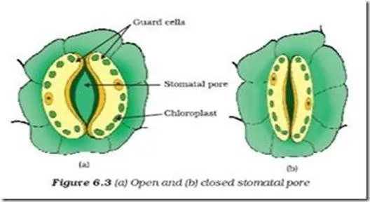
Structure
•
The inner wall of guard cells are thicker than the outer
wall.
•
The guard cells are surrounded by subsidiary cells which
in turn are surrounded by epidermal cells.
Types of Stomata ( on the basis of location)
•
Sunken type : in xerophytic plants the stomata are located in groove or
pits that’s why it is called sunken stomata. These stomata adapted to
minimize the rate of transpiration or conserve the water,
•
Hypostomatic type: these are located on the under surface of the
epidermis of bifacial leaves.
•
Epistomatic: it is just in opposite of hypostomatic, i.e. stomata are
located only on the upper surface of the leaves as in nymphia (lotus)
•
Amphistomatal type: when stomata occures on the both surface it is
called amphistomatic ( amphis= Both).
Types of stomata on the basis of structure
•
1. Ranunculaceous or Anomocytic:
•
Type A — (Anomocytic = irregular celled). In
this type the stoma remains surrounded by a
limited number of subsidiary cells which are
quite alike the remaining epidermal cells. The
accessory or subsidiary cells are five in
number.
contd
•
Cruciferous or Anisocytic:
Type B – (Anisocytic =
unequal celled). In this type stoma remains
surrounded by three accessory or subsidiary cells
of which one is distinctly smaller than the other
two.
•
3. Rubiaceous or Paracytic:
•
Type C – (Paracytic = parallel celled). In this type,
the stoma remains surrounded by two subsidiary
or accessory cells which are parallel to the long
axis of the pore and guard cells.
Contd..
•
4. Caryophyllaceous or Diacytic:
•
Type D – (Diacytic = cross celled)-In this type the stoma
remains surrounded by a pair of subsidiary or accessory cells
and whose common wall is at right angles to the guard cells.
Contd..
•
5. Graminae type:
The gramineous stoma possesses guard
cells of which the middle portions are much narrower than
the ends so that the cells appear in surface view like dump-
bells. They are commonly found in Gramineae and
Cyperaceae family of monocotyledons.
•
6. Coniferous Stomata:
•
They are sunken and appear as though suspended from the
subsidiary cells arching over them. In their median parts the
guard cells are elliptical in section and have narrow lumina. At
their ends they have wider lumina and are triangular in
section. The characteristic of these guard cells is that their
walls and those of the subsidiary cells are partly lignified and
partly non-lignified
Functions of Stomata
•
They are used for the exchange of gases in between the plant
and atmosphere. To facilitate this function, each stoma opens
in a sub-stomatal chamber or respiratory cavity.
•
Evaporation of water also takes place through stomata.
•
The physiology of opening and closing is linked with:
•
A.
the rate of photosynthesis within the guard cells.
•
B.
The translocation of sugar from guard cells to subsidiary
cells and vice versa
•
C.
K ion translocation in the guard cells .
St. Edmund Campion, an English Catholic Jesuit priest and martyr, dedicated his life to serving the persecuted Catholics in England during a time of religious turmoil. He bravely undertook a secret mission to minister to those in fear and persecution, ultimately sacrificing his life for his beliefs. Canonized as a saint in 1970, Campion's legacy of courage and faith continues to inspire. Learn more about his remarkable journey and the impact he had on religious freedom and devotion.

Download Presentation

Please find below an Image/Link to download the presentation.
The content on the website is provided AS IS for your information and personal use only. It may not be sold, licensed, or shared on other websites without obtaining consent from the author.If you encounter any issues during the download, it is possible that the publisher has removed the file from their server.
You are allowed to download the files provided on this website for personal or commercial use, subject to the condition that they are used lawfully. All files are the property of their respective owners.
The content on the website is provided AS IS for your information and personal use only. It may not be sold, licensed, or shared on other websites without obtaining consent from the author.
E N D
Presentation Transcript
By : Dr. H.B. Jaruhar Assist. Prof., Dept. of Botany , S.P.D. College, Garhwa Stomata structure and Function Programme name: B. Sc. Botany Course name: Morphology and Anatomy Title: Stomata .
STOMATA These are minute openings or pores present in the epidermal layers of leaves. A stoma ( sing) is surrounded by a pair of kidney shaped living Guard cells filled with chloroplast,
Structure The inner wall of guard cells are thicker than the outer wall. The guard cells are surrounded by subsidiary cells which in turn are surrounded by epidermal cells.
Types of Stomata ( on the basis of location) Sunken type : in xerophytic plants the stomata are located in groove or pits that s why it is called sunken stomata. These stomata adapted to minimize the rate of transpiration or conserve the water, Hypostomatic type: these are located on the under surface of the epidermis of bifacial leaves. Epistomatic: it is just in opposite of hypostomatic, i.e. stomata are located only on the upper surface of the leaves as in nymphia (lotus) Amphistomatal type: when stomata occures on the both surface it is called amphistomatic ( amphis= Both).
Types of stomata on the basis of structure 1. Ranunculaceous or Anomocytic: Type A (Anomocytic = irregular celled). In this type the stoma remains surrounded by a limited number of subsidiary cells which are quite alike the remaining epidermal cells. The accessory or subsidiary cells are five in number.
contd Cruciferous or Anisocytic: Type B (Anisocytic = unequal celled). In this type stoma remains surrounded by three accessory or subsidiary cells of which one is distinctly smaller than the other two. 3. Rubiaceous or Paracytic: Type C (Paracytic = parallel celled). In this type, the stoma remains surrounded by two subsidiary or accessory cells which are parallel to the long axis of the pore and guard cells.
Contd.. 4. Caryophyllaceous or Diacytic: Type D (Diacytic = cross celled)-In this type the stoma remains surrounded by a pair of subsidiary or accessory cells and whose common wall is at right angles to the guard cells.
Contd.. 5. Graminae type: The gramineous stoma possesses guard cells of which the middle portions are much narrower than the ends so that the cells appear in surface view like dump- bells. They are commonly found in Gramineae and Cyperaceae family of monocotyledons. 6. Coniferous Stomata: They are sunken and appear as though suspended from the subsidiary cells arching over them. In their median parts the guard cells are elliptical in section and have narrow lumina. At their ends they have wider lumina and are triangular in section. The characteristic of these guard cells is that their walls and those of the subsidiary cells are partly lignified and partly non-lignified
Functions of Stomata They are used for the exchange of gases in between the plant and atmosphere. To facilitate this function, each stoma opens in a sub-stomatal chamber or respiratory cavity. Evaporation of water also takes place through stomata. The physiology of opening and closing is linked with: A. the rate of photosynthesis within the guard cells. B. The translocation of sugar from guard cells to subsidiary cells and vice versa C. K ion translocation in the guard cells .


")