South East England General Histopathology EQA Scheme

South East England General Histopathology EQA Scheme holds a case discussion round meeting on Tuesday, 28th November 2023, focusing on case reviews, meeting etiquette, and educational exercises. Attendees gain CPD points, and meeting participation is crucial for feedback and learning.

Download Presentation

Please find below an Image/Link to download the presentation.
The content on the website is provided AS IS for your information and personal use only. It may not be sold, licensed, or shared on other websites without obtaining consent from the author.If you encounter any issues during the download, it is possible that the publisher has removed the file from their server.
You are allowed to download the files provided on this website for personal or commercial use, subject to the condition that they are used lawfully. All files are the property of their respective owners.
The content on the website is provided AS IS for your information and personal use only. It may not be sold, licensed, or shared on other websites without obtaining consent from the author.
E N D
Presentation Transcript
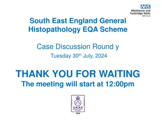
South East England General Histopathology EQA Scheme Case Discussion Round w Tuesday 28thNovember, 2023 THANK YOU FOR WAITING The meeting will start at 12:00pm
Meeting Etiquette 4 3 2 1 Mute your mic if you re not speaking Wait for the Chair person to call on you before you unmute your mic Use the raise hand Or chat feature to raise questions or share ideas If your camera is on, everyone can see you Remember Everyone can see your chat comments 6
Agenda 1. Welcome & Introduction of Scheme Staff 2. Meeting Terms of Reference 3. Case and Preliminary Score Review a) Case 901-911 b) Educational Cases 912-913 4. Questions / comments
This meeting is held between the end of case consultation and results being issued and now replaces the additional final week of the case consultation. This meeting is an educational exercise; an opportunity to explain the reasons behind scoring and merging or why cases were excluded. For clarity, this is not an opportunity to alter merging decisions, as participants have that opportunity during the Case Consultation period. An additional CPD point will be awarded to those who attend, and it will be added to the annual certificate. Please note you have to stay for >50% of the meeting to gain this point (attendance times are monitored automatically by Teams) We always welcome any feedback good or bad you may have about today.
CaseConsultation 158 responses received for round w 92 responses received for consultation 58% QUORATE Thank-you for submitting responses and consultation on time you have made completion of this round much easier for all Basic Rules regarding Case Consultation and Merging Diagnostic categories: If you are exempt from a category, your consultation response to that case is not counted Each case must have received a consultation response from at least 50% of those that answered it For a merge to be automatically accepted, more than 50% of consultation respondents must agree Between 40-50% agreement, the merge will be accepted only with the agreement of the Organiser (i.e. clinically valid). The consensus CAN be over-ridden if there are clinically valid reasons for doing so. These are recorded, and reviewed at the AMR.
Case 901 Endocrine Specimen: Thyroid Submitted Diagnosis: Papillary thyroid carcinoma, encapsulated classic type Submitted Clinical Macro Immuno Image link Preliminary Results Final Merge Results M25.Left thyroid lobe large cystic nodule, THY3F Thyroid lobe 84gr, 74x57x51mm, replaced by a solid / cystic tan nodule with calcifications and thick fibrous capsule. None provided Click here to view digital image 1. Papillary Thyroid carcinoma 9.97 2. Papillary variant of Follicular carcinoma 0.03 73% agreed to no merges
Case 902 Respiratory Specimen: EBUS-TBNA Submitted Diagnosis: Granulomatous inflammation suggestive of sarcoidosis Clinical Macro Immuno Image link Preliminary Results Final Merge Results F73. Mediastinal Lymphadenopathy? Lymphoproliferative disorder? Carcinoma 25ml reddish collection fluid ZN & PAS negative Click here to view digital image 1. Granulomatous Inflammation 6.08 - sarcoid mentioned 2. Granulomatous Inflammation 3.74 - sarcoid NOT mentioned 3. Pneumocystis 0.07 4. Lymphoproliferative disorder 0.07 5. Atypical cells ? Malignant 0.04 (slightly favour SCC) 90% agreed to merge 1 & 2
Case 903 Miscellaneous Specimen: Submandibular mass Submitted Diagnosis: Chronic sclerosing sialadentitis (Kuttner tumour) Clinical Macro Immuno Image link Preliminary Results Final Merge Results M71. left submandibular gland mass 58% agreed to merge 1, 2 & 4. However there has been a CLINICAL OVER-RIDE in this case and diagnoses 7 & 10 will also be merged with the group Cream tan tissue measuring 25 x 20 x 14mm Germinal centres are Bcl2(-) and appear non- colonized. Click here to view digital image 1. Lymphoepithelial sialadenitis / LESA / Sjogren s 3.14 Syndrome 2. Chronic sclerosing sialadenitis 1.66 / Kuttner's tumour 3. IgG4 sialadenitis 1.87 4. Chronic sialadenitis 2.82 5. Mantle cell Lymphoma 0.09 6. Hashimoto s Thyroiditis 0.06 7. Lymphoepithelial lesion (Mikulicz disease) 0.23 8. MAL Toma 0.01 9. Reactive lymphoid infiltrate 0.06 10. Active chronic inflammation ? calculi 0.06 Plasma cells are polyclonal. IgG:lgG4 ratio is <40%
Case 904 Breast Specimen: Breast Submitted Diagnosis: Grade 2, invasive ductal carcinoma Clinical Macro Immuno Image link Preliminary Results Final Merge Results 46% agreed to merge 1 &3. F69. P1, M3/4, U4. Ill-defined mass right breast. 12g. 40 x 35 x 12mm. Spiculate tumour 11mm. ER and PR +, Click here to view digital image 1. Invasive ductal carcinoma 8.20 2. Invasive lobular carcinoma 0.27 3. Breast carcinoma 1.39 4. Invasive solid papillary carcinoma 0.13 5. Neuroendocrine carcinoma 0.01 Her 2 - Please note 3 is the updated terminology
Case 905 Lymphoreticular Specimen: Axillary Mass Submitted Diagnosis: Rosai Dorfman Disease Clinical Macro Immuno Image link Preliminary Results Final Merge Results 78% agreed to no merges F31. Left axillary mass ?Lymphoid ?Adnexal tumour. Fibrofatty tissue with attached skin ellipse measuring 35x33x30mm. Large histiocytes positive for S100, Cyclin D1, and OCT2 and negative for CD1a. CD3 and CD20 show an appropriate mixture of T cell and B cell lymphocytes. CD21 and BCL2 confirm the reactive nature of the lymphoid follicles. Click here to view digital image 1. Rosai-Dorfmann disease 9.71 2.Acute on chronic xanthogranulomarous 0.07 inflammation 3. LCH 0.08 4. Kikuchi Disease 0.07 5. Reactive lymphoproliferative disorder 0.07 On slicing there is a possible tumour measuring 23x16mm which is poorly defined and has a pale white/ cream appearance. Imaging and cytology inconclusive PAS, Grocott and ZN are
Case 906 Gynae Specimen: Products of conception Submitted Diagnosis: Complete hydatidiform mole Clinical Macro Immuno Image link Preliminary Results Final Merge Results F27. Products of conception; miscarriage 5/40. Spongy pieces of tissue 41gms with some vesicles. None provided Click here to view digital image 1. Complete hydatidiform Mole 6.96 2. Partial Hydatidiform Mole 2.00 3. Hydatidiform Mole - subtype not mentioned 0.91 4. POC. No definite molar changes 0.06 5. Hydropic abortus 0.07 51% agreed to merge 1, 2 & 3 Scan indicates ?Molar pregnancy
Case 907 GU Specimen: Kidney Tumour Submitted Diagnosis: Papillary Renal Cell Carcinoma Clinical Macro Immuno Image link Preliminary Results Final Merge Results F62. Right lower pole kidney tumour - partial nephrectomy. 35mm cream coloured tumour with haemorrhagic areas CK7+, CK19+, Vimentin+, Click here to view digital image 1. Papillary Renal Cell carcinoma 9.66 2. Renal Cell carcinoma 0.07 3. Papillary carcinoma. Rule out mets 0.07 (due to IHC) 4. Renal collecting duct carcinoma 0.18 5. Metanephric adenoma 0.01 6. Tubulocystic carcinoma 0.01 59% agreed to merge 1 & 3 CD10 -ve
Case 908 GI Specimen: Appendix Submitted Diagnosis: Endometriosis, no inflammatory process. Clinical Macro Immuno Image link Preliminary Results Final Merge Results F35. Vermiform appendix US proven appendicitis. Appendix 41 long, 8mm diam. Attached fibrofatty tissue 15 x 6 x 4 mm. None provided Click here to view digital image 1. Endometriosis 9.83 2. Benign 0.06 3. Goblet cell carcinoma 0.06 4. Reactive lymphoid hyperplasia 0.01 (inc eosinophils and fat) 5. Cryptosporidosis 0.01 6. MALToma 0.01 7. Eosinophilic appendicitis 0.01 8. Normal / No significant histological 0.01 abnormality 80% agreed to no merges
Case 909 Skin Specimen: Skin Submitted Diagnosis: Apocrine hidrocystoma. Clinical Macro Immun o Image link Preliminary Results Final Merge Results M31. Cyst right jawline. A rounded nodule measuring 30 x 22 x 15mm. No surface skin. Slicing reveals a multiloculated thin walled cyst containing tan fluid. None provided Click here to view digital image 1. (Apocrine) hydrocystoma / apocrine cyst 9.01 2. Papillary eccrine adenoma 0.13 3. Benign (oncocytic) cyst 0.54 4. Intraductal carcinoma 0.13 5. Hidradenoma 0.06 6. Branchial cleft 0.13 53% agreed to merge 1 & 3 Sliced and all embedded.
Case 910 Skin Specimen: Skin Submitted Diagnosis: Pigmented spindle cell naevus of Reed. Clinical Macro Immuno Image link Preliminary Results Final Merge Results M32. Skin type I. Previous sun bed user. Darkly pigmented lesion left leg. None provided None provided Click here to view digital image 1. Naevus - dysplasia not mentioned 0.49 2. Dysplastic naevus - severity not mentioned 3.24 3. Dysplastic naevus - mild/moderate 3.51 4. Dysplastic naevus - Severe 0.93 5. Atypical naevus 0.13 6. (features of a ) Spitz naevus 0.26 7. Melanoma / Melanoma in situ 0.28 8. Dysplastic neavus / melanoma in situ 0.19 9. Solar Lentigo 0.07 10. Reed naevus 0.90 51% agreed to merge 2, 3 & 4. However as the submitted diagnosis is different, THIS CASE WILL BE EXCLUDED FROM PERSONAL SCORING
Case 911 Miscellaneous DIGITAL ONLY Specimen: Submandibular biopsy Submitted Diagnosis: Salivary gland pleomorphic adenoma Clinical Macro Immuno Image link Preliminary Results Final Merge Results F38, Right submandibular lesion. ??Intra-glandular - lobulated irregular borders. ??LN Single core of tissue 15mm long x 1mm in diameter None provided Click here to view digital image 1. Pleomorphic adenoma 9.88 / benign mixed tumour 2. Sialometaplasia 0.06 3. Verrucous hyperplasia 0.06 99% agreed no merges ?SMG lesion. Nil else in neck - no LN. Painful post 1 x biopsy.
Case 912 (EDUCATIONAL) Specimen: Buccal Clinical Macro Immuno Image link Suggested Diagnosis (Top 5) Submitted Diagnosis F81. Longstanding lichenoid lesion on buccal mucosa An ellipse of mucosa 19 x 9 x 7mm with a roughened 'warty' surface. None provided Click here to view digital image 1. Verrucous carcinoma x 52 2. Pseudoepitheliomatous hyperplasia x 22 3. Well differentiated squamous cell carcinoma x 20 4. Verrucous squamous cell carcinoma x 17 5. Proliferative verrucous leukoplakia x 11 Verrucous Hyperplasia
Case 913 EDUCATIONAL Specimen: Skin from back Clinical Macro Immuno Image link Suggested Diagnosis (Top 5) Submitted Diagnosis F19. Cystic lesion of left upper back deep to skin Received as an encapsulated lesion without discernible skin measuring 34x27x20mm. Focally positive for MUC4. Negative for S100, SOX10, AE1/3, Desmin, Myogenin SMA, CD34, CD31, ERG, STAT6 Click here to view digital image 1. Low grade fibromyxoid sarcoma x 124 2. Fibromyxoid sarcoma x 4 3. Low Grade Myxofibrosarcoma x 2 4. Myopericytoma (myofibroma) x 2 5. ANGIOLEIOMYOMA x 2 Low grade fibromyxoid sarcoma Surface is inked green, serially sliced. Serial slicing reveals a white whorled appearance, no areas of haemorrhage or necrosis are seen.
4. Questions Comments Suggestions Feedback Thank you for attending. This presentation can be found on the EQA website from next week.


")