Shock: Definitions, Types, and Management Principles
Shock is a condition characterized by inadequate tissue perfusion, leading to a range of systemic effects. This article outlines the definitions, signs and symptoms, general principles of management, and specific types of shock, including hypovolemic, cardiogenic, neurogenic, anaphylactic, and septic shock.

Download Presentation

Please find below an Image/Link to download the presentation.
The content on the website is provided AS IS for your information and personal use only. It may not be sold, licensed, or shared on other websites without obtaining consent from the author.If you encounter any issues during the download, it is possible that the publisher has removed the file from their server.
You are allowed to download the files provided on this website for personal or commercial use, subject to the condition that they are used lawfully. All files are the property of their respective owners.
The content on the website is provided AS IS for your information and personal use only. It may not be sold, licensed, or shared on other websites without obtaining consent from the author.
E N D
Presentation Transcript
Outlines Definitions Signs and symptoms of shock Classification General principles of management Specific types of shock
Definitions Shock: is a disturbance of circulation leading to inadequate perfusion of vital tissues and with a wide variation of systemic effects. Normal tissue perfusion: requires three intact mechanisms: 1- A functioning pump (heart) 2- Adequate volume (blood and plasma) 3- An intact vascular system
Signs and symptoms 1- confusion and delirium. 2- scared appearance. 3- rapid and shallow respiration. 4- rapid and weak pulse. 5- cold and pale skin. 6- nausea and vomiting. 7- reduced blood pressure.
Classification of Shock by cause
Hypovolemic shock Caused by blood or plasma loss due to: A- Burns. B- Bleeding peptic ulcer. C- Ruptured varices. D- Extensive fractures. E- Crushing accidents. F- External or internal injuries.
Cardiogenic shock Inability of heart to pump needed blood: Causes: 1- myocardial infarction. 2- cardiac arrhythmia. 3- pulmonary embolism. 4- myocardial contusion. Myocardial Infarction
Neurogenic shock Caused by failure of nervous system to control diameter of blood vessels. Causes: 1- severe pain. 2- spinal injury.
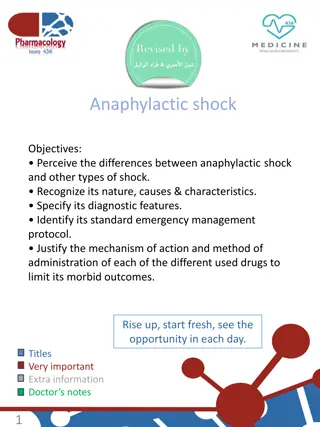
Anaphylactic shock Allergic shock. Functioning blood vessels is disturbed by sensitivity to injection of a foreign substance. Causes: 1- insect stings. 2- certain medicines (antibiotics)
Septic shock Occurs in severe infections. Exotoxins of gram +ve bacteria (staph aureus) Endotoxins of gram ve bacteria (E coli) Exotoxin (cell-protein-heat liable) Endotoxin (cell wall lipid heat stable)
General principles of management
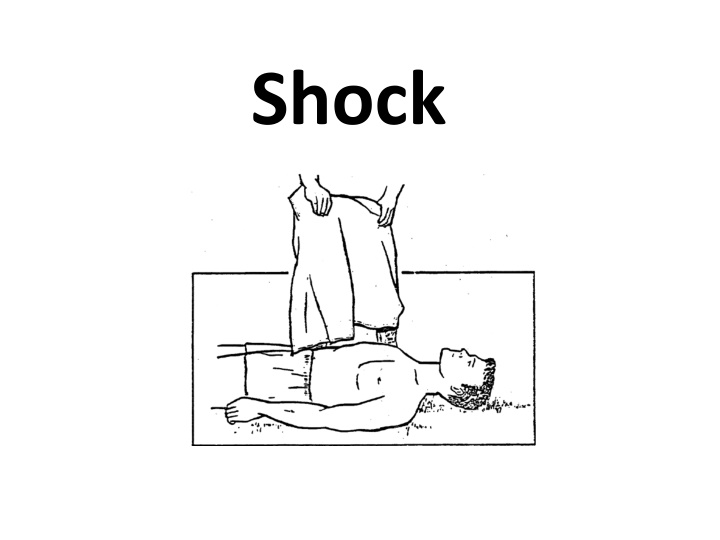
1- Carry standard CPR first aid to maintain an open airway, monitor breathing, and prevent further blood loss. 2- oxygen to assist respiration. 3- position: depending on problem Lower extremity elevation (improve circulation). Supine position in fracture of extremities
Position an unconscious casualty Semi prone Position on his side with his head turned so fluids can drain from his mouth. Maintains airways open and provide best drainage of vomiting.
Cont. 4- control bleeding. 5- keep normal temperature: cover patient with blanket, remove wet clothings. 6- start I.V line. 7- drug therapy according to physician s order. 8- monitor all vital signs. 9- avoid movement (head , spine, neck injury)
Specific types of shock Hypovolemic shock: Replace volume therapy by I.V fluids In massive hemorrhage, more than one I.V line is required.
Cardiogenic shock Continuous oxygen to combat hypoxia. Correct hypovolemia. Appropriate therapy. Measure urine volume (catheter). Monitor arterial blood gases.
Septic shock I.V antibiotic therapy. Continue appropriate therapy after culture and sensitivity results. Anaphylactic shock Ensure open airways. Oxygen administration. Monitor blood pressure. Start I.V line. Hydrocortisone administration. Antihistaminic.
THANK YOU