Regulation of Cdc14 Activity in Budding Yeast during Anaphase
The FEAR (Cdc14 early anaphase release) network and the MEN (mitotic exit) network play vital roles in controlling Cdc14 activity in budding yeast during anaphase. These networks regulate Cdc14 through its association with the inhibitor Net1 (Cfi1) and facilitate Cdc14 activation through sequential processes involving protein degradation, phosphorylation, and dephosphorylation. The coordinated action of these networks ensures proper progression through mitosis and cell division.
Download Presentation

Please find below an Image/Link to download the presentation.
The content on the website is provided AS IS for your information and personal use only. It may not be sold, licensed, or shared on other websites without obtaining consent from the author.If you encounter any issues during the download, it is possible that the publisher has removed the file from their server.
You are allowed to download the files provided on this website for personal or commercial use, subject to the condition that they are used lawfully. All files are the property of their respective owners.
The content on the website is provided AS IS for your information and personal use only. It may not be sold, licensed, or shared on other websites without obtaining consent from the author.
E N D
Presentation Transcript
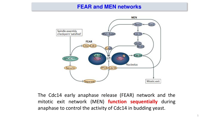
FEAR and MEN networks The Cdc14 early anaphase release (FEAR) network and the mitotic exit network (MEN) function sequentially during anaphase to control the activity of Cdc14 in budding yeast. 1
FEAR and MEN networks Both networks regulate Cdc14 through its cell cycle dependent association with the inhibitor Net1 (Cfi1), which sequesters Cdc14 in the nucleolus. 2
FEAR pathway Esp1 protease of cohesin proteins The FEAR network mediates the initial activation of Cdc14 during early anaphase, when Cdk1 activity is still high. This network is activated by APC/CCdc20 induced proteasomal degradation of securin (also known as Pds1 in budding yeast), leading to the activation of separase (also known as Esp1 in budding yeast) After its release from securin, separase inhibits the phosphatase PP2A Cdc55 by direct binding, independently of its proteolytic activity. This drop in PP2A Cdc55 phosphatase activity allows Cdk1 and the Polo-like kinase Cdc5 to phosphorylate Net1, which then releases active Cdc14 from the nucleolus into the nucleoplasm. Cdc14 promotes anaphase spindle elongation, by dephosphorylating the spindle midzone proteins anaphase spindle elongation 1 (Ase1), required for spindle elongation and stabilization 3
Overview of mitotic exit pathways: APC/C, FEAR, MEN, and RAM Mitotic exit is controlled by a series of systems that both activate (green pointed arrows) and inhibit (red block arrows) one another as cells pass from M into G1 phase: the first to act is the anaphase promoting complex/cyclosome (APC/C) in complex with the protein Cdc20, which initiates degradation of the mitotic cyclins as well as the material that keeps replicated metaphase chromosomes from separating; the second and third mechanisms to act are the Cdc fourteen early anaphase release (FEAR) pathway and the mitotic exit network (MEN). These pathways control Cdc14, which dephosphorylates mitotic CDK substrates, and also directly regulate processes important for productive cell division. Activation of the FEAR and MEN systems convert the APC/C to a different form, a complex with the protein Cdh1; A fourth pathway acts later by driving localization and activation of the transcription factor Ace2, which turns on expression of separation genes, as well as other mechanisms that promote septum destruction. This system also functions in cell morphogenesis control and is thus referred to as the regulation of Ace2 and morphogenesis (RAM) network. 4
FEAR and MEN networks Two regulatory networks the Cdc14 early anaphase release (FEAR) network and the mitotic exit network (MEN) function sequentially during anaphase to control the activity of Cdc14 in budding yeast. Both networks regulate Cdc14 through its cell cycle dependent association with the inhibitor Net1 (Cfi1), which sequesters Cdc14 in the nucleolus. 5
MEN pathway Movement of one of the SPBs into the daughter is permissive for MEN activation A key component of this pathway is the Ras-like small GTPase Tem1, which is positively and negatively regulated by the guanine nucleotide exchange factor (GEF) Lte1 and the bipartite GAP Bfa1 Bub2, respectively MEN components concentrate at the spindle pole body (SPB), the budding yeast centrosome equivalent; the localization of Tem1, Cdc15 and Mob1-Dbf2 to the SBP is critical for the system s activation in response to proper spindle position Tem1 activation depends on correct spindle orientation and elongation, as this brings spindle pole localized Tem1 in proximity to its activator, Lte1, which concentrates at the bud cell cortex 6
MEN pathway Cdc14 activation is sustained by the MEN pathway. Tem1 then activates two kinases, Cdc15 and (Mob1) Dbf2, which either directly or indirectly increase Net1 phosphorylation, leading to sustained activation of Cdc14 and complete reversal of mitotic phosphorylation. The MEN pathway also induces the release of Cdc14 to the phosphorylation of a nuclear localization signal on Cdc14, thereby allowing it to remove Cdk1 phosphorylations on Sic1 and Cdh1. Cdk1 substrate cytoplasm through 7
The polo-like kinase Cdc5: a regulator of both FEAR and MEN The highly conserved polo-like kinase Cdc5 is critical for mitotic exit, and thus for the initiation of mother/daughter separation The amount of Cdc5 present in cells is strongly linked with mitotic progress: it accumulates in S phase, reaches maximum levels in late M phase, and is rapidly de- graded in G1. Like other kinases of this family, Cdc5 has an essential C-terminal domain known as the polo box domain (PBD), which mediates association with S-(pS/pT)-(P/X) motifs in which the central residue is phosphorylated Cdc5 controls both the FEAR pathway and the MEN 8
The APC/C is inactive from late G1 phase until early mitosis, which allows its main substrates to accumulate (securin, polo kinase and S-phase and mitotic cyclins) After cells have entered mitosis, the APC/C is activated by cyclin-Cdk1- dependent phosphorylation enhancing the ability of Cdc20 to bind the APC/C complex APCCdc20 begins the destruction of mitotic cyclins, their complete degradation and persistent instability in the subsequent G1 phase requires the APC/C to associate with the Cdh1 subunit. This complex, known at APCCdh1, completes the inactivation of mitotic cyclin CDK Cdh1 binds APC/C as cells exit mitosis and in interphase One mechanistic reason for the transition between APCCdc20 and APCCdh1 is that activity of APCCdc20 is optimally maintained when cyclin CDK levels are high, while APCCdh1 function is actually antagonized by mitotic CDK phosphorylation Once mitosis is complete, APCCdh1 helps define the G1 state: it turns off mitotic exit by targeting the polo-family protein kinase Cdc5 for destruction in late M or early G1 and destabilizes mitotic cyclins throughout G1 When APC is active, it is imperative that it should not target securin and mitotic cyclins for destruction until all the chromosomes are attached to the mitotic apparatus (SAC, spindle assembly checkpopint) 9
MEN pathway The activation of Cdc14 by the FEAR network is only transient Decreasing Cdk1 activity during anaphase progression cannot sustain high levels of Net1 phosphorylation Cdc14 activation is therefore sustained by the MEN pathway 11
Cellular Cellular reorganization reorganization during during mitotic mitotic progression progression The activity of important mitotic kinases and phosphatases
Model for regulatory networks of PP2A phospatase during vertebrate mitotic exit Phosphorylation levels of cyclin-dependent kinase 1 (CDK1) substrates are governed by the balance of CDK1 and PP2A B55 activities -endosulphine (ENSA) and cyclic AMP-regulated phosphoprotein 19 (ARPP19) are protein phosphatase inhibitor that specifically inhibits protein phosphatase 2A (PP2A) during mitosis Activation of the APC/CCDC20 leads to CDK1 inactivation Then, the activity of Greatwall kinase decreases This results in decreased phosphorylation of ENSA and ARPP19, relieving their inhibition of PP2A B55 Green indicates high kinase or phosphatase activity, whereas pink indicates low activity 13
Phosphatase Phosphatase PP2A PP2A- -B55 B55 during during mitotic mitotic progression progression CDK1-counteracting phosphatases that are distinct from CDC14 have been identified in animal cells Different phosphatases of the PP1 and PP2A families contribute to the reversal of mitotic CDK1-mediated phosphorylation events with several regulatory mechanisms PP2A protein complexes are phosphatases that are abundant in cells and are involved in many processes, including cell growth, differentiation, apoptosis, cell motility, the DNA damage response and cell cycle progression. 14
Phosphatase Phosphatase PP2A PP2A- -B55 B55 during during mitotic mitotic progression progression PP2A active form is composed of one catalytic subunit, one scaffold subunit and one of the many regulatory subunits that provide substrate specificity About 15 regulatory subunits in vertebrates have been classified into four different subunit families B55, B56, B and B In vitro, B55-type regulatory subunits confer specificity of PP2A complexes towards a CDK1 substrate consensus sequence (SerPro or ThrPro), suggesting that PP2A B55 isimportant for mitotic exit in animal cells Drosophila melanogaster larval neuroblasts with mutated B55 show perturbed chromosome segregation. 15
Building new Building new interphase interphase cells cells The structural re-organization that is driven by mitotic phosphoregulation is the breakdown and reassembly of the nuclear envelope in vertebrate cells The nuclear envelope breakdown involves CDK1-dependent phosphorylation of lamin proteins (neclear lamin), which leads to the disassembly of the nuclear lamina (nuclear membranes) At the same time, phosphorylation of nucleoporins mediates disassembly of nuclear pore complexes. During mitotic exit, and PP2A phosphatase is required for timely nuclear envelope reassembly, at the level of lamin and/or nucleoporin dephosphorylation Reassembly of functional nuclei after mitosis also requires chromatin decondensation, which depends on phosphatatase An other example of how substrate phosphorylation controls cellular reorganization during the progression through mitosis is the disassembly and reassembly of the Golgi apparatus, which is driven by mitotic phosphorylation of Golgi stacking proteins and the matrix protein 130 kDa cisGolgi matrix (GM130) Dephosphorylation of GM130 induces Golgi reassembly during mitotic exit and depends on the ubiquitously localized phosphatase PP2A-B55 16
Counteracting Counteracting CDK1 CDK1- -cyclin B cyclin B activity activity CDK1 cyclin B activity is regulated by several mechanisms, including transcriptional control, phosphorylation and intracellular localization The formation of CDK1 cyclin B complexes initiates during interphase through increased synthesis of cyclin B Before mitotic entry, the WEE1 kinase restrains the activity of CDK1 cyclin B by inhibitory phosphorylation at Thr14 and Tyr15 on CDK1 On entry into mitosis, the conserved dual-specificity phosphatase CDC25 (which in mammals has three potentially redundant isoforms, CDC25A, CDC25B and CDC25C) removes these phosphorylations After bipolar attachment of all chromosomes to the mitotic spindle, APCCDC20 targets cyclin B for destruction by the proteasome. In animal cells, this leads to almost complete CDK1 inactivation during early anaphase 17
